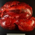
Cystic adrenal masses are uncommon and may be discovered incidentally or may be symptomatic. , Adrenal cysts are classified as pseudocysts, endothelial cysts, epithelial cysts, and parasitic cysts. Pseudocysts (typically the result of hemorrhage into an adrenal neoplasm) are the most common clinically recognized form of adrenal cyst (see Case 43). Cystic adrenal neoplasms must be differentiated from simple adrenal cysts—findings on computed tomography (CT) and magnetic resonance imaging (MRI) are key in making this distinction. Herein we share a case of a patient with an incidentally discovered simple adrenal cyst.
Case Report
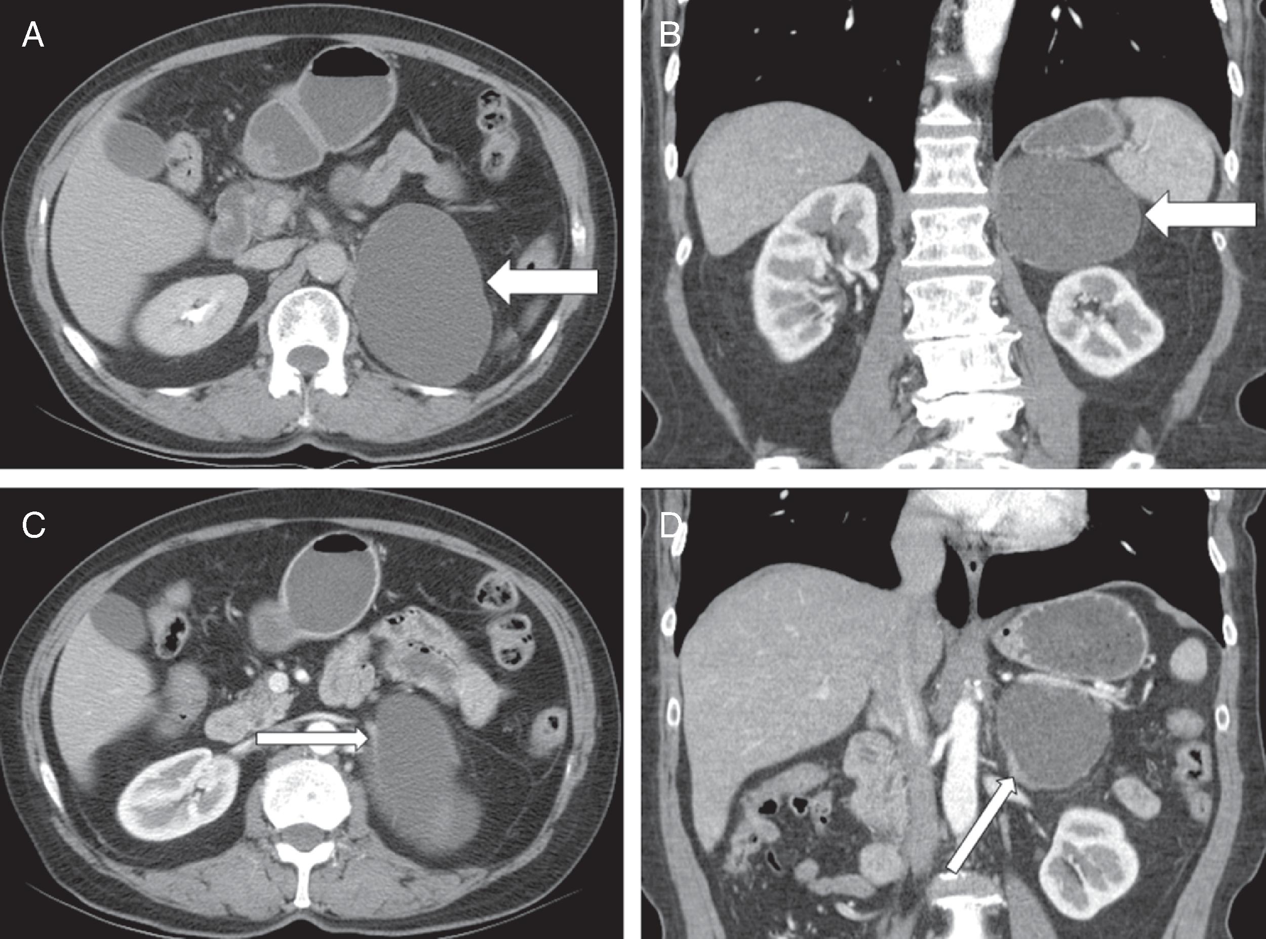
The patient was a 72-year-old woman referred for evaluation of an incidentally discovered left adrenal cyst. The patient had noticed left upper quadrant fullness for a few years. An abdominal CT scan was obtained and showed a well-circumscribed, thin-walled cystic mass (6.0 × 6.7 × 9 cm) in the left suprarenal region ( Fig. 71.1 ). The CT images were consistent with a primary benign adrenal cyst. There was no prior abdominal imaging for comparison. The patient’s sister had been recently diagnosed with pancreatic carcinoma, and our patient was concerned that her left adrenal cyst could be related. She was very active—playing tennis and mowing the lawn. She had a history of normal blood pressure. She had no symptoms related to the apparent adrenal cyst except for the left upper quadrant fullness. The patient took no regular medications. On physical examination she appeared well. Her body mass index was 27.4 kg/m 2 , blood pressure 145/64 mmHg, and heart rate 66 beats per minute. Examination of the abdomen showed no visible asymmetry and the cyst was not palpable.

Stay updated, free articles. Join our Telegram channel

Full access? Get Clinical Tree


