Segmentectomy


Segmentectomy


CRITICAL ELEMENT


  • Patient Selection for Segmentectomy

Recommendation: Segmentectomy should be an option in only select node-negative patients and is dependent on being able to achieve an adequate parenchymal margin.

Type of Data: Retrospective.

Strength of Recommendation: Weak.


Rationale

Sublobar resections are a treatment modality primarily used for early-stage non-small cell lung cancer (NSCLC) in patients with limited pulmonary reserve, where a larger anatomic resection would not be tolerated. Sublobar resections are also currently under prospective randomized evaluation in two independent trials as an alternative treatment strategy for healthy patients with small, peripheral, node-negative tumors. Lobectomy is currently the recommended treatment for stage I and II NSCLC according to the American College of Chest Physicians (ACCP) evidence-based guidelines for the diagnosis and management of lung cancer1 and the National Comprehensive Cancer Network (NCCN) guidelines.2 These recommendations are based primarily on results from the only randomized trial completed to date which compared lobectomy to sublobar resection for stage I NSCLC and noted a threefold increase in local recurrence and trend toward worse survival in those undergoing sublobar resection.3

A sublobar resection for NSCLC is defined as either an anatomic segmentectomy or a wide wedge resection. Segmentectomy is generally considered to be the oncologically superior of the two approaches, typically with wider margins and greater numbers of harvested lymph nodes than wedge resection.4 Segmentectomy is therefore the preferred sublobar approach for primary resection of NSCLC when technically and medically
feasible. A systematic evaluation of the mediastinal lymph nodes is recommended for all sublobar resections performed for NSCLC.

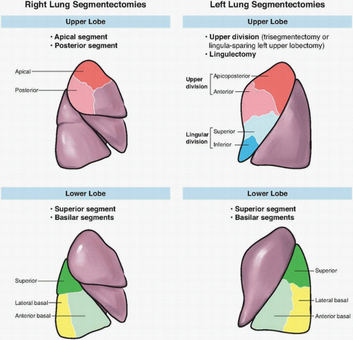
Surgeons undertaking segmentectomy need to be familiar with segmental anatomy. There are 19 pulmonary segments, 10 on the right and 9 on the left, with the anterior basal and medial basal on the left considered a single segment. Although segmental resections have been described for nearly all bronchial segments, generally accepted segmental resections are outlined in Figure 10-1.5,6 Suggested criteria for use of segmentectomy include small tumors (<3 cm), location in the peripheral one-third of the lung, and no endobronchial involvement. Such conditions allow for adequate surgical margins.7,8 Careful review of the chest computed tomography (CT) is suggested preoperatively. This allows the surgeon to ascertain if the tumor is confined within the segmental boundary and appropriate for sublobar techniques.5,6,9,10 It is also helpful for determining the adequacy of margins and identification of aberrant anatomy. Flexible bronchoscopy is indicated to ensure absence of endobronchial disease and delineating bronchial anatomy prior to division.
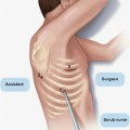
10 General anesthesia with single lung ventilation is beneficial to maintain a quiet operative field for open
approaches and is mandatory to obtain the pneumothorax required for minimally invasive approaches. Lateral decubitus positioning with flexion is recommended. Thoracotomy, video-assisted thoracoscopic surgery (VATS), or robotic-assisted approaches are all acceptable for sublobar resections, and the approach should not alter the technical aspects of the procedure.6 The optimal approach depends on the patient and tumor characteristics and the surgeon’s expertise. The efficacy of minimally invasive techniques for wedge resection has been long accepted.11 Numerous nonrandomized trials and single institution case series have demonstrated equivalent short- and long-term oncologic outcome for segmental resections by VATS and open techniques.12,13,14,15,16,17,18,19 Although no prospective randomized trials comparing the different approaches have been completed, limited evidence suggests VATS may be better tolerated with reduced pain and length of stay and greater postoperative independence.19,20,21,22,23,24 The greatest benefit from minimally invasive approaches appears to be in those patients with severe medical comorbidity. Data from the Society of Thoracic Surgery General Thoracic Surgery Database suggest significant reductions in perioperative morbidity and mortality with minimally invasive approaches to lung resections in high-risk population.25,26 The robotic approach is the most novel and has the least evidence for efficacy for segmental resections, but a recent series reports safety and feasibility for the approach.27


FIGURE 10-1 Pulmonary segments amenable to anatomic resection.


Intraoperative Technical Suggestions

Segmentectomy is an anatomic resection, defined by removal of one or more of the pulmonary parenchymal segments and their corresponding bronchovascular supply.2,3,6,7 By definition, segmentectomy requires individual ligation of the segmental vein, artery, and bronchus and parenchymal division through the intersegmental fissure.

Surgeons should begin with the hilar dissection and removal of hilar and segmental lymph nodes to allow for identification of the segmental bronchovascular structures and to provide adequate length for their individual ligation. As stressed previously, frozen section assessment of these lymph nodes is essential, as this procedure is only indicated for node-negative patients. The segmental vein is typically divided first, especially in minimally invasive approaches, because it facilitates exposure of the segmental artery and bronchus.3,6,7 The techniques for identification and the order of division of the artery and bronchus vary with the individual segment being resected.

The parenchyma is typically divided last with a number of the techniques being utilized for anatomical demarcation. Techniques include following the line of the intact adjacent vein,9 providing gentle traction on the divided segmental vein to delineate plane,5 partial ventilation of remaining lung after division of the segmental bronchus,5,9 and partial ventilation of the lung, prior to stapling, then reisolation with the stapler or clamp across the bronchus to leave resected segment inflated.10 Parenchymal division can be completed with a stapling device, electrocautery, or by using energy-based coagulative fusion technology (LigaSure, Valley Labs, Boulder, CO). There are limited data comparing the different techniques of parenchymal division, but there appear to be equivalent results with the various techniques, and no clear recommendation in favor of one technique can be made.28 The parenchymal division plane should be inspected for large air leaks and a pleural drainage tube is recommended. The most important aspect of parenchymal division is maintaining a minimum distance of 1 cm or larger diameter of the tumor should be maintained between the tumor and the parenchymal division line for oncologic efficacy.25,26,27 The evidence for decreased local control with narrow resection margins is outlined in Table 10-1.


TABLE 10-1 Evidence for Decreased Local Control with Narrow Resection Margins


Author


Year


Type of Study (Prospective/Retrospective, Case Series/Cohort/Case Control/RCT/Modeling Study/Propensity Core Matches Analysis—e.g., Retrospective Case Series, Like Most of Our Literature, or Prospective RCT)


Number and Type of Resection


Definition of Close Margin


Local Recurrence Rate


Key Finding


Significant Potential for Bias (Yes/No)


Close


Wide


El-Sherif32


2007


Retrospective case series


55 wedge 26 segment


<1 cm


14.6%


7.5%


Margins <1 cm are at high risk for locoregional recurrence.


Wedge more frequently associated with a close margin than segmentectomy


Schuchert8


2007


Retrospective case series


182 segment


M/T ratio ≤1


25%


6.2%


M/T ratio <1 is associated with higher rate of recurrence.


Recurrences not separated between locoregional and systemic


Sienel33


2007


Retrospective case series


49 segment


<1 cm


23%


0%


Local recurrence following segmentectomy is associated with resection margins <1 cm.


Sawabata34


2012


Retrospective case series


37 wedge


M/T ratio ≤1


38.5%


0%


All local recurrences occurred in cases of lung with M/T <1.


Not all patients with stage I disease


Mohiuddin35


201


Retrospective case series


367 wedge


<1.5 cm


HR 0.41 (ref margin 0.5 cm)


Risk of local recurrence can be decreased with margins ≥1.5 cm.


All tumors <2 cm


HR, heart rate; M/T, margin distance/tumor width ratio; RCT, randomized controlled trial.


Intraoperative touch prep of the specimen to assess the bronchial margin has been suggested29 as a means of ensuring negative margins and improving oncologic efficacy. A slide is simply run over the specimen after it is removed from the patient but before the specimen is cut by the pathologist. A negative microscopic margin in combination with negative marginal cytology decreases the risk for local recurrence following sublobar resection.30,31 This strategy was evaluated in the recent prospective trial by the American College of Surgeons Oncology Group (ACOSOG) Z40302, which examined the use of intraoperative brachytherapy following sublobar resection, and significant logistical hurdles were encountered, which appear to limit its widespread use of intraoperative touch preps.

Only gold members can continue reading. Log In or Register to continue

Stay updated, free articles. Join our Telegram channel

Jul 22, 2016 | Posted by in ONCOLOGY | Comments Off on Segmentectomy

Full access? Get Clinical Tree

Get Clinical Tree app for offline access