Figure 16.1
Elderly Caucasian male with large eroded lesion on the neck
Differential Diagnosis
Basal cell carcinoma
Squamous cell carcinoma
Amelanotic melanoma
Keratoacanthoma
Sebaceous carcinoma
Merkel cell tumor
Biopsy Results
Sebaceous carcinoma: lesion extends to the deep margin.
Diagnosis
Sebaceous carcinoma
Microscopic Features
Under the microscope, sebaceous carcinoma appears as irregular lobular arrangements of cells of various sizes and with varying levels of differentiation. Wolfe et al. categorized sebaceous carcinomas based on their grade of differentiation. Well-differentiated cells with foamy cytoplasm were categorized as Grade 1 (Fig. 16.2) [1]. Undifferentiated cells with little cytoplasm were categorized as Grade 4. Nuclear atypia is observed with oval nuclei, prominent nucleoli, and high mitotic rates [2]. Sebaceous carcinoma classically demonstrates intra-epithelial or Pagetoid spread.
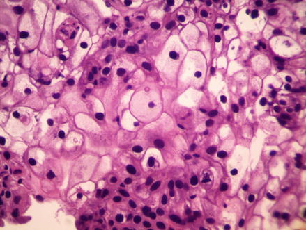
Figure 16.2
H&E. 400× magnification. Sheets of tumor cells with highly variable shapes and sizes. Some have a foamy cytoplasm while others have scanty eosinophilic cytoplasm. There is also variability of the nuclei
Discussion
Sebaceous carcinoma is a rare and extremely aggressive tumor. There are approximately 200 cases reported in the literature. In the past, these tumors have been divided into periocular and extraocular cases. The majority of sebaceous carcinomas, approximately 75 %, occur around the orbit. They arise from the Meibomian glands, Zeis glands, and the sebaceous glands of the eyebrow [2]. They are found more commonly on the upper eyebrow area. Sebaceous carcinomas found on both the upper and lower eyelids portend a poor prognosis [4]. Sebaceous carcinomas are often seen in Muir-Torre syndrome, so it is important to screen patients so that this diagnosis can be ruled out.
Related posts:
 Zosteriform Cutaneous Metastasis
Metastatic Cutaneous Adenocarcinoma
Adenocystic Carcinoma
Balloon Cell Nevi and Balloon Cell Melanomas: What Are They?
The Keystone Design Perforator Island Flap: An Easy Option for the Lower Limb, But How Does It Actually Work?
Rotation Flaps of the Scalp: Study of the Design, Planning and Biomechanics of Single, Double and Triple Pedicle Flaps
Zosteriform Cutaneous Metastasis
Metastatic Cutaneous Adenocarcinoma
Adenocystic Carcinoma
Balloon Cell Nevi and Balloon Cell Melanomas: What Are They?
The Keystone Design Perforator Island Flap: An Easy Option for the Lower Limb, But How Does It Actually Work?
Rotation Flaps of the Scalp: Study of the Design, Planning and Biomechanics of Single, Double and Triple Pedicle Flaps
Stay updated, free articles. Join our Telegram channel

Full access? Get Clinical Tree


