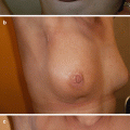
Fig. 6.1
Mammography in a 52-year-old woman with three sisters diagnosed with breast cancer at ages 45, 49 and 55 years old. Digital mammography was performed annually for the past 5 years. Within 9 months since the last mammogram, the patient presented with a palpable mass. Mammography revealed a mass measuring 2.7 × 1.6 cm, with indistinct borders (rightmost panels). Histopathological examination showed a triple-negative breast cancer with one positive sentinel node
6.2 Breast MRI
Breast MRI provides the highest diagnostic sensitivity of the three modalities for screening high-risk women with evidence suggesting that an additional 30% of cancers would have been diagnosed as interval cancers between screening rounds if a multimodality approach had not been employed [16]. ◘ Table 6.1 summarises the results of the main studies [9, 11, 15–21] comparatively examining the sensitivity/specificity of mammography, breast US and MRI (see ◘ Table 6.1).
Table 6.1
Results of the main studies comparatively examining the sensitivity/specificity of mammography, breast US and MRI
Sensitivity % | Specificity % | |||||||||
|---|---|---|---|---|---|---|---|---|---|---|
Studies | Country | Sample size | Age range | No of cancers | MRI | Mammography | Ultrasound | MRI | Mammography | Ultrasound |
Warner (2004) | Canada | 236 | 25–65 | 22 | 77 | 36 | 33 | 95 | 99 | 96 |
Kriege (2004) | The Netherlands | 1909 | 25–70 | 50 | 80 | 33 | Not examined | 90 | 95 | Not examined |
Kuhl (2005) | Germany | 529 | 30+ | 43 | 91 | 33 | 40 | 97 | 97 | 91 |
Leach (2005) | United Kingdom | 649 | 35–49 | 35 | 77 | 40 | Not examined | 81 | 93 | Not examined |
Lehman (2005) | USA – international consortium | 390 | 25+ | 4 | 100 | 25 | Not examined | 95 | 98 | Not examined |
Weinstein (2009) | USA | 609 | 25–80 | 20 | 71 | 33 | 17 | 79 | 94 | 88 |
Kuhl (2010) | Germany | 687 | 25–71 | 27 | 93 | 33 | 37 | 98 | 99 | 98 |
Sardanelli (2011) | Italy | 501 | 25+ | 52 | 91 | 50 | 52 | 97 | 99 | 98 |
Riedl (2015) | Austria | 559 | 22–83 | 40 | 90 | 38 | 38 | 89 | 97 | 97 |
6.2.1 Sensitivity, Specificity and Cancer Detection Rates with MRI Screening
The overall sensitivity of MRI in women at high familial risk varies between different studies from 71% to 100%. Importantly the sensitivity is not modified by the density of the breast [24]. A meta-analysis of 11 studies showed a sensitivity of 77% for the performance of MRI alone and 94% when MRI was combined with mammography, compared to 39% for mammography alone [22]. One of the explanations that has been given regarding the variation between published studies is the unusual MRI imaging features in this group. Frequently, these tumours demonstrate benign kinetic features and often present with non-mass like enhancement [23]. Additionally, these lesions appear with smooth, non-infiltrative borders, without architectural distortion or space-occupying effects on T1- or T2-weighted images. Consequently, the specificity of MRI in these populations varies between 79% and 95%. These particular features give an explanation for the lower detectability of familial breast cancer and emphasises that the significance of this type of finding should not be underestimated when identified [23].
The cancer detection rate reported for MRI alone in different prospective cohort studies ranges from 8.2 to 15.9 per 1000 [9, 11, 16, 18, 22]. Different studies demonstrate similar or increased detection rates in BRCA1 and BRCA2 mutation carriers and their first-degree relatives compared to women with a family history of breast/ovarian cancer with no documented mutation [24]. Additionally, the detection rate of MRI varied among different age groups. Chiarelli and colleagues identified a higher cancer detection rate for MRI alone in women who were over 50 years old compared to women younger than 50 years old [25]. A number of studies have suggested that in high-risk women, MRI has an increased sensitivity in identifying multifocal and multicentric disease, compared to breast ultrasound and mammography [11, 24].
The ability of MRI to detect breast cancers, virtually uninfluenced by the density of breast parenchyma in familial high-risk women [21], led the National Institute for Health and Care Excellence [5] and the GC-HBOC [26] to recommend annual MRI alone, and not in combination with mammography, for familial high-risk women between the ages of 30 and 39, who do not have a prior diagnosis of the disease. It has been recommended by some that the starting age for MRI screening should be adjusted according to the age of diagnosis in the youngest affected relative, with MRI screening commencing 5 years earlier than this age, and according to the type of mutations (BRCA1, BRCA2 or TP53) as age-specific risks vary [27]. UK NICE guidelines advise MRI screening should commence at age 20 in TP53 gene carriers compared to age 30 in BRCA gene carriers [5]. Most recommendations suggest continuing intensified surveillance, including MRI, at least until the age of 50. Nevertheless, Sardanelli and colleagues found that the effectiveness of MRI continues even after the age of 50 [24]. However, cost issues must also be taken into account by health funding agencies when developing guidelines.
6.2.2 Interval Cancer Rates in MRI
The rates of interval cancers in familial high-risk patients undergoing MRI surveillance may be as high as 40% [11]. Annual MRI surveillance is associated with a significant increase in the incidence of smaller size cancers in BRCA2 carriers. However, this was less frequently observed in BRCA1 carriers, where some women presented with palpable interval cancers, between 6 and 12 months after a normal annual screening MRI [28]. This observed difference might be due to the faster rate of tumour growth in BRCA1 carriers, given the fact that BRCA1-associated cancers are frequently triple negative and of basal phenotype, which are usually high grade and with a very high proliferation index [22]. Interestingly, Tilanus-Linthorst and colleagues [29] reported a significant difference in the doubling time of tumours between BRCA mutation carriers and noncarriers. The reported doubling time for carriers was 45 days compared to 84 days for noncarriers [29]. Therefore, some high-risk screening programmes recommend 6-month surveillance with CBE and/or breast ultrasound in addition to regular annual screening with MRI or alternating MRI and mammography every 6 months, so BRCA1 carriers are offered a shorter interval between screening rounds.
6.2.3 Decline in Mortality Rate: Survival Rate
Despite the higher sensitivity of MRI compared to mammography, it remains unclear whether MRI leads to decreased mortality from breast cancer. Up until now, no randomised trial has been designed to directly compare mortality reduction offered by mammography versus MRI; the existing evidence is only indirect and further research is urgently needed.
The estimated indirect benefit that has been achieved from MRI surveillance is derived from downstaging of breast cancer, where the small size and noninvasiveness of breast cancers have been used as reasonable proxy indices for cancer survival [11]. In BRCA1-associated breast cancer diagnosed in an MRI-based surveillance programme, the 10-year survival rate for cancers less than 1 cm was 93%, compared to 58% for cancers measuring from 1 to 2 cm [30]. Documented evidence has shown that BRCA1 tumours are more aggressive and that size is not an ideal criterion for improved survival since these tumours tend to metastasize early and small tumours may often already have positive lymph nodes [31]. According to earlier studies, BRCA1 patients had a 73–74% 5-year survival [32, 33]. Nevertheless, a more recent study reported a 10-year survival equal to 81%; data suggested that once prognostic factors are taken into account, the outcome among carriers is similar to noncarriers [34].
Apart from the high cost, the most important problems regarding breast MRI are its low specificity, high false-positive recall rates and low sensitivity for detecting DCIS; nevertheless, the latter seems of minor importance since DCIS is rare in BRCA 1 carriers. With continual improvements in MRI technology, specificity has improved. In addition, mammography and second look ultrasound substantially increases the specificity of MRI [35]. Modern MRI units, with high field strength, with a minimum standard of at least 1.5 Tesla, a dedicated bilateral breast coil, quality control visits on a regular basis and experienced radiologists are some of the requirements for high-quality MRI screening. In addition, substantial reporting expertise is needed and some health systems recommend double reading to ensure quality. Furthermore MRI examination is not feasible in certain women due to claustrophobia or contraindications, such as pacemakers, metallic implants, morbid obesity (many scanners have a weight limit), inability to lie prone for up to an hour or renal dysfunction precluding the use of contrast agents [36]. Cost prohibits its use for many health economies in the world.
In a recently published work, Kuhl and colleagues introduced an ultra-fast, 3-min, breast MRI for cancer screening. The results of this study showed that ultra-fast breast MRI substantially reduces the time of image acquisition as well as it decreases the reading time and cost of the exam, displaying a comparable sensitivity and specificity to that of conventional MRI in the screening setting [37].
6.3 Mammography
Randomised controlled trials have shown that in general population mammography is the only screening modality that reduces breast cancer-specific morbidity and mortality [38, 39]. The sensitivity of mammography varies depending on the pattern of breast tissue and can range from as high as 98% in fatty breasts to as low as 30–40% in women with dense breasts [40].
Although screening mammography has been suggested in women with high familial breast cancer risk under the age of 50, its efficacy has been disappointing in BRCA carriers [15, 25]. Several studies have demonstrated low sensitivity in BRCA mutation carriers, leading to a high rate of interval cancers, ranging from 29% to 50%, while 40–56% of patients had nodal involvement at the time of diagnosis, and 20–78% of invasive tumours were larger than 1 cm in size [9, 41].
The lower performance of mammography in this group of women compared to the general population has been attributed to several factors; one of them is the early onset of disease associated with these mutations, at a time in a woman’s life when breast density is high. This adversely affects mammographic sensitivity. It should be declared that there is a concern if the benefit of the reduction in the mortality rate among younger carriers outweighs the potential increased risks caused by radiation injury, since implicated genes (such as BRCA1/BRCA2) are involved in the DNA repair mechanism [42]. This is a cause for special concern in TP53 gene carriers, and guidelines generally recommend MRI screening only for such women.
Another study by Kriege and colleagues [15] highlighted the higher sensitivity of mammography compared to MRI for detecting DCIS in women with a familial or genetic predisposition. In contrast, mammography had a lower sensitivity for the detection of invasive cancers (40.0%) compared to the sensitivity of MRI (71.1%); however, the specificity and positive predictive value of MRI in this study were lower than those of mammography.
Consequently, surveillance solely with X-ray mammography in young women with a high familial risk is not adequate, and additional modalities such as MRI or breast ultrasound with high-frequency linear transducers are recommended. The results from published studies are encouraging, as the implementation of a multimodality approach has demonstrated a high performance level in detecting the disease at an earlier stage [43].
Additionally the results of the digital mammographic imaging screening trial (DMIST) suggest that digital mammography could overcome some of the limitations of screen-film mammography [44]. In digital mammography, the X-ray transmission can be varied to enhance the visualisation and conspicuity of subtle anatomical changes that have developed on the background of dense breast parenchyma. Studies have shown the higher sensitivity of digital versus conventional mammography in the detection of microcalcification and subtle masses that have developed in the contour of the breast tissue if the image contrast is adjusted [45]. Therefore, whenever possible, digital mammography should be implemented rather than conventional screen-film mammography for intensified surveillance of women with high familial risk [25, 46].
Due to the limitations of X-ray mammography in this setting, alongside the increased availability and great improvement in MRI, a new strategy for the surveillance of high-risk women younger than 40 has been widely adopted. In 2013 the German Consortium of Hereditary Breast and Ovarian Cancer (GC-HBOC) followed later by the United Kingdom (NHS breast screening programme, NHSBSP) modified their screening guidelines, which suggested not to perform mammography in women under the age of 40 without a prior diagnosis of breast cancer and instead undertake high-quality breast MRI screening [5].
The evolution of digital mammography has opened the pathway for the development of digital breast tomosynthesis (DBT), which provides a series of thin slices covering the entire breast parenchyma and therefore improves breast imaging. Currently, however, no studies have demonstrated the value of replacing digital mammography with DBT in women at high familial risk, and further studies are warranted.
6.4 Breast Ultrasound
Supplemental screening with handheld breast ultrasound after mammography in women with dense breasts increases the detection rate for cancer by 2.7–4.6 per 1000 [47]. Importantly, cancers identified with US have been noted to be particularly small invasive cancers with negative lymph nodes. However, breast ultrasound screening in familial high-risk women, in combination with MRI, has been shown to have only a limited value for the detection of the disease [25, 48].
Handheld ultrasound was not designed for screening but for assisting in the differential diagnosis of a palpable or a mammographically detected lesion. Factors hampering the use of ultrasound as a screening modality include operator dependence, variability between different operators, small field of view with the risk of not scanning the entire breast, shortage of qualified personnel to conduct and interpret the exams, lack of standardisation scanning protocols and false-positive findings [49].
Stay updated, free articles. Join our Telegram channel

Full access? Get Clinical Tree


