Sarcomas


Sarcomas


Samuel Singer

Torsten Nielsen

Cristina R. Antonescu


Soft tissue sarcomas are life-threatening mesenchymal neoplasms that account for approximately 1% of all human cancer. They pose a significant therapeutic challenge because more than 50% of patients with newly diagnosed sarcoma eventually die of disease. Soft tissue sarcomas also pose significant diagnostic challenges as there are more than 50 histologic subtypes with unique molecular, pathologic, clinical, prognostic, and therapeutic features. Figure 26.1 shows the histologic appearance of the major subtypes.

The expansion in the molecular genetic and cytogenetic characterization of soft tissue sarcoma has improved classification and has divided sarcomas into two broad groups: those with simple karyotypes and those with highly complex karyotypes. Figure 26.2 shows the molecular alterations found in some of the subtypes in each group. The first group consists of sarcomas with simple genetic alterations (translocations or specific activating mutations) and with near-diploid, simple karyotypes. These alterations include translocations in myxoid/round-cell liposarcoma and synovial sarcoma, APC or β-catenin mutations in desmoid tumors, and KIT or PDGFRA activating mutations in gastrointestinal stromal tumors (GIST). Translocation-associated sarcomas typically occur in young adults, with highest incidence in the 30s and 40s. For most translocation-associated sarcomas, oncogenesis results from transcriptional deregulation induced by fusion genes. The second group consists of sarcomas with aberrant, highly complex genomes. Examples include dedifferentiated and pleomorphic liposarcoma, leiomyosarcoma, pleomorphic malignant fibrous histiocytoma, and myxofibrosarcoma. The peak incidence for these complex sarcoma types is in the 50s and 60s. Although these complex sarcoma subtypes commonly have alterations in cell-cycle genes TP53, MDM2, RB1, and INK4a and defects in specific growthfactor signaling pathways, the critical subtypespeci fic molecular alterations that drive sarcomagenesis largely remain to be discovered. This information will be essential for the development of therapeutics that can selectively target the driver genetic alterations required for sarcoma survival. This idea is best illustrated by the development of imatinib, a small molecule that inhibits ABL, KIT, and PDGFRA tyrosine kinases. The discovery of activating mutations in KIT and PDGFRA, specifically in GIST, led to rapid clinical development of imatinib for GIST, in which it proved to be an effective, low toxicity therapy. This success illustrates how targeting a sarcomaspecific oncogenic mechanism can lead to dramatic responses.

Table 26.1 outlines the diagnostic histologic characteristics and molecular and cytogenetic abnormalities of the major soft tissue sarcoma subtypes.


TRANSLOCATION-ASSOCIATED SARCOMAS


Myxoid/Round Cell Liposarcoma

Myxoid liposarcoma typically presents in the thigh or other deep soft tissues in adult patients (peak age 30-50 years). The diagnosis can usually be made with confidence based on characteristic morphology: myxoid matrix, plexiform vasculature, and lipoblasts. These features may, however, be partially lost in its high-grade form, termed round cell liposarcoma. The great majority of myxoid/round cell liposarcomas carry a balanced translocation, t(12;16)(q13;p11),1 fusing FUS (also known as TLS) with DDIT3 (aka CHOP, GADD153).2 In rare cases, EWSR1 substitutes for its homologue FUS. At least nine FUS-DDIT3 transcript variants have been reported,3,4 and several are known to be capable of inducing a sarcoma phenotype in model systems.5,6 The translocations fuse 5′ exons of FUS (encoding transcriptional regulatory domains that interact with the RNA polymerase II complex7) to the full coding sequence of DDIT3, a leucine-zipper transcription factor with roles in cell cycle control8 and adipocytic differentiation.9 The fusion oncoprotein complexes with cofactors including C/EBPβ to deregulate gene expression, although few direct targets have been validated to date.10,11 The net result is activation of critical pathways including those related to the angiogenic factor interleukin (IL)-8, early adipose differentiation (PPARγ), growth factor signaling (insulinlike growth factor [IGF], the protooncoprotein RET), and cell-cycle control (cyclinD-CDK4).10,12,13,14


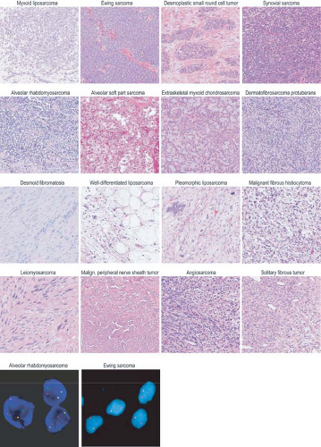
FIGURE 26.1 Sarcoma subtypes discussed in the text. Upper panels, hematoxylin and eosin-stained paraffin sections. Malign., malignant. Lower panels are fluorescence in situ hybridization images showing (left) alveolar rhabdomyosarcoma with fusion of probes for PAX3 (red) and FOXO1 (green) and (right) Ewing sarcoma with break-apart of probes flanking the EWS breakpoint region, EWSR1.


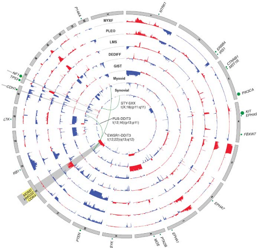
FIGURE 26.2 Nucleotide and copy number alterations in soft tissue sarcoma. The outer ring indicates chromosomal position. The second through fifth rings represent four subtypes with complex karyotypes (as labeled; MYXF, myxofibrosarcoma; PLEO, pleomorphic liposarcoma; LMS, leiomyosarcoma; DEDIFF, dedifferentiated liposarcoma). The three inner rings represent subtypes with simple karyotypes (Myxoid, myxoid/round cell liposarcoma). The plots show the statistical significance of genomic aberrations, with amplification in red and deletion in blue. Green curves indicate the chromosomal breakpoints of pathognomonic translocations in myxoid/round-cell liposarcoma and synovial sarcoma. Genes harboring somatic nucleotide alterations are indicated with green circles whose size is proportional to their frequency of occurrence. (Courtesy of Barry S. Taylor, Computational Biology Center, Memorial Sloan-Kettering Cancer. Adapted from ref. 19.)

Clinically, evidence for FUS-DDIT3 translocations from reverse transcription polymerase chain reaction (RT-PCR)15 or fluorescence in situ hybridization (FISH)16 can help confirm the diagnosis and may be useful for small biopsies dominated by a round cell component. Fusion subtype, however, appears to have little prognostic value beyond what is known from stage and grade. In general, molecular markers in myxoid liposarcoma have been difficult to test for independent prognostic significance, given the difficulty of assembling large series with long follow-up.17 Nevertheless, p53, IGF1R/IGF2, and RET overexpression may be adverse factors.13,18 Such findings support IGF/Akt/mTOR and Ras-Raf-ERK/MAPK pathway inhibitors as potential targeted therapies in myxoid liposarcomas. In addition, mutations in PIK3CA, which were found in 18% of myxoid/round cell liposarcomas, were associated with worse outcome.19


TABLE 26.1 CYTOGENETIC AND MOLECULAR ABNORMALITIES IN SOFT TISSUE SARCOMAS


Disease


Diagnostic Morphology or Immunohistochemistry


Cytogenetic Event


Molecular Abnormality


Molecular Diagnostica


Myxoid/round cell liposarcoma


Lipoblasts, plexiform vasculature, myxoid atrix


t(12;16)(q13;p11)
t(12;22)(q13;q12)


FUS-DDIT3 (>90%)
EWSR1-DDIT3 (<5%)


DDIT3 breaks (FISH)16,187


Ewing sarcoma family tumor


Small, blue, round cells; CD99 and FLI1 expression; lack of lymphoid biomarker expression


t(11;22)(q24;q12)
t(21;22)(q22;q12)
Alternative events: fusions of 22q12 with 7p22, 17q22, 2q33; inv 22q12; t(16;21) (p11;q22)


EWSR1-FLI1 (>80%)
EWSR1-ERG (10-15%)
Other ETS family partners: ETV1, ETV4, FEV, PATZ1 (˜5%) FUS-ERG (<1%)


EWSR1 breaks (FISH)36 or RT-PCR


Desmoplastic small, round cell tumor


Small, blue, round cell islands in dense stroma; positive for keratin, desmin, vimentin, and WT1


t(11;22)(p13;q12)


EWSR1-WT1 (>75%)


EWSR1 breaks (FISH)36


Synovial sarcoma


Biphasic histology, positive for TLE164


t(X;18)(p11;q11) (>90%)


SYT-SSX1 (66%), SYT-SSX2 (33%), SYT-SSX4 (<1%)


SYT breaks (FISH)188


Alveolar rhabdomyosarcoma


Small, blue cells expressing desmin, myogenin, myoD1


t(2;13)(q35;q14)
t(1;13)(p36;q14)


PAX3-FOXO1 (˜80%)
PAX7-FOXO1 (˜20%)
PAX3-NCOA1 (<1%) PAX3-NCOA2 (<1%)


PAX3/7 typespecific FISH or RT-PCR189


Alveolar soft-part sarcoma


Nested polygonal cells in vascular network; positive for TFE378


t(X;17)(p11;q25)


ASPSCR1-TFE3 (>90%)


ASPSCR1-TFE3 RT-PCR190


Dermatofibrosarcoma protuberans


Bland spindle cells, storiform and honeycomb growth in subcutis, positive for CD34


Rings derived from t(17;22) (>75%)
t(17;22)(q22; q13.1)86,87,191 (10%)


COL1A1-PDGFB


Embryonal rhabdomyosarcoma


Spindle cells and rhabdomyoblasts, positive for desmin and myogenin


Trisomies 2q, 8 and 20 (>75%)


LOH at 11p15 (>75%)


Extraskeletal myxoid chondrosarcoma


Bland epithelioid cells arranged in reticular pattern in myxoid stroma


t(9;22)(q22;q12)


EWSR1-NR4A3 (75%)


EWSR1 breaks (FISH); RT-PCR95-97 RT-PCR95-97


t(9;17)(q22;q11)
t(9;15)(q22;q21)
t(3;9)(q12;q22)


TAF15-NR4A3 (<10%)
TCF12-NR4A3 (<10%) TFG-NR4A3 (<5%)


Endometrial stromal tumor


Bland spindle cells, positive for CD10 and ER


t(7;17)(p15;q21)


JAZF1-SUZ12 (30%)


Clear cell sarcoma


Nested epithelioid cells with clear or amphophilic cytoplasm, positive for S100 and HMB-45


t(12;22)(q13;q12)
t(2;22)(q34;q12)


EWSR1-ATF1 (>75%)
EWSR1-CREB1 (<5%)


EWSR1 breaks (FISH)36,192


Infantile fibrosarcoma


Monomorphic spindle cells, herringbone pattern


t(12;15)(p13;q25)


ETV6-NTRK3 (>75%)


FISH, RT-PCR


Inflammatory myofibroblastic tumor


Myofibroblastic cells with lymphoplasmacytic infíltrate, positive for ALK


t(1;2)(q25;p23)
t(2;19)(p23;p13)
t(2;17)(p23;q23)


ALK-TPM34 ALK-TPM ALK-CLTC


ALK breaks (FISH)


Gastrointestinal stromal tumor


Spindle (70%), epithelioid (20%) or mixed (10%) morphology, positive for CD117 (KIT), DOG1, and CD34


Monosomies 14 and 22 (>75%)
Deletion of 1p (>25)


KIT or PDGFRA mutation (>90%)193,194


PCR mutation analysis


Desmoid fibromatosis


Bland myofibroblastictype cells, fascicular growth, nuclear positivity for β-catenin


Trisomies 8 and 20 (30%)


APC inactivation by mutation/deletion (10%)
CTNNB1 (β-catenin) mutations (85%)


IHC for β-catenin expression


Well-differentiated/dedifferentiated liposarcoma


Atypical multinucleated stromal cells, lipoblasts, positive for MDM2, CDK4


12q13-15 rings and giant markers


MDM2 and CDK4
amplification (>85%)


MDM2 amplification (FISH)


Pleomorphic liposarcoma


Pleomorphic spindle and giant cells, pleomorphic lipoblasts


Complexb (>90%)


None


Myxofibrosarcoma and pleomorphic MFH


Pleomorphic spindle and giant cells, storiform growth, variable myxoid stroma


Complexb (>90%)


SKP2 amplification


None


Leiomyosarcoma


Elongated fusiform cells with eosinophilic cytoplasm, in intersecting fascicles, positive for desmin and smooth muscle actin


Complexb (>50%) Deletions of 1p


RB1 point mutations/deletions


None


Malignant peripheral nerve sheath tumor


Monomorphic spindle cells, high mitotic count, geographic necrosis


Complexb (90%)


None


NF1 mutation, loss or deletion (>50%)


None


FISH, fluorescence in situ hybridization; RT-PCR, reverse transcription polymerase chain reaction; LOH, loss of heterozygosity; IHC, immunohistochemistry; MFH, malignant fibrous histiocytoma.


a Refers to molecular tests that can be run on formalin-fixed paraffin-embedded material for molecular confirmation of diagnosis: quantitative RT-PCR of transcripts,189 or FISH to interphase genomic DNA.195

b Complex karyotypes containing multiple numerical and structural chromosomal aberrations.


The dense microvasculature and high levels of IL-810 and vascular endothelial growth factor (VEGF)20 expression seen in this tumor may underlie its observed sensitivity to radiotherapy21 and trabectedin,22 suggesting a value for antiangiogenic therapies. Trabectedin may also function by disrupting the binding of FUS-DDIT3 to target promoters.11


Ewing Sarcoma

Ewing family tumors appear most commonly in adolescents and young adults; primary sites can
be either bone or soft tissues. A range of aggressive small, blue, round cell tumors with variations in clinical and morphologic features have been subsumed under the general term Ewing sarcoma family tumor following the recognition of common pathognomonic chromosomal translocations.23,24 EWSR1, the common 5′ translocation partner, is fused to one of several possible ETS family transcription factor genes (usually FLI1). In the fusion protein, EWSR1 provides, at minimum, its 264 amino acid N-terminal transcriptional regulatory domain, and the ETS factor provides its C-terminal DNA-binding domain. In the process, EWSR1 loses its RNA recognition domain, and the ETS factor loses its native transactivation domain. Several direct transcriptional targets for the fusion oncoprotein are supported by strong evidence. Some of these targets are up-regulated (ID2,25 PTPL1,26 MK-STYX,27 DAX128) and some repressed (CIP1,29 TGFBR2,30 IGFBP331) in Ewing sarcoma, but gene repression events, mediated by cofactors, appear to predominate overall.32 The net result is activation of pathways driving proliferation and cell survival (including IGF signaling33), with concurrent repression of pathways promoting mesenchymal differentiation.34,35

Molecular confirmation of an EWSR1 translocation can be critical for patient management because many of the clinical, morphologic, and immunophenotypic features of Ewing sarcoma are shared with entities such as mesenchymal chondrosarcoma and small cell osteosarcoma. Commercially available EWSR1 split-apart FISH probes are valuable ancillary diagnostic tools (Fig. 26.1).36 RT-PCR alternatives are complicated by the need to cover the many alternative gene and exonic fusion sites, but RT-PCR offers the advantage of identifying the specific exon fusion involved, which can have independent prognostic relevance.37

Several existing agents (including IGF/mTOR and histone deacetylase inhibitors38,39,40) that target recently identified, translocation-induced mechanisms and pathways are currently being tested in clinical trials for Ewing sarcoma, and novel strategies to directly inhibit the oncoprotein itself are in active development.41


Desmoplastic Small Round Cell Tumor

In desmoplastic small round cell tumor, the same 5′ portions of EWSR1 involved in Ewing sarcomas are fused to WT1,42,43 a tumor suppressor deleted in Wilms tumor.44 The 56-kDa EWSR1-WT1 chimeric protein includes the last three of the four WT1 DNA-binding zinc finger domains. Despite some similarities to Ewing sarcoma family tumors, desmoplastic small round cell tumors show little response to conventional chemotherapy. Prognosis is dismal, and new therapies are needed.45 Several transcriptional targets of the EWSR1-WT1 chimeric oncoprotein have been identified.43 PDGFA expression is directly induced by EWSR1-WT1,46 explaining the desmoplastic background and possibly the recent observation of a partial response to sunitinib.47 IL2RB is also induced, and its downstream JAK/STAT signaling pathway appears active in this tumor,43,48 representing a potential target for novel treatment approaches.


Synovial Sarcoma

Synovial sarcoma differs from most translocation-associated sarcomas in that the genes involved in its defining translocation, t(X;18)(p11;q11), encode epigenetic regulators, not transcription factors with direct DNA-binding activity.49 The translocation fuses the widely expressed SYT (aka SS18) gene with an SSX gene normally expressed only in testis.50 In the fusion oncoprotein, SYT, which interacts with components of the SWI/SNF chromatin remodeling complex,51 retains all but the last eight amino acids from its C-terminal transcription activation domain. The SSX partner (SSX1, 2 or 4) retains only its C-terminal 78-residue repressor domain, which confers nuclear localization in association with Polycomb group proteins that mediate chromatin condensation and epigenetic gene silencing.52 The resulting chimeric oncoprotein dysregulates transcription at the level of epigenetic modifications53 and, when forcibly expressed in a mesenchymal stem cell background54 or conditionally expressed in mice,49,55 recapitulates synovial sarcoma. Direct targets of SYT-SSX include the tumor suppressors COM156 and EGR157; both genes are repressed in synovial sarcoma, with the EGR1 promoter undergoing SYT-SSX-dependent histone methylation.57 Thus, transcriptional reactivation agents such as histone deacetylase inhibitors are worth investigation in this disease, as they may reactivate mesenchymal differentiation and reverse some effects of the SYT-SSX oncoprotein.58,59

The synovial sarcoma oncoprotein may also function by disrupting normal interactions between transcription factors and their DNA-binding sites, such as interactions between SLUG/SNAIL and the E-cadherin promoter, leading to transcriptional activation.60 Subtle differences in such interactions may underlie the propensity of SYT-SSX1 translocations, compared to SYT-SSX2 translocations, to be associated with biphasic histology and poorer outcome, although prognostic differences are controversial.61,62 The need for diagnostic molecular translocation testing, however, may be obviated by assays for TLE1, a transcriptional corepressor63 that is highly expressed in synovial sarcoma and serves as a sensitive and specific biomarker.64 Other oncogenic pathways that are activated, directly or indirectly, in synovial sarcoma include IGF2,54,65,66 suggesting
potential value for IGF/Akt/mTOR inhibitors in this disease.


Alveolar Rhabdomyosarcoma

In alveolar rhabdomyosarcoma, an aggressive cancer of older children and adolescents, the transcriptional activation domain of FOXO1 (aka FKHR) from 13q14 is fused to the DNA-binding domain of paired box transcription factor PAX3 (2q35) or PAX7 (1p36).67,68 Until recently, about 20% of cases were thought to be translocationnegative, but recent work has proven that such tumors in fact represent histologic variants of embryonal rhabdomyosarcoma.69 Furthermore, those cases with translocations involving PAX3 have a considerably worse prognosis than those involving PAX7.70 Thus, molecular confirmation of the diagnosis by FISH (Fig. 26.1) and/or RT-PCR is required to optimize patient care.71 Either translocation results in high-level nuclear expression of a chimeric transcription factor that abnormally activates PAX targets, many of which are genes involved in neurogenesis that are not expressed in normal skeletal muscle.72 In addition, PAX3-FOXO1 directly induces PDGFRA; small-molecule (imatinib) and antibody inhibitors of this receptor tyrosine kinase are effective in mouse models.73 Another probable direct target of PAX3-FOXO1, based on comparison of primary tumor and mouse model expression profiles, is the cell-cycle regulator SKP2,74 perhaps helping explain why alveolar rhabdomyosarcoma is responsive to conventional cytotoxic chemotherapy.


Alveolar Soft-Part Sarcoma

Alveolar soft-part sarcoma has a clinical presentation and pathognomonic molecular event with many similarities to other translocation-associated sarcomas.75 In this disease, the 5′ half of the widely-expressed ASPSCR1 (aka ASPL) gene on 17q25 is fused to exon 3 or 4 of TFE3 on Xp11, the latter retaining its transcriptional activation, basic helix-loop-helix, and leucine zipper domains.76 Interestingly, the same fusion is present in some renal cell carcinomas.77 Although alveolar soft-part sarcoma has distinctive histology, translocation detection by RT-PCR or by immunohistochemistry for TFE3 can serve as a diagnostic adjunct.78,79 The disease lacks validated prognostic biomarkers. Direct targets of ASPSCR1-TFE3 are not yet identified, although gene expression profiling and tissue microarray studies have highlighted prominent activation of c-Met signaling and angiogenesis pathways.80,81,82 Antiangiogenic targeted therapy is effective in xenograft models83 and has yielded partial responses in patients with metastatic disease.84


Dermatofibrosarcoma Protuberans

The cytogenetic hallmark of dermatofibrosarcoma protuberans (DFSP) is supernumerary ring chromosomes that contain material from chromosomes 17 and 2285,86,87 or, less commonly, an unbalanced der(22)t(17;22)(q21-23;q13). The molecular consequence of both types of aberration is the overexpression of the platelet-derived growth factor-beta (PDGFB) gene on chromosome 22, through fusion with the collagen gene COL1A1 on chromosome 17.88,89 The same fusion gene is also seen in two histologic variants: giant cell fibroblastoma and Bednar tumor (pigmented DFSP). FISH and comparative genomic hybridization (CGH) studies have indicated that increased COL1A1-PDGFB copy number is associated with fibrosarcomatous transformation of DFSP, although the copy number increase is not an invariable feature of these cases.90,91

May 27, 2016 | Posted by in ONCOLOGY | Comments Off on Sarcomas

Full access? Get Clinical Tree

Get Clinical Tree app for offline access