Figure 9.1
Preoperative Halo graft markings
The skin graft is harvested from the ‘halo’ area i.e., the outer area geometrically called the annulus as shown. I use a No. 22 blade and take shavings of partial thickness skin. The split-skin graft fragments are placed over the recipient defect i.e., the inner circle where the lesion was excised (Fig. 9.2). I dress the graft with a foam dressing (Allevyn, Smith & Nephew, Christchurch, New Zealand), which is placed over the whole wound. 2- layer compression bandages are applied using cotton-wool and crepe and the patient is fully mobilized. The graft dressing is done at 1 week. The wound is usually completely healed in 2–3 weeks. (If the wound appears inflamed or is potentially infected, I use Allevyn Ag i.e., silver-impregnated Allevyn dressing)
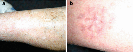

Figure 9.2
Halo graft markings postoperatively showing (a) minimal scarring or contour defect and (b) with less contour defect than a conventional graft
The Geometry
Annulus = A space contained between the circumferences of two circles, one within the other.
If the outer radius is 1.141 times the inner radius, the annulus area becomes 3.14 which equates to π. In other words the outer radius needs to be approximately 1.4 times the inner radius to make the annulus area the same as the area of the inner (recipient) circle.
Case Study 1
A 55-year old female was referred to me with a 2.5 cm BCC on her left leg, overlying the tendo-achilles region. Given the location and orientation of the lesion, primary closure was not feasible. A ‘halo’ split-skin graft was planned. In this technique the area to be excised is marked out (in this case an approximate 3 cm diameter circle). The outer ‘annulus’ area was marked at 1.4 times the inner radius i.e., diameter of the outer circle was 4.2 cm and the radius 2.1 cm). Split skin grafts were harvested from the annulus area and laid on the defect like a jigsaw. Dressings were done in the usual manner and the wound was fully healed at 13 days (Figs. 9.1, 9.2, and 9.3). The patient was mobilized with soft cotton-wool and crepe bandages for compression. The wound was kept dry until the first graft dressing at 7 days.


Figure 9.3
Halo grafts laid onto the defect
Case Study 2
A 61-year old female was referred to me with a 3.1 cm BCC on her left leg, on the lateral aspect of her calf. Given the location and orientation of the lesion primary closure was not feasible. A ‘halo’ split-skin graft was planned. In this technique the area to be excised is marked out (in this case an approximate 4 cm diameter circle). The outer ‘annulus’ area was marked at 1.4 times the inner radius i.e., diameter of the outer circle was 5.6 cm and the radius 2.6 cm). Split skin grafts were harvested from the annulus area and laid on the defect like a jigsaw. Dressings were done in the usual manner and the wound was fully healed at 18 days. Even though the split skin grafts were laid on subcutaneous fat, the wound were fully healed (defined by not needing further dressings) in 18 days (Figs. 9.1b, 9.2b, and 9.3b). The patient was mobilized with soft cotton-wool and crepe bandages for compression. The wound was kept dry until the first graft dressing at 5 days (Table 9.1).
Patient no. | Defect diam.(Cms) | NMSC | Male/female | Final dressing post-op day |
|---|---|---|---|---|
1 | 3.1 | BCC | F | 17 |
2 | 2.7 | other | M | 16 |
3 | 3 | BCC | M | 17 |
4 | 2.5 | other | F | 13 |
5 | 3 | SCC | M | 21 |
6 | 3.1 | BCC | F | 18 |
7 | 4 | BCC | M | 15 |
8 | 3.5 | SCC | F | 19 |
9 | 3.5 | SCC | M | 17 |
10 | 4.5 | SCC | M | 20 |
11 | 2.5 | SCC | F | 14 |
12 | 3.9 | Other | M | 17 |
13 | 2.5 | BCC | M | 16 |
14 | 4.5 | SCC | F | 20 |
15 | 2.5 | SCC | M | 14 |
16
Stay updated, free articles. Join our Telegram channel
Full access? Get Clinical Tree
 Get Clinical Tree app for offline access

|

