Renal Cell Carcinoma
INTRODUCTION
The incidence of renal cell carcinoma (RCC) in the United States has been rising steadily, with an estimated 65,150 cases and 13,680 deaths in 2013 (1). RCC is the 6th most common malignancy in men and the 8th in women with a male-to-female ratio of 1.6:1. The majority of patients are now identified through incidental findings on imaging studies. Fortunately, the prognosis of early stage disease is excellent. However, 25% of patients have advanced disease at initial presentation, and metastatic disease is generally considered incurable. The median survival for patients with metastatic disease historically was 12–15 months, with 5-year survival of 10% (2). However, this appears to have changed over the past decade, as advances in the understanding of RCC biology combined with the development of novel targeted therapies have improved and redefined management of advanced RCC.
ETIOLOGY AND PATHOGENESIS
Numerous environmental, lifestyle, and genetic factors have been linked to the development of RCC, including cigarette smoking and obesity. Different pathologic types of RCC have been identified including clear cell, papillary, chromophobe, collecting duct, medullary, and unclassified RCC (Table 39-1) (2). High-grade variants with spindle cell differentiation are described as having sarcomatoid growth patterns and typically confer a poor prognosis (3). There are important biologic, prognostic, and therapeutic distinctions between these different diseases.
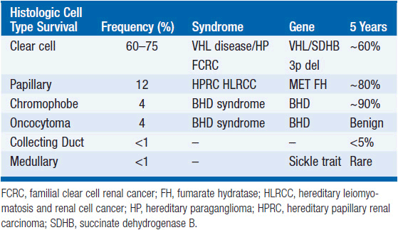
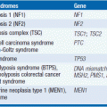
Several hereditary syndromes predispose patients to RCC (4). Insight into Von Hippel Lindau (VHL) disease has formed the foundation of our understanding of clear cell RCC, which accounts for 75% of renal cancer and >90% of metastatic RCC. The VHL tumor suppressor gene is located on chromosome 3, and in the hereditary form of clear cell RCC, one allele is inherited as an abnormal gene. Development of RCC occurs when the second allele is altered by deletion, hypermethylation, or mutational inactivation. Biallelic gene alteration leads to loss of function of the tumor suppressor protein and is observed in nearly 100% of the hereditary cases as well as >75% of sporadic cases of clear cell renal carcinoma. The VHL protein normally functions to inhibit cellular growth and is involved in regulating the expression of several proangiogenic genes. Under normal oxygen conditions, the VHL protein targets the hypoxia-inducible factor (HIF) family of transcription factors for ubiquitination and degradation. Biallelic loss or inactivation of VHL leads to constitutive upregulation of intracellular HIF proteins with subsequent increased expression of downstream gene targets including vascular endothelial growth factor (VEGF), transforming growth factor (TGF), and others.
The pathogenesis of non-clear cell RCC is entirely different. Various other mutations have been linked to development of papillary or chromophobe subtypes of RCC. These include activating mutations of the MET proto-oncogene at 7q31.3, mutations of the Birt-Hogg-Dube gene on chromosome 17, mutations in the fumarate hydratase gene, and others (5).
An additional important intracellular pathway identified in RCC is the mammalian target of rapamycin (mTOR) pathway, and agents that inhibit mTOR have proven efficacy in RCC as well (6). Genetic mutations in this pathway have not been identified in renal cancer.
CLINICAL PRESENTATION
A majority of patients diagnosed with RCC are asymptomatic at the time of presentation. The classic triad of hematuria, abdominal pain, and flank/abdominal mass is observed in fewer than 5% of patients. Patients with RCC may present with a wide range of signs and symptoms including hematuria, anemia, pain, or constitutional symptoms such as weight loss. Paraneoplastic syndromes associated with RCC include polycythemia, nonmetastatic liver enzyme abnormalities (Stauffer syndrome), hypercalcemia, and others.
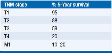
The cure rate for localized RCC following definitive local therapy is dependent primarily on stage, with >90% long-term disease-free survival for stages 1 and 2, but closer to 50% 5-year disease-free survival for stage 3 (see Tables 39-2 and 39-3). Of those who undergo surgical resection of apparently localized disease, about 33% will eventually develop disease recurrence. RCC can metastasize to nearly any anatomic location but most commonly metastasizes to the lungs, mediastinum, retroperitoneum, adrenals, bone, pancreas, liver, or CNS.

For diagnostic evaluation of suspected RCC, either CT or MRI of the abdomen is highly sensitive and specific. They have largely replaced intravenous pyelograms. Initial evaluations should also include the following:
• Family history screening for familial syndromes, especially if <50 years of age.
• Chest x-ray or chest CT scan.
• Consider MRI evaluation for involvement of renal vein and inferior vena cava.
• Note that bone scintigraphy and positron emission tomography (PET) scan are insensitive for RCC, and are not routinely recommended.
PROGNOSIS
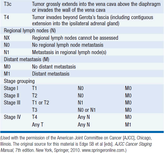
Prognosis in RCC is closely related to T-N-M staging (Table 39-3). In advanced RCC, prognosis can be stratified according to well-characterized poor risk features. A common classification system in patients undergoing initial therapy for metastatic RCC uses five clinical criteria (Table 39-4) (7):
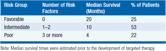
• Impaired Karnofsky performance status
• Elevated serum lactate dehydrogenase
• Low hemoglobin
• Less than 1-year interval from initial RCC diagnosis to start of systemic therapy
• Elevated serum calcium
It is important to note that prognostic system was developed prior to the availability of targeted therapies. Prognosis for patients beyond initial therapy can also be stratified based on clinical risk factors and has important implications with regard to subsequent therapy (8).
TREATMENT
 LOCALIZED DISEASE
• Radical nephrectomy has long been standard of care for localized RCC, and consists of removal of Gerota’s fascia, kidney, ureter, ipsilateral adrenal gland, and adjacent hilar lymph nodes (LNs).
• Nephron-sparing surgery (partial nephrectomy) is a preferred alternative in selected patients for whom it is feasible. Multiple case series support long-term outcomes that include cancer control, safety, and feasibility.
• Percutaneous ablation (radiofrequency ablation [RFA] or cryotherapy) may be an alternative in selected patients who cannot tolerate definitive surgical resection. Single-center case series have shown excellent outcomes and good tolerability with RFA in tumors <4 cm.
 ADJUVANT THERAPY
Several studies have evaluated the role of adjuvant therapy for patients with RCC. To date, none has demonstrated benefit. In one study, 283 patients with T3-T4a or N1 disease were randomized to observation or interferon (IFN)-α following nephrectomy and lymphadenectomy (9). After a 10-year median follow-up, there was no difference in overall survival or relapse-free survival. Another randomized study with high-dose interleukin-2 (IL2) compared to observation was terminated early after an interim analysis for futility (10). With the advent of effective targeted therapies, a new generation of adjuvant clinical trials is being carried out in patients at high risk of recurrence. These trials are designed to evaluate efficacy and feasibility of sunitinib, sorafenib, pazopanib, everolimus, and others (11). While results are awaited, the standard of care remains clinical trial participation or active surveillance.
 METASTATIC DISEASE
RCC is relatively resistant to cytotoxic chemotherapy and to radiation therapy with a median survival that was measured in months until recently. The natural history of advanced RCC is substantially varied, as some cases progress rapidly while others exhibit spontaneous tumor regressions. Immunotherapy with IFN or IL2 has been studied extensively and was the mainstay of systemic therapy prior to the development of targeted agents.
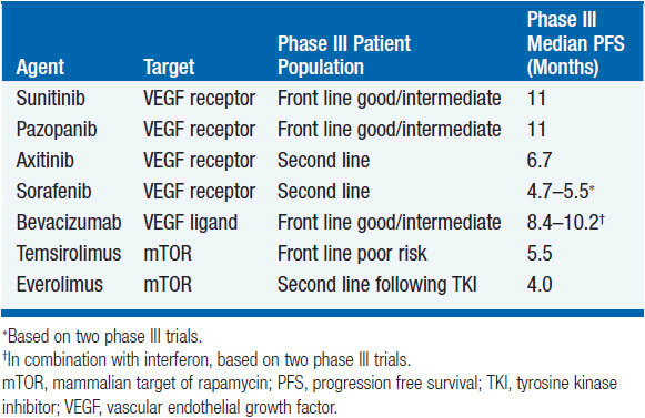
In the wake of new biologic insights, particularly for clear cell RCC, a wave of important therapeutic advances has transformed management of metastatic disease and resulted in improved outcomes for the majority of patients. Between 2005 and 2012, seven new targeted agents were approved by the U.S. FDA for the treatment of advanced RCC (Table 39-5). Nevertheless, curative treatments are still lacking and clinical trial participation should be encouraged whenever possible.
Related posts:
Stay updated, free articles. Join our Telegram channel

Full access? Get Clinical Tree


