■ Obliteration of the oral cavity. This is achieved when all oral cavity mucosal surfaces are in contact with one another when the mouth is closed. This goal is important because it should decrease the likelihood of food getting lost in a dead space in the oral cavity. Additionally, it should improve the handling of secretions by bringing the revascularized free tissue transfer in contact with the remaining native mucosa.
■ Maintain premaxillary contact. This is an extension of the goal of obliteration of the oral cavity. In terms of speech generation, premaxillary and palatal contact is important for maintaining the precision of articulation for a number of speech sounds. Generally, reduced precision of linguadental, alveolar, palatal, and velar sounds will occur if adequate contact is not achieved. The surgeon needs to ensure that some of the volume of the reconstructive flap is concentrated anteriorly to allow for the obliteration of the oral cavity.
■ Maintain the finger function of the tongue. This is the ability of the tongue to sweep and clear the buccal, labial, and alveolar sulci and protrude past the coronal plane of the incisors.
■ Maintain movement of secretions from the anterior to the posterior aspect of the oral cavity.
■ Optimize sensation of the remaining native tissue and the revascularized free tissue transfer.
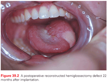
In general, these goals are best met with local tissue and revascularized autogenous tissue reconstruction. Traditional regional flaps, such as the pectoralis flap, are less commonly used because they are associated with higher gastrostomy tube rates.33 There are published studies that suggest that autogenous revascularized free tissue transplantation is a disadvantage.34 These data are not generally representative of present day reconstruction because, in this historic cohort, free flaps were used for large defects and skin grafts were used for smaller defects. The differences related more to the size of the defect than the reconstructive approach.
For oral cavity rehabilitation, the speech pathologist will perform an oral motor assessment. An assessment includes an evaluation of oral sphincter competence, the patient’s ability to handle secretions, tongue to premaxillary/palatal contact, anterior–posterior movement of the tongue, location of sensate tissue, and identification of areas where food will collect (i.e., dead space). A clinical swallow examination is used to assess swallowing function with a focus on the oral phase of the swallow. The patient’s ability to remove the bolus from an eating utensil (e.g., a spoon), create a labial seal, manipulate the bolus, control the bolus, and clear the bolus is assessed. The challenge for the speech pathologist is to modify the treatment plan and strategies used to compensate for the changing reconstruction during the first year of rehabilitation.
During the immediate postoperative period, the reconstruction will frequently be bulky and edematous; with radiation, the reconstruction will become smaller and the native tissue will undergo fibrosis. The objective throughout the first year is to maximize and maintain mobility of the tongue, focusing on the use of the remaining native tissue. In this patient group, use of liquid washes to add moisture and to aid in bolus passage with dry and solid consistencies should be considered the norm and not a failure of oral rehabilitation.
Maintaining the remaining native dentition is important for communication, swallowing, and for general health; therefore, including a dentist as part of the treatment team is critical. The best approach is prevention, which involves a reduction of radiation dose to the mandible when possible, the removal or restoration of carious teeth prior to treatment, regular fluoride trays before and after treatment, and the treatment of inflamed gingival tissue. Should a patient develop osteoradionecrosis, surgical excision and reconstruction is the mainstay, because hyperbaric oxygen therapy has been shown to lack efficacy in controlled clinical trials.35
The maxillofacial prosthodontist makes important contributions to the rehabilitation of the patient with an oral cavity defect. Dental rehabilitation with dental prostheses is important for function and cosmesis. When introducing dental prosthetics, it is important to consider the patient’s ability to masticate and prevent bolus loss. The introduction of a dental prosthesis can impair bolus control by covering sensate tissue, preventing glossal–labial contact, and decreasing the functional oral opening. It is also important to ensure that the patient can perform a tongue sweep of the labial sulci to clear food residue, especially if a lower (mandibular) dental prosthesis is introduced. If the patient is unable to perform this maneuver, then use of a digit may be required to clear food particles while eating. Even if the patient is a good candidate for a dental prosthesis, implants may be required to assist in the retention of a lower dental prosthesis. Implants can be placed in the native mandible or in an osseous free flap for the purpose of supporting and retaining a dental prosthesis. An individual knowledgeable of the use of implants in radiated bone is important because the rate of implant failure is high and there is a risk of osteoradionecrosis.36 Prostheses can also be useful for the rehabilitation of soft tissue deficits. For example, if the patient does not have good palatal–maxillary contact, a palatal drop prosthesis can be fashioned, facilitating the obliteration of dead space within the oral cavity, which allows the tongue to contact the prosthetically reconstructed palate. This may result in improved clarity of speech sounds and, therefore, overall speech intelligibility. In addition, the palatal drop prosthesis may assist in improved bolus manipulation, control, and oral transfer.
Rehabilitation After Partial Laryngeal Procedures
Both the communication and swallowing functions can be adversely affected with a partial surgical resection of the larynx. Supraglottic laryngectomy, hemilaryngectomy, and supracricoid laryngectomy all result in some degree of compromised phonatory function. Following these surgical procedures, the swallowing function is generally adversely affected in the short term, but improvement can be anticipated with the process of healing and the implementation of swallowing therapy. Postoperative dysphagia after partial laryngeal procedures is common due to a decrease in sensation and alteration of normal laryngeal anatomy. As a result, the patient is at risk for penetration and aspiration secondary to the compromise in airway protection that completion of these procedures brings about. Postoperatively, the patient will be trained on swallowing strategies to improve laryngeal closure in an attempt to prevent aspiration. In the early stages of recovery, liquids are usually the most difficult consistency to consume due to reduced sensation in and around the laryngeal complex and incomplete laryngeal closure, thus reducing airway protection. Therefore, consumption of a modified diet (thickened liquids and purees) with or without the use of alternative means of nutrition is not uncommon until adequate airway protection can be achieved. Implementation of swallowing maneuvers, such as the supra- and supersupraglottic swallow maneuvers, are helpful in facilitating airway protection.19–21
Rehabilitation After Laryngectomy or Laryngopharyngectomy
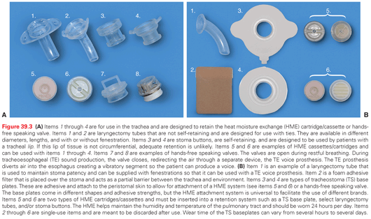
After a total laryngectomy, the patient has a tracheostoma in the lower neck and a separated digestive tract. The stoma and lungs require management to prevent stomal stenosis, prevent stomal trauma, enhance humidification, and reduce tracheal crusting. There are a variety of products that prevent tracheostomal stenosis, protect the stoma from digital trauma, and enhance humidification (Fig.39.3A, B). Many of these tracheal stomal products are designed to be used with or without tracheoesophageal (TE) prostheses.
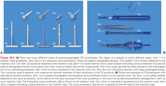
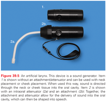
Rehabilitation of speech after a total laryngectomy has improved. Options for alaryngeal communication are TE voice, voice generated by an artificial larynx, and esophageal voice. TE voice has become the gold standard for voice rehabilitation after a total laryngectomy. The challenge with prosthetic rehabilitation is customized solutions, because one size does not fit all. There are many different types of TE voice prostheses (Fig. 39.4A, B). An experienced speech pathologist is essential for long-term patient compliance. The artificial larynx is a device that produces mechanical sound. This sound is transferred into the oral cavity via the placement of the device to the cheek or neck. Additionally, there is an option for an intraoral adapter, allowing for direct transmission of sound into the oral cavity. This device can be used short term until a patient achieves functional esophageal or TE speech. In some cases, it is used long term as the primary means of alaryngeal communication. This device can be difficult to use. Training of and practice by the patient are essential to become adept for daily communication requirements (Fig. 39.5). Another alaryngeal speech option is esophageal speech, which does not utilize devices or implants. It involves trapping air in the pharynx distal to the cricopharyngeus with a subsequent controlled release of air through the pharyngoesophageal segment to produce sound. Learning esophageal speech is time intensive and can take up to a year or longer to achieve a functional result. In some cases, fluent sound is never realized. As a result, esophageal speech is not commonly used. However, voice production with a TE voice prosthesis, in many instances, can be achieved on the day of insertion.
Stay updated, free articles. Join our Telegram channel

Full access? Get Clinical Tree


