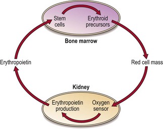
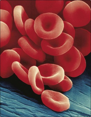
2 Red cells The mature red cells of the blood transport the respiratory gases, oxygen and carbon dioxide (CO2). Oxygen is carried from the lungs to the tissues, where it is exchanged for CO2. Red cells are equipped to perform this function for 120 days during which they make a 300 mile journey around the microcirculation. Prior to discharge from marrow sinuses into the peripheral blood, red cells shed their nuclei. This gives the advantages of reduced weight and transformation into a biconcave disc with increased deformability compared with the more rigid spheroidal nucleated precursor (Fig 2.1). Fig 2.1 Scanning electron microscope picture of mature red cells showing clearly the characteristic biconcave shape. (Copyright Dennis Kunkel Microscopy Inc.) The blood volume comprises the mass of red cells and the plasma. Plasma volume is regulated by stretch receptors in the heart and kidney which influence secretion of antidiuretic hormone (ADH) and aldosterone. Erythropoiesis is regulated chiefly by the growth factor erythropoietin. Erythropoietin Unlike other growth factors, erythropoietin is mainly synthesised by the peritubular endothelial cells of the kidney. Production is triggered by tissue hypoxia (lack of oxygen). Cells can sense hypoxia via mediators such as the transcription factor HIF (hypoxia-inducible factor). HIF activates genes vital in the adaptive response to hypoxia including the erythropoietin gene. Erythropoietin molecules bind to specific membrane receptors on primitive erythroid cells in the bone marrow and induce maturation. The increase in red cells released into the blood stops when normal oxygen transport is restored – this feedback circuit is illustrated in Figure 2.2. Fig 2.2 Feedback circuit in production of erythropoietin. Structure The mature red cell is around 7.8 µm across and 1.7 µm thick. Its biconcave shape allows maximum flexibility and an umbrella shape is adopted to traverse the smallest capillaries which have diameters of only 5 µm. The ability of red cells to recover from the recurrent stresses of the turbulent circulation hinges on the design of the membrane. The red cell membrane is composed of a collapsible lattice of specialised proteins (the ‘cytoskeleton’) and an outer lipid bilayer (Fig 2.3 Only gold members can continue reading. Log In or Register to continue You may also needMegaloblastic anaemiaIron deficiency anaemiaLaboratory haematology I – Blood and bone marrowPolycythaemiaThe thalassaemiasOther leukaemiasMyelomaVon Willebrand disease and other inherited coagulation disorders Share this:Click to share on Twitter (Opens in new window)Click to share on Facebook (Opens in new window)Click to share on Google+ (Opens in new window) Related Tags: Haematology An Illustrated Colour Text Jun 12, 2016 | Posted by admin in HEMATOLOGY | Comments Off on Red cells