Radiologic Imaging Concerns
Jeffrey A. Bennett
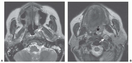
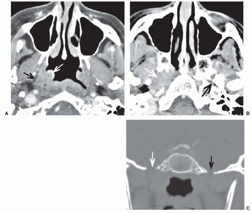
Cancer of the nasopharynx is staged differently than other pharyngeal primaries in that more emphasis is placed on tumor extension to other spaces than on size. Therefore, it is critical that imaging clearly shows the full extent of the primary lesion. As with other mucosal tumors, endoscopy can show the mucosal extent of the tumor very well, but deep tumor invasion requires imaging. Extension to the nasal cavity or oropharynx as well as parapharyngeal invasion classifies the tumor as T2 (Fig. 22-6). A T3 lesion involves bony structures and/or the paranasal sinuses. The tumor is upstaged to T4 if there is intracranial extension, involvement of the cranial nerves, the infratemporal fossa, the hypopharynx, the orbit, or the masticator space (Fig. 22-7). Because of the importance of detecting soft tissue invasion, intracranial extension, and perineural spread, magnetic resonance imaging (MRI) is preferred to computed tomography (CT) for imaging of nasopharyngeal cancers.1,2,3,4 However, care must be taken with fat suppression as this often produces artifacts at air-bone interfaces at the skull base. CT is not without benefit as it is very good at showing cortical bone erosion.
Usually, the diagnosis of nasopharyngeal cancer is known at the time of imaging, but occasionally it is found incidentally when imaging patients with unilateral serous otitis media and Eustachian tube dysfunction, otalgia, sore throat, or cranial neuropathies. When a tumor is discovered, it is essential to describe its pattern of spread and full extent so that the patient can be correctly staged and treated appropriately. Often both CT and MRI are obtained and these certainly give complementary information. Usually, the neck is scanned with CT to evaluate lymphadenopathy as the addition of a neck MR adds significant time and expense to the MR study. Suggested protocols are given in Figures 22-8 and 22-9.
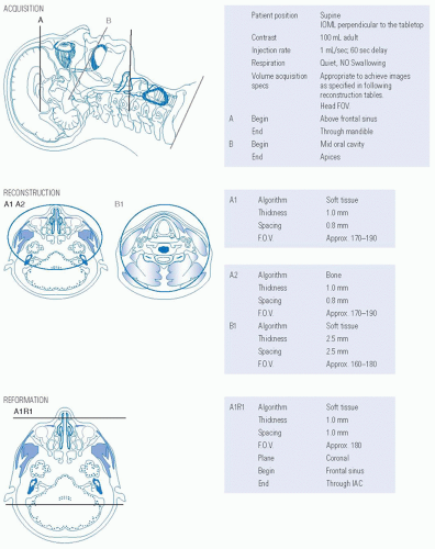 FIGURE 22-8. Protocol for CT maxillofacial detail and neck with intravenous contrast.
Stay updated, free articles. Join our Telegram channel
Full access? Get Clinical Tree
 Get Clinical Tree app for offline access

|
