General Principles and Management


General Principles and Management


Tim J. Kruser

Nitin A. Pagedar

Henry T. Hoffman

Paul M. Harari


INTRODUCTION

Cancers of the hypopharynx and the cervical esophagus share a number of overlapping features with regard to risk factors, epidemiology, management, and treatment outcomes. The anatomy of this region is served by a rich lymphatic network; therefore, presentation with nodal metastases is commonplace. In addition, patients with hypopharynx and cervical esophageal tumors frequently present with advanced-stage disease and harbor medical comorbidities that may limit the feasibility of delivering aggressive cancer care. Therefore, despite therapeutic advances in recent years, oncologic outcomes in these tumors remain relatively poor. Although organ-preservation surgery may be feasible in selected early-stage tumors, surgical management more commonly requires radical surgery such as laryngopharyngectomy or pharyngolaryngoesophagectomy. These procedures result in significant functional impairment, as well as cosmetic changes, and therefore tumors of the hypopharynx and the cervical esophagus are commonly managed with radiation or chemoradiation in an effort to control tumor while preserving organ function.

Cancers of the hypopharynx and the cervical esophagus are commonly considered together, as they share many features, and tumor frequently extends from one anatomic region into the other. Cancers of the hypopharynx accounted for 5.2% of upper aerodigestive malignancies from 2000 to 2008.1 Similarly, cervical esophageal cancer is rare, with only 2 % to 10% of esophageal carcinomas occurring in the cervical location.2 In a recent world-wide study of >4,500 esophagectomy patients, upper esophageal tumors accounted for only 4.1% of the cohort.3 The relative rarity of these tumors lends itself to grouping them together in order to effectively study epidemiology, management strategies, and outcomes. However, randomized control studies specific to tumors of the hypopharynx and/or the cervical esophagus are rare; studies of these cancers are often retrospective, incorporating heterogeneous management strategies spanning many years, and in some cases decades. The relative rarity of these tumors leads to a paucity of large-scale clinical trials data with which to base treatment guidelines.

Cancers of the hypopharynx and the cervical esophagus are commonly associated with tobacco use.4,5 In addition, patients are typically elderly, with dysphagia and weight loss as a result of their tumor burden and/or a poor baseline diet.5,6 These matters complicate management decisions and delivery of effective therapy. Given the complexity of these tumors, the surrounding structures, and frequent patient comorbidities, management of patients warrants multidisciplinary management with evaluation by head and neck, reconstructive, and thoracic surgeons, medical oncologists, radiation oncologists, speech/swallow therapists, nutritionists, and social workers. Despite advances in the treatment of hypopharyngeal and cervical esophageal cancer, the overall outcome in these patients remains poor. Efforts to improve cure rates and reduce treatment-related morbidity, including the incorporation of molecularly targeted agents, remain vital for the future.


Incidence, Etiology, and Epidemiology

In the United States, cancer of the hypopharynx and cervical esophagus account for <10% of the tumors of the upper aerodigestive tract.7 Approximately 1,800 cases of hypopharyngeal cancers are diagnosed each year in the United States.8 Tumors of the cervical esophagus account for 5.3% of all esophageal cancers,7 which equates to roughly 900 of the nearly 17,000 esophageal cancer cases per year in the United States.9 Squamous cell carcinoma (SCC) accounts for 95% of cancers in the hypopharynx and cervical esophagus, and the causative factors for SCC in these locations are shared.

The mean age of patients presenting with hypopharyngeal cancer is 65, with three-fourth of cases occurring in men. More than 90% of patients will report a tobacco history.10 Alcohol consumption potentiates the carcinogenic effects of cigarette smoke; in combination, heavy tobacco and alcohol users are at
up to 100 times elevated risk of developing tumors in the upper aerodigestive tract as compared with nonsmoking, nondrinking individuals. In addition to potentiating the effects of tobacco, a moderate to high consumption of alcohol can put nonsmoking individuals at a higher risk for hypopharyngeal and cervical esophageal cancers (Table 19.1).7,11 Alcohol appears to have a stronger neoplastic effect in the hypopharynx than in the larynx, and dark liquor may carry a higher risk than light liquor.12 In addition to tobacco and alcohol, gastroesophageal reflux, obesity, and achalasia can lead to higher rates of esophageal cancer.


TABLE 19.1 Odds Ratios for Oral and Pharyngeal Cancers Associated with Alcohol Consumption. Odds Ratios (or) Adjusted for Smoking, Age, and Race


Males


Females


Drinks per We


OR


95% CI


OR


95% CI


<1


1.0


1.0


1-4


1.2


0.7-2.0


1.2


0.7-1.9


5-14


1.7


1.0-2.7


1.3


0.8-2.1


15-29


3.3


2.0-5.4


2.3


1.2-4.5


>30


8.8


5.4-14.3


9.1


3.9-21.0


Source: Adapted from Blot WJ, McLaughlin JK, Winn DM, et al. Smoking and drinking in relation to oral and pharyngeal cancer. Cancer Res. 1988;48:3282-3287, with permission.


As tobacco and alcohol are the major causative agents for tumors of the hypopharynx and cervical esophagus, field cancerization is a common phenomenon in these patients. The wide mucosal exposure to carcinogens results in a field of diseased mucosa characterized by high-grade dysplasia, giving rise to the index hypopharyngeal or cervical esophageal tumor. Therefore, these patients have high rates of synchronous and/or metachronous upper aerodigestive primary tumors. Approximately 10% of patients may be found to have synchronous primary tumors,13 and up to 20% will develop a metachronous second primary tumor. Hypopharyngeal cancer patients have a worse overall survival than other malignancies of the head and neck due to the tendency for advanced disease at presentation.10 In addition, even among survivors of the initial presenting lesion, survivorship is poorer in the following years; the fact that metachronous malignancies are more common in these patients than in other head and neck squamous cell carcinoma (HNSCC) patients contributes to this observation (Fig. 19-1). Counseling patients on smoking cessation can diminish treatment-related side effects, can minimize the risk of subsequent malignancies of the upper aerodigestive tract, and should be emphasized during the initial evaluation of these patients.

Occupational factors may also play a causative role in the formation of cancers of the hypopharynx and the cervical esophagus. Two European studies have shown that masons, iron workers, railway workers, cement industry workers, and rock diggers demonstrate high rates of cancer in the larynx and the pharynx,14,15 suggesting that coal dust, steel dust, iron compounds, and fumes may contribute to carcinogenesis in these anatomic regions. In addition to occupational exposures, dietary factors can influence cancer rates in the hypopharynx and the cervical esophagus. High consumptions of salted foods and nitrosamines have been linked to esophageal SCC.16 Although the incidence of esophageal SCC are approximately 2.5 to 5/100,000 for men in most areas of the world, rates in China, Singapore, Iran, former USSR, Chile, Brazil, Puerto Rico, Switzerland, France, and South Africa have been demonstrated to be much higher.17 Dietary and occupational factors, as well as infection with the human papillomavirus (HPV) (see below), have been posited to account for these differences. One such example of a dietary risk for neoplasia is Plummer-Vinson syndrome, which is characterized by iron-deficiency anemia, hypopharyngeal webs, weight loss, and dysphagia. The syndrome is accompanied by an elevated risk of cancers in the postcricoid region.7 This risk has been ameliorated by the addition of iron to flour in the northern Midwest United States and Scandinavian countries, where this syndrome was problematic.


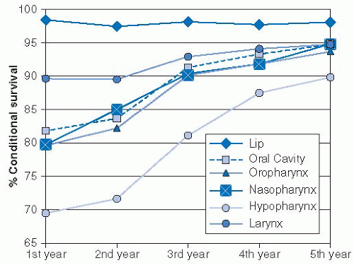
FIGURE 19-1. Conditional 5-year survival of head and neck cancers. A nearly 30% difference exists in survival through the first year, with hypopharynx cancers faring the poorest. Hypopharyngeal cancer patients continue to have lower rates of survival in years 2 to 5; secondary malignancies and comorbidities contribute to this phenomenon.

Source: Reprinted with permission from Cooper JS, Porter K, Mallin K, et al. National Cancer Database report on cancer of the head and neck: 10-year update. Head Neck. 2009;31:748-758.

The relationship between HPV infection and head and neck cancer has become well recognized in recent years, especially for cancers of the oropharynx. Epidemiologic studies have strongly suggested that HPV has a causative role in SCC of the base of tongue and the tonsil, with up to 60% to 70% of oropharyngeal SCC patients demonstrating evidence for HPV infection.18 HPV positivity is a strongly positive prognostic factor in oropharyngeal SCC patients undergoing chemoradiation.19 Whether HPV is similarly carcinogenic and/or prognostic in tumors of the hypopharynx and the cervical esophagus is less clear. Approximately 20% to 25% of hypopharynx cancer tumor specimens have detectable HPV DNA.20,21 Additionally, the detection of antibodies against the HPV16 E6 and E7 antibodies is associated with an elevated risk of hypopharyngeal cancer,22 suggesting a causal relationship. A large analysis of HPV rates in esophageal SCC showed the HPV can be detected by in situ hybridization in 23% of tumors, and in 15% by polymerase chain reaction.17 A wide geographic variance in HPV detection was noted, with low detection rates in Europe and the United States and higher rates noted in high-risk areas for esophageal SCC such as China, Japan, and South Africa. However, while HPV-positive oropharyngeal patients are more likely to be younger and nonsmokers as compared with their
HPV-negative counterparts, HPV-positive and HPV-negative esophageal cancer patients have been shown to have similar clinical risk factors.23 Therefore, although current data are suggestive of a carcinogenic role for HPV in the hypopharynx and the cervical esophagus, prospective studies demonstrating that persistent HPV infections result in malignant transformation, such as those performed in uterine cervix cancer, may be necessary to confirm this suspicion.


RELEVANT ANATOMY

The hypopharynx (sometimes referred to as the laryngopharynx) and the cervical esophagus are contiguous structures that collectively span from the hyoid bone to the thoracic inlet (Fig. 19-2). The hyoid bone demarcates the inferior border of the oropharynx, and the hypopharynx extends from the hyoid bone superiorly to the inferior border of the cricoid cartilage distally (approximately the level of the sixth cervical vertebral body). The cervical esophagus continues inferiorly from the lower edge of the cricoid cartilage to the thoracic inlet, approximated by the suprasternal notch.


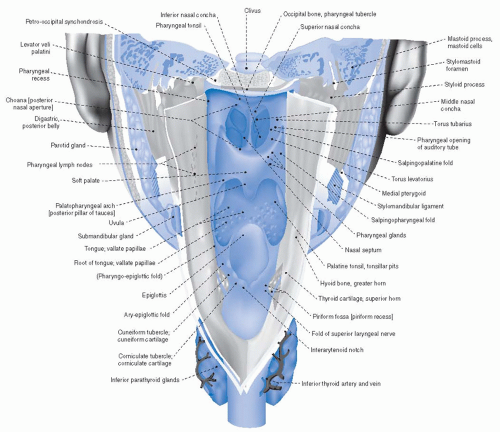
FIGURE 19-2. Posterior view of the larynx and hypopharynx as viewed by a posterior midline incision through the posterior pharyngeal wall, cricopharyngeus, and cervical esophagus.

Source: From Sobotta, Atlas der Anatomie des Menschen © Elsevier GmbH, Urban & Fischer Verlag München, with permission.

The hypopharynx is composed of three primary anatomic subsites: the bilateral pyriform sinuses, the posterior pharyngeal wall, and the postcricoid region. Anteriorly, the aryepiglottic fold forms the border between the postcricoid hypopharynx and the supraglottic larynx; tumors of the aryepiglottic fold can be labeled of hypopharyngeal or of supraglottic larynx origin, depending on where the majority of the tumor bulk lies. The postcricoid region is composed of the mucosa overlying the cricoid cartilage and is bound by the posterior arytenoid cartilages and aryepiglottic folds superiorly and the cricopharyngeus inferiorly (Fig. 19-3).

The pyriform sinuses are bilateral conical structures with the open-ended bases superiorly at the level of the pharyngoepiglottic
fold, and the medial, anterior, and lateral walls narrowing inferiorly to form the apices, which extend slightly below the cricoid cartilage. The pyriform sinuses are open posteriorly and contiguous with the posterior pharyngeal walls. The internal branch of the superior laryngeal nerve pierces the thyrohyoid membrane, which surrounds the superior aspects of the pyriform sinuses laterally. Tumor involvement of the sensory component of this nerve, which synapses superiorly with the nerves of the external auditory canal in the jugular ganglion, can result in referred otalgia. The remaining sensation in this region is provided by nerve fibers of the glossopharyngeal nerve. The recurrent laryngeal nerve provides motor function to the cricopharyngeus muscle and the posterior cricoarytenoid muscles of the larynx, with remaining primary motor supply to the hypopharynx provided by the pharyngeal plexus.


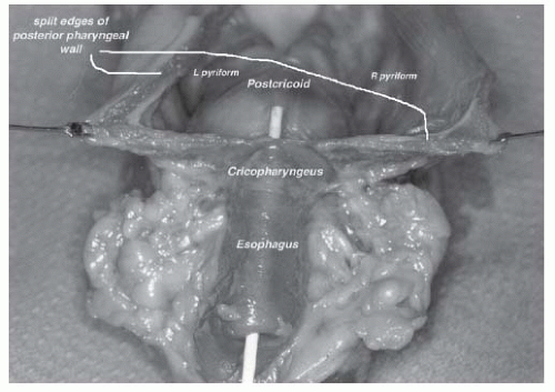
FIGURE 19-3. This anatomic specimen (with the posterior pharyngeal wall incised) allows a posterior view of the hypopharynx, demonstrating the three primary anatomic subsites (posterior pharyngeal wall, postcricoid region, and pyriform sinuses) of the hypopharynx.

The posterior pharyngeal wall is predominantly composed of squamous mucosa overlying the middle and the inferior pharyngeal constrictor muscles. The posterior pharygneal wall is separated from the prevertebral fascia by the retropharyngeal space; the mucosa overlying this region is thin and provides little barrier to direct tumor infiltration from the posterior wall to the prevertebral fascia. The distal fibers of the inferior constrictor coalesce to join the cricopharyngeus muscle, which serves as the junction between the hypopharynx and the cervical esophagus.

The cervical esophagus is lined by nonkeratinizing squamous mucosa overlying two muscular layers: an inner circular layer and an outer longitudinal layer. These muscles are striated in the upper third of the esophagus, whereas a gradual transition to smooth muscle occurs through the course of the thoracic esophagus. The cervical esophagus lies just posterior to the trachea, extending from the cricopharyngeus (C6 level) for about 4 to 5 cm to the thoracic inlet (suprasternal notch). Measured from the incisors (as commonly reported in endoscopic procedures), this ranges from 14 to 16 cm (cricopharyngeus) to approximately 20 cm. The tracheoesophageal grooves run laterally between these structures, and the recurrent laryngeal nerves and the paratracheal nodes are located in these grooves. The lateral lobes of the thyroid gland approximate the cervical esophagus and lie between the cervical esophagus medially and the carotid sheaths more laterally (Fig. 19-4). Vascular supply to the cervical esophagus is from branches of the inferior thyroid artery and veins, as well as ascending vessels along the thoracic esophagus. The sympathetic trunks and branches of the recurrent laryngeal nerves provide innervation.

There is a rich network of lymphatics that surround the hypopharynx and the cervical esophagus. From the hypopharynx, primary lymphatic drainage is to the jugulodigastric nodes (levels II-IV). Spinal accessory (level V) nodes are also at risk. Anterior lymphatic channels within the hypopharynx may drain from the pyriform sinuses directly through the thyrohyoid membrane along the path of the superior laryngeal artery. When tumors invade the posterior pharyngeal wall, lymphatic vessels passing through the inferior constrictor muscles place the retropharyngeal nodes at risk for involvement, including the most cephalad retropharyngeal nodes at the base of skull (nodes of Rouvière).

Lymphatic drainage from the cervical esophagus may mirror that of the hypopharynx and can pass superiorly toward the nodes surrounding the internal jugular vein. Lymphatics may also drain within the paratracheal and esophageal grooves to involve paratracheal lymph nodes. These lymph nodes are in the central compartment (level VI) and lie between the medial edge of the common carotid laterally, the trachea medially, inferior to the cricoid, and superior to the suprasternal notch. These paratracheal nodes are not usually included in a standard neck dissection and deserve consideration in hypopharyngeal and cervical esophageal tumors.24 The paratracheal lymph nodes are contiguous with lymph nodes of the upper mediastinum inferiorly; the anatomic and radiographic border between these two regions is poorly defined. Nonetheless, caudal drainage from the hypopharynx and cervical esophagus can involve these lymph node basins, which may also warrant attention from surgeons and radiation oncologists.25


NATURAL HISTORY, PATHOLOGY, PATHOGENESIS, AND PATTERNS OF SPREAD


Natural History

Tumors of the hypopharynx and the cervical esophagus tend to present with advanced disease, usually including nodal metastases. A retrospective study from Washington University demonstrated that 87% of patients with pyriform sinus tumors and 82% of posterior pharyngeal wall tumors presented with either stage III or IV disease.26 Similarly, a study of 107 patients with cervical esophageal tumors demonstrated that 79% of patients had stages III to IV disease.27 Therefore, despite stepwise advances in the diagnosis and treatment of these tumors, outcomes remain disappointing. Hypopharynx cancers fare poorly as compared with patients with HNSCC in other tumor sites, with 5-year survival rates approximating 30% (Fig. 19-5).8 Likewise, single institution reports have noted 5-year overall survival rates for cervical esophageal patients to range from 12% to 28%,13,28,29 and cervical esophageal patients have demonstrably inferior outcomes to tumors located in the thoracic esophagus (Fig. 19-6).3 A thorough understanding of the patterns of spread is essential to surgeons and oncologists in combating these aggressive tumors.


Pathology

Historically, approximately 95% of tumors of the hypopharynx or the cervical esophagus are SCC. Recent analysis of the Surveillance, Epidemiology, and End Results (SEER) database from 2000 to 2008 support this claim, as 93.9% of hypopharynx cancers were SCC, whereas lymphoma, sarcoma, adenocarcinoma, and adenoid cystic carcinoma accounted for approximately 0.5% of cases each.1 Similarly, the National Cancer Data Base (NCDB) Benchmark Reports evaluated 17,654 cases of hypopharyngeal cancer in the United States between the years of 2000 and 2008. More than 90% of cases were SCC.30


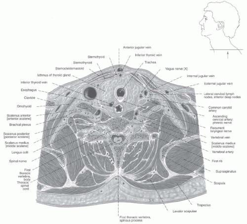
FIGURE 19-4. Transverse section detailing relationship of the cervical esophagus and adjacent structures at the level of the first thoracic vertebrae.

Source: Adapted from Sobotta, Atlas der Anatomie des Menschen © Elsevier GmbH, Urban & Fischer Verlag München, with permission.


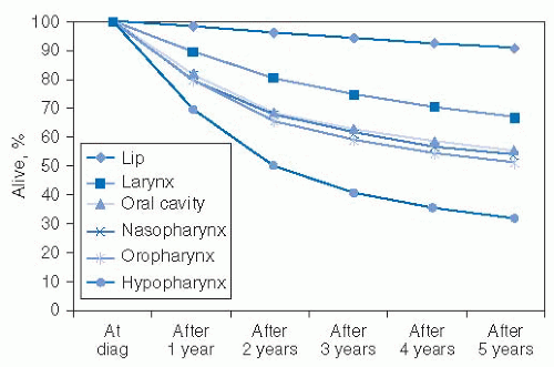
FIGURE 19-5. Five-year survival by mucosal site for head and neck cancers from 1990 to 1999 cases.

Source: Reprinted with permission from Cooper JS, Porter K, Mallin K, et al. National cancer database report on cancer of the head and neck: 10-year update. Head Neck. 2009;31:748-758.


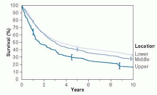
FIGURE 19-6. Survival according to upper extent of cancer in the esophagus.

Source:Reprinted with permission from Rice TW, Rusch VW, Apperson-Hansen C, et al. Worldwide esophageal cancer collaboration. Dis Esophagus. 2009;22:1-8.


Primary Tumor Location, Spread

Within the hypopharynx, tumor involvement of more than one subsite can cloud the ability of clinicians and/or radiologists to assign the tumor to a specific primary subsite. Within the Surveillance, Epidemiology, and End Results database from 2000 to 2008, 83% of tumors with known subsite arose within the pyriform sinus. The posterior pharyngeal wall accounted for 9% of primary tumors; the postcricoid was less commonly the primary site, accounting for 4% of tumors.1 Cancers in the hypopharynx and cervical esophagus are prone to submucosal spread, though less so in the pyriform sinus than in other locations. Serial sectioning of surgical specimens has shown that 60% of hypopharynx cancers demonstrate significant subclinical spread, which can contribute to local failures following definitive management. Typical patterns of submucosal spread in such patients can include a range of 10 mm superiorly and 20 mm inferiorly.31 Skip lesions from the hypopharynx therefore commonly involve the cervical esophagus. Cervical esophageal tumors are similarly prone to submucosal spread, especially caudally; up to 23% of cervical esophageal tumors may have clinical evidence of thoracic esophageal involvement at the time of diagnosis.27

Cancers originating in the pyriform sinus can infiltrate laterally to invade the posterolateral portion of the thyroid cartilage and/or the superior lobe of the thyroid gland. The medial wall is the most common site of involvement for pyriform sinus tumors, and tumor invasion in this location can result in invasion of the cricoarytenoid joint and/or muscle or the recurrent laryngeal nerve and may cause the vocal cord to become fixed. Superior extension may involve the aryepiglottic folds and arytenoids, and the paraglottic space is also at risk for involvement. Posterior pharyngeal wall extension is common as the tumor grows laterally.

Tumors primarily of the postcricoid area commonly extend inferiorly to involve the cervical esophagus, and circumferential growth frequently occurs, with invasion of the cricoid cartilage and cricoarytenoid muscle and resultant vocal cord fixation. These tumors are commonly extensive at presentation and portend a worse prognosis in comparison to tumors from other hypopharyngeal locations.32 Tumors of the posterior pharyngeal wall tend to expand in the craniocaudal dimension, tracking submucosally in a superior fashion to the oropharynx or inferiorly to the cricopharyngeus and the cervical esophagus. Deep infiltration can involve the retropharyngeal space with access to the lymphatics in that region and subsequently into the prevertebral fascia.

Cervical esophageal tumors commonly extend into adjacent tissues at presentation.13 Direct anterior extension can result in tumor involvement of the trachea, and involvement of the larynx and/or recurrent laryngeal nerve may result in vocal cord paralysis in approximately 10% of patients. Lateral extension into the thyroid gland is not uncommon, and possible posterior infiltration into the prevertebral fascia warrants careful evaluation on upfront diagnostic imaging. Thus, the extensive pattern of primary tumor infiltration in hypopharyngeal and cervical esophageal tumors can present considerable challenge in the effort to achieve clear surgical margins or achieve full dosimetric coverage with radiotherapy.


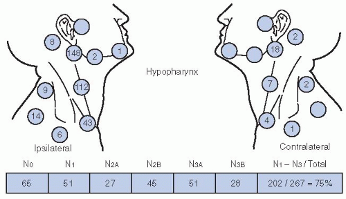
FIGURE 19-7. Nodal distribution patterns for a series of 267 patients with hypopharynx cancer as summarized by admission records at the MD Anderson Hospital.

Source: Redrawn with permission from Lindberg R. Distribution of cervical lymph node metastases from squamous cell carcinoma of the upper respiratory and digestive tracts. Cancer. 1972;29:1446-1449.


Lymphatic Spread

A rich lymphatic network surrounds the hypopharynx and the cervical esophagus, and lymph node involvement occurs in the majority of tumors by the time of diagnosis. More than 50% of patients will present with clinically positive cervical lymph nodes at diagnosis. An additional 30% to 40% of NO necks are found to show pathologic involvement when dissected electively,33 rendering 65% to 80% of patients ultimately node positive.

The primary lymph node drainage of the cervical esophagus is to the paratracheal lymph node bed, and surgical series consistently demonstrate these lymph nodes to be most commonly involved, with approximately 40% to 70% of cases demonstrating metastatic involvement on pathologic analysis.13,34,35 The general lymph drainage of the cervical esophagus is directed upward, and therefore levels III and IV of the neck are at the next highest risk, with levels II and V at lower but not negligible risk. Lymph node risk is associated with the length of the primary tumor, with lesions >5 cm showing lymph node involvement in 90% of cases.36

In patients with hypopharyngeal cancer, 50% to 80% of patients will have metastatic lymph node involvement. Jugular chain nodes (levels II-IV) as well as the retropharyngeal nodes are all at high risk for metastatic involvement. Lymphatics from the hypopharynx can drain anteriorly along with the lymphatics of the supraglottic larynx through the thyrohyoid membrane to primarily drain into levels II and III. Posterior lymphatics more commonly drain tumors of the inferior hypopharynx, penetrate the superior constrictor muscle, and drain to the lateral retropharyngeal and the paratracheal lymph nodes. Cross-draining lymphatics are present, and bilateral cervical node metastases are common (Fig. 19-7). In addition to the jugular chain lymph nodes typically addressed during neck dissection procedures, paratracheal lymph nodes are also at risk in hypopharynx cancer, with surgical specimens demonstrating involvement in 20% to 25% of cases37,38 and warrant surgical consideration.


Distant Metastases

Cancers of the hypopharynx have higher rates of distant metastatic disease than do primary tumors of other HNSCC subsites.39 Approximately 16% of hypopharyngeal cancer patients present with distant metastases at diagnosis.40 Distant metastatic rates rise with time, especially in patients with residual or recurrent locoregional disease following initial therapy; rates of ultimate distant metastases have been reported up to 60% in older reports.39 Data regarding initial metastatic presentation and ultimate metastatic risk with cervical esophageal cancer is less robust. Nonetheless, metastatic patterns of tumors from the hypopharynx and the cervical esophagus are similar, with the lung most commonly involved, followed in incidence by the mediastinal lymph nodes, the liver, and bone.


CLINICAL PRESENTATION

Patients with cancers of the hypopharynx and the cervical esophagus initially develop nonspecific symptoms, which are commonly attributed to presumed infectious or gastrointestinal causes, leading to frequent delays in diagnosis. Therefore, the majority of patients will present with advanced local and/or regional disease. Presentations of hypopharyngeal and cervical esophageal tumors are similar, although cervical esophageal tumors tend to present earlier, as the lumen of the cervical esophagus is smaller than that of the hypopharynx. Dysphagia is the most common presenting symptom, present in up to 85 % of cases, although a palpable neck mass is the most common physical finding.13 Other common symptoms at presentation include odynophagia, weight loss, and neck pain. The majority of patients will note more than one of these symptoms by the time of diagnosis.41 Rarely, early-stage tumors may be diagnosed in the evaluation of new-onset reflux symptoms.10

Other presenting symptoms can include unilateral ear pain (referred otalgia), classically seen in patients with tumors of the pyriform sinus. This results from involvement of the superior laryngeal nerve (a branch of the vagus nerve), which synapses with the auricular branch of cranial nerve × (the nerve of Arnold) in the jugular ganglion. Hoarseness can result from tumor infiltration into laryngeal structures or from involvement of the recurrent laryngeal nerve by tumors in the cervical esophagus or the postcricoid regions. Weight loss is common and is often substantial and can reflect poor nutritional status that may result from clinically significant dysphagia, alcohol dependency, and/or tumor-mediated cachexia/anorexia.


DIAGNOSTIC EVALUATION AND CLINICAL STAGING


Workup and Evaluation

Patients with tumors of the hypopharynx and/or the cervical esophagus should be evaluated in a multidisciplinary fashion, with input from surgeons, medical oncologists, radiation oncologists, speech-and-swallow specialists, social workers, dentists, nutritionists, and often specialists in alcohol and/or tobacco cessation. A detailed history should be elicited, with the nature and duration of symptoms documented, as well as the degree of weight loss. Assessment of dysphagia to both solids and liquids should be noted. Changes in voice quality and the complaint of referred otalgia are frequently present. Exposure in pack-years of cigarette smoking should be noted, as should the use of smoke-free tobacco (chewing tobacco, snuff). Alcohol history should be elicited, with particular attention to current use, which may predispose patients to withdrawal.

The initial physical examination should indirect visualization of the full laryngopharyngeal axis. Given the high rate of second head and neck mucosal primaries, thorough attention to any other mucosal irregularities is warranted. Tumors of the pyriform sinus can be notoriously difficult to visualize; having the patients puff their cheeks (blow against closed lips) during nasopharyngolaryngoscopy can aid in visualization of the pyriform sinuses. In addition to the primary tumor (size, location, involved structures), particular attention should be paid to the mobility of the vocal cords with phonation, as this impacts tumor staging. Furthermore, the initial examination should include an assessment of dentition, particularly if (chemo)radiation is likely to be employed as a treatment modality. Unlike many tumors of the head and neck region, where indirect visualization may suffice, direct visualization under anesthesia is warranted in most tumors in these locations to fully delineate the extent of tumor spread and for biopsy confirmation of malignancy. An upfront thorough assessment of dentition allows for dental extractions, if needed, to be performed simultaneously while the patient is under anesthesia for tumor mapping under direct visualization (Table 19.2).

Physical exam should include assessment of cervical lymphadenopathy, providing a description of the size, location, number, mobility, and texture of suspicious lymph nodes. As all patients with biopsy-proven malignancies of the hypopharynx and/or the cervical esophagus warrant advanced radiographic imaging, fine-needle aspirate of enlarged lymph nodes to further confirm metastatic lymphadenopathy is rarely of value and should not be routinely performed.

Panendoscopy should be complemented by advanced imaging to assess the extent of the primary tumor and rigorously assess cervical lymph node chains for presence and/or extent of regional lymph node involvement. High-resolution computed tomography (CT) with contrast from the base of skull to below the clavicles is usually the imaging study of choice, although MRI can be utilized. A low threshold should be utilized in extending a CT through the lungs distally, as tumors of the hypopharynx and cervical esophagus have significant rates of pulmonary metastases at presentation. For the rare early-stage, lymph nodenegative hypopharyngeal tumor, chest x-ray may suffice to rule out distant pulmonary metastases. Cervical esophageal tumors in particular commonly involve the lymph nodes of the upper mediastinum25 and may have thoracic esophageal involvement,27 and therefore CT imaging should routinely include the thorax in these patients.18 FDG-PET (or PET-CT, with accompanying CT slice co-registration) is increasingly utilized in the upfront staging of tumors of the hypopharynx and the cervical esophagus. PET-CT allows for detection of distant metastases and provides information that may aide in the determination of lymph node malignant involvement. Furthermore, PET-CT imaging may influence radiation planning, in terms of field design and/or prescribed dose.42 A recent prospective study of PET imaging in HNSCC tumor management included 46 patients with hypopharyngeal cancer. Therapeutic decisions were made with physicians blinded, and then unblinded, to FDG-PET imaging data. Tumor/node/metastases (TNM) staging was altered in 100 of 233 enrolled patients (43%), whereas PET altered the management of 32 patients (13.7%) (Table 19.3). This study, and similar experiences with PET imaging in HNSCC management support a role for PET-CT imaging in the upfront evaluation of patients with hypopharyngeal and cervical esophageal tumors. Worldwide, approximately 75% of oncologists utilize FDG-PET imaging in a proportion of esophageal cancer patients for staging purposes, with routine use slightly more common in North America than in Europe or Asia.43

Patients with cancers of the hypopharynx and/or the cervical esophagus commonly present with significant weight loss,
often as a result of progressive tumor-associated dysphagia and odynophagia. An assessment should be made of the patient’s nutritional status, with attempts to correct nutritional deficits prior to the initiation of aggressive therapy. A nutritionist should be involved as a part of the multidisciplinary team. A percutaneous enteral gastrostomy (PEG) tube is commonly utilized. In surgical patients, a PEG tube is commonly placed at the time of surgery to allow for maintenance of nutrition during the postoperative recovery period. Whether upfront PEG tube placement should occur routinely in patients planning to undergo (chemo)radiation or should happen only in patients who demonstrate significant difficulties with maintaining weight and hydration during radiation is an area of debate. Patients with tumors of the hypopharynx and cervical esophagus tend to have more dysphagia, odynophagia, and prediagnosis weight loss than other HNSCC patients. Therefore, in our experience, the benefits of prophylactic PEG tube placement in terms of improved weight preservation and maintenance of hydration appear warranted for the majority of patients. Therefore, we routinely place PEG tubes during the pretreatment evaluation phase prior to chemoradiation.


TABLE 19.2 Workup and Pretreatment Evaluation for Patients with Tumors of the Hypopharynx and Cervical Esophagus


History


  • Nature/duration of symptoms


  • Degree of weight loss


  • Dysphagia (to solids? also to liquids?)


  • Referred otalgia?


  • Change in voice quality?


  • Tobacco history (cigarettes, smoke-free)


  • Alcohol history—current heavy use?


Physical examination


  • Assessment of dentition (extractions necessary)


  • Cervical lymphadenopathy description


  • Indirect nasopharyngolaryngoscopy, with particular attention to other mucosal irregularities (second primaries) and cord mobility


  • Examination under anesthesia for biopsy, tumor mapping, with opportunity for dental extraction simultaneously


Imaging


  • CT with contrast (preferred) or MRI of the neck (skull base to below clavicles)


  • CXR (early-stage HP tumors only)


  • CT of the chest (advanced HP tumors; all CE tumors)


Additional specialty input


  • Dental assessment


  • Nutrition assessment; consideration of PEG tube placement


  • Speech and swallow assessment


  • Tobacco and alcohol cessation


  • Social work input (transportation, finances, other support)


TABLE 19.3 Impact of FDG-PET on Staging and Management in 233 HNSCC Patients


Endpoint


Impact of FDG-PET


Discordant TNM stage


100/233(43%)


PET accurately upstaged


30/233(13%)


PET accurately downstaged


17/233(7.3%)


PET innacurately changed stage


13/233(5.6%)


No confirmed gold standard TNM


40/233(17%)


Impact on patient management


No significant change


188/233(80.7%)


Change within the same treatment modality


12/233(5.2%)


Change in treatment intent and/or modality


20/233 (8.6%)


Source: Adapted from Lonneux M, Hamoir M, Reychler H, et al. Positron emission tomography with [18F]fluorodeoxyglucose improves staging and patient management in patients with head and neck squamous cell carcinoma: a multicenter prospective study. J Clin Oncol. 2010;28:1190-1195, with permission.


In addition to input from a nutritionist, a formal evaluation by speech-and-swallow therapists before initiation of therapy to assess baseline dysphagia is valuable. If swallowing dysfunction is noted, adaptive techniques may be taught to the patient to improve caloric intake and minimize aspiration risks. Although informal bedside assessment may be performed of swallow function, a more definitive fluoroscopic barium swallow study (videopharyngogram or oropharyngeal motility study [OPMS]) is usually preferable. Close posttreatment surveillance with a speech-and-swallow specialist familiar with the patient is important following definitive therapy.

All patients should undergo dental evaluation, with routing cleaning and Panorex imaging to assess for periapical abscesses, unerupted third molars, or other pathology prior to initiation of therapy. For patients who receive (chemo)radiation, long-term xerostomia may result and places patients at risk for accelerated dental decay. Therefore an assessment of teeth in poor repair that may warrant extraction is warranted, especially for any teeth that
occupy a region likely to receive high-dose radiation. Upfront removal of such teeth in poor repair is preferable to subsequent removal following radiation, as manipulation of the teeth and sockets following radiation places patients at risk for osteoradionecrosis. Dental extractions should precede radiation by 10 to 14 days to allow for adequate healing. For patients who have teeth that will remain in place through radiation should receive custom fluoride carrier trays. Long-term use following radiotherapy of fluoride can minimize dental decay in the setting of postradiotherapy xerostomia.

Patients with hypopharyngeal and cervical esophageal cancers commonly have ongoing issues with alcohol addiction and tobacco dependency. Engagement with specialists for counseling and other methods to aide tobacco and alcohol cessation can be valuable. Medications such as the nicotine patch can be prescribed by surgeons and oncologists to assist patients with tobacco cessation. Additionally, many of these patients with benefit from input from social workers, as they often face issues such as financial difficulties, lack of transportation for appointments, and limited family/social support. A case manager or social worker can help these patients navigate the medical system and cope with these concerns through their cancer management.


Staging

In 2009, the American Joint Committee for Cancer (AJCC) updated their staging system, releasing the 7th edition.44 While the AJCC staging system provides a platform for grouping patients, in order to help guide discussions between physicians and for assessment of outcomes, patient factors include comorbidities, age, and motivation for organ preservation must be taken into account when considering management of an individual patient. For tumors of the hypopharynx, no significant changes were instituted in the 7th edition, with the exception that tumors extending to the esophagus are now T3 (previously T4a). T1 tumors are <2 cm in greatest dimension and limited to one subsite. T2 tumors measure between 2 and 4 cm, and/or invade more than on subsite of the hypopharynx or an adjacent site. Extension to the esophagus or fixation of the hemilarynx renders a tumor T3, as does size >4 cm. Tumor that invades either the hyoid bone, the thyroid gland, the central compartment soft tissue (prelaryngeal strap muscles and subcutaneous fat), or the thyroid and/or the cricoid cartilage is deemed moderately advanced local disease (T4a). Tumors are very locally advanced (T4b) if they invade the prevertebral fascia, involve mediastinal structures, or encase a carotid artery. Nodal staging and stage grouping is identical for hypopharyngeal cancer patients as to other HNSCC sites (excluding the nasopharynx).

For tumors of the cervical esophagus, major changes were implemented between the 6th (2002) and 7th (2009) editions of the AJCC staging based on a worldwide analysis of 4,627 patients with tumors of the esophagus and esophagogastric junction treated with surgery alone.3,45 While the previous staging system was based only on tumor/node/metastases criteria, these analyses resulted in histology, grade, and location being incorporated into the stage grouping for 7th edition. T staging of esophageal tumors is based on depth of invasion and remains largely unchanged from the 6th edition of the AJCC staging system. As the vast majority of cervical esophageal tumors are SCC, the tumor (T) and the nodal (N) definitions for esophageal SCC are depicted in Table 19.4. It should be noted that only 4.1% of the tumors comprising the data set used for derivation of this staging system were located in the upper one-third of the esophagus, which would also include upper thoracic esophageal tumors.

Tis (in situ) is characterized by noninvasive high-grade dysplasia. Tla tumors invade the lamina propria or muscularis mucosae, with invasion of the submucosae staged T1b. T2 tumors invade muscularis propria, whereas T3 tumors invade adventitia. T4 tumors (invading adjacent structures) were split into T4a and T4b in the 7th AJCC system. T4a tumors are resectable tumors invading the pleura, pericardium, or diaphragm. T4b tumors are unresectable, invading other structures including the aorta, the trachea, and the vertebral body.


TABLE 19.4 T and N Staging for Esophageal Squamous Cell Carcinoma


Tumor Level of Invasion


T Descriptor


Noninvasive, high-grade dysplasia


Tis (in situ)


Invading the lamina propria or muscularis mucosal layers


T1a


Involving submucosa


T1b


Involving the muscularis propria


T2


Involving the adventitia


T3


Involving the diaphragm, pleura, or pericardium (resectable)


T4a


Involving aorta, trachea, vertebral body, other adjacent tissues (unresectable)


T4b


Metastatic Lymph Node Burden


N Descriptor


1-2 lymph nodes


N1


3-6 lymph nodes


N2


>7 lymph nodes


N3


Source: Adapted from Edge SB, Byrd DR, Compton CC, et al. AJCC Cancer Staging Handbook. New York, NY: Springer; 2010, with permission.


Nodal (N) staging also changed between the 6th and 7th AJCC staging systems for esophageal tumors. Previously lymph node staging was dependent on the location of lymph nodes, with lymph nodes in the cervical, scalene, internal jugular, periesophageal, and supraclavicular regions termed “regional” for cervical esophageal tumors. Currently, metastases in 1 to 2 regional lymph nodes renders a patient N1, metastases to 3 to 6 regional lymph nodes is N2, and involvement of 7 or more regional lymph nodes is N3. Regional lymph nodes are now designated as any nodes from the cervical nodes to the celiac nodes for all esophageal cancers, whereas previously cervical lymph nodes were deemed metastatic for thoracic esophageal tumors.

Grade and location now are also incorporated into AJCC staging for esophageal SCC, impacting the stage grouping in early-stage tumors (Table 19.5). Grade that cannot be assessed (grade x) and grade 1 tumors (well-differentiated) are grouped together, whereas grade 2 (moderately differentiated), grade 3 (poorly differentiated), and undifferentiated (grouped as grade 3 squamous) are grouped together. Furthermore, anatomic location impacts stage grouping. Tumors of the cervical esophagus are defined in this system as being bordered by the hypopharynx superiorly and by the thoracic inlet (sternal notch) superiorly, or from 15 to 20 cm from the incisors by endoscopy.45


MANAGEMENT STRATEGIES AND TREATMENT PLANNING GUIDELINES

Historically, surgery has been a mainstay in management of cancers of the hypopharynx and cervical esophagus. This has typically involved sacrifice of the larynx, either via laryngo-pharyngectomy
or pharyngolaryngoesophagectomy. However, in recent years, increasing efforts toward organ preservation have become more prevalent. As tumors of the hypopharynx and cervical esophagus tend to present with locally advanced and/or node-positive disease, these efforts at organ preservation most commonly involve chemoradiation for locally advanced disease (Fig. 19-8). However, more advanced surgical techniques may allow for organ preservation in those patients who present with early-stage disease amenable to larynx-sparing surgery. Nonetheless, radical surgery remains an important component of management and should be considered as an alternative to organ-sparing approaches, depending on the motivation of the patient for organ preservation and the patient’s medical comorbidities. Active rehabilitation therapy in order to maximize eventual speech and swallow function must accompany all definitive management, regardless of the specific treatment approach. Rehabilitation and posttreatment surveillance are discussed at the end of this section.


TABLE 19.5 Stage Grouping for Esophageal Squamous Cell Carcinoma


Stage


T


N


M


G


Location


0


Tis (HGD)


0


0


1


Any


IA


1


0


0


1


Any


IB


1


0


0


2-3


Any


2-3


0


0


1


Lower


IIA


2-3


0


0


1


Upper, middle


2-3


0


0


2-3


Lower


IIB


2-3


9


0


2-3


Upper, middle


1-2


1


0


Any


Any


IIIA


1-2


2


0


Any


Any


3


1


0


Any


Any


4a


0


0


Any


Any


IIIB


3


2


0


Any


Any


IIIC


4a


1-2


0


Any


Any


4b


Any


0


Any


Any


Any


3


0


Any


Any


IV


Any


Any


1


Any


Any


Source: From Rice TW, Rusch VW, Ishwaran H, et al. Cancer of the esophagus and esophagogastric junction: data-driven staging for the seventh edition of the American Joint Committee on Cancer/International Union Against Cancer Cancer Staging Manuals. Cancer. 2010;116:3763-3773, with permission.


Hypopharyngeal Cancer Management

Stages I to II Disease (T1-2, NO). A large analysis of 853 hypopharyngeal cancer patients revealed that approximately 84% of patients will present with either T3-4 and/or node-positive disease,46 whereas a review of 1,210 hypopharyngeal cases diagnosed in 2003 through the NCDB identified only 150 (12.3%) of cases to be stages I to II.30 Therefore, stages I to II hypopharyngeal cancer is a relatively rare clinical scenario. In general, curative radiation therapy is preferred as the definitive treatment approach for patients with early-stage hypopharynx tumors, as it provides a good chance for organ preservation. Alternatively, open or endoscopic surgery (partial laryngopharyngectomy) with ipsilateral or bilateral neck dissection may be performed (Table 19.6). More recently, transoral laser microsurgeries have been performed with promising results in selected early-stage lesions.

Radiation Therapy. Radiation is commonly favored in stages I to II hypopharynx cancers, as surgery in this region often results in chronic aspiration due to injury/sacrifice of the superior laryngeal nerve. Additionally, given that concerning pathologic features (positive margins, node positivity, pathologic T3 or T4 status, perineural invasion, lymphovascular invasion) often prompt postoperative (chemo)radiation following surgery, definitive radiation may allow patients to forego the toxicities associated with multiple treatment modalities. However, for patients who receive definitive radiation, it is important for the head and neck surgeon to remain involved in the patient care. Endoscopic biopsy for detailed assessment of tumor extent may be necessary prior to definitive radiation, and the surgeon should remain involved in the multidisciplinary oncologic follow-up after treatment.

Radiotherapy for stages I to II hypopharygeal cancer classically consists of a 6-to-7-week course, with treatment delivered 5 days per week.47,48,49,50,51,52 Lymphatic coverage from the skull base to the clavicle is routine for patients undergoing radiation for hypopharyngeal cancer. Conventional field design utilizes opposed lateral fields covering the primary tumor and upper neck regions matched to an anterior field covering low-neck and supraclavicular neck lymphatic regions. The superior border of the opposed lateral radiation fields should include jugular chain lymphatics to the base of skull, whereas inferiorly fields should extend to the lower aspect of the cricoid cartilage, unless more inferior coverage is mandated by extensive caudal tumor extension. Anteriorly, fields should flash skin at the level of the thyroid cartilage, and posterior borders should extend behind vertebral spinous processes, or further if needed to cover metastatic level V lymph nodes. Wedges or custom compensators should be used for the lateral fields to account for varying tissue thickness of the head and neck to maximize dose homogeneity. When the spinal cord reaches a dose of ≤45 Gy, shrinking-field techniques should be used to maintain the spinal cord dose below tolerance. Posterior neck electrons supplement dose to the posterior cervical lymph nodes, whereas 54 to 60 Gy should be delivered to the remaining lymphatics via lateral fields before final mucosal reductions are made to deliver 66 to 70 Gy to the primary tumor plus a margin (for T1 tumors) or 70 Gy for T2 tumors. Three-dimensional planning with intensity-modulated radiation therapy (IMRT) is now commonly utilized for radiotherapy in hypopharynx cancer and will be discussed further in the sections discussing management of locally advanced disease.

Only gold members can continue reading. Log In or Register to continue

Stay updated, free articles. Join our Telegram channel

Jun 21, 2016 | Posted by in ONCOLOGY | Comments Off on General Principles and Management

Full access? Get Clinical Tree

Get Clinical Tree app for offline access