Radiologic Imaging Concerns
Jeffrey A. Bennett
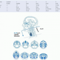
Imaging of the hypopharynx is typically performed in a patient with persistent, often unilateral sore throat, dysphagia, glomus sensation, or otalgia after a tumor has been found by endoscopy. The endoscopist is usually able to map out the mucosal extent of the tumor, but the relationship of the tumor to the esophageal verge, postcricoid area, or piriform sinus apex may be difficult to determine. The submucosal extent of disease also cannot be determined by endoscopy. Computed tomography (CT) with intravenous contrast is the usual first imaging study performed. There is no longer a role for fluoroscopy with oral contrast in primary diagnosis, but video fluoroscopy is still performed, mainly in posttreatment patients for analysis of swallowing and to look for the presence of aspiration or a fistula. Suggested CT and magnetic resonance imaging (MRI) protocols are given in Figures 19-17, 19-18 and 19-19.
As in other areas of the head and neck, the purpose of imaging is to determine the full extent of the primary tumor and to look for perineural spread in this location along recurrent laryngeal nerve, or superior laryngeal neurovascular bundle, or lymphatic spread, typically not only to levels II through VI, but also to the retropharyngeal nodes (Fig. 19-20).1,2 In addition, the possibility for acute airway obstruction should be assessed. The airway may be at risk in the posttreatment setting as well, if there is radionecrosis of the cricoid cartilage.
CT and MRI can both provide information on spread of tumor to the larynx, the oropharynx, the paraglottic space fat, or the preepiglottic fat. Figures 19-21 and 19-22 showpiriform sinus primary tumors with spread into the larynx. Figure 19-23 shows a postcricold tumor extending to the esophageal verge. Figures 19-24 and 19-25 show primary cervical esophageal tumors.
CT and MRI are both somewhat limited in the detection of early cartilage invasion, as there is variability in the ossification of the cartilage, but CT might be slightly better because of the ability to acquire very thin, < 1 mm thick slices. MRI of this region is often degraded by motion artifact. Where MRI provides an advantage is in its superior soft-tissue differentiation in determining whether tumor abuts or invades the esophagus or the trachea.
Since esophageal cancers can be synchronous with other head and neck squamous cell carcinomas, the radiologist should search for other mucosal-based tumors when an esophageal tumor is found. One study by Wang et al.3 showed 69 of 315 patients (21.9%) had synchronous head and neck squamous cell cancer and esophageal neoplasia. Drinking alcohol was a significant risk factor. Another study4 found the risk of synchronous esophageal and head and neck squamous cell cancers to be rare for the oral cavity, but were more common in the larynx, the hypopharynx, and the oropharynx.
Stay updated, free articles. Join our Telegram channel

Full access? Get Clinical Tree


