Radiation Therapy Technique
William M. Mendenhall
The treatment technique depends on the location of the tumor, the number of lesions (1 or ≥2), and whether the tumor is malignant. The vast majority of tumors are single and benign.
Temporal bone tumors may be treated with stereotactic radiosurgery or fractionated radiation therapy. Stereotactic radiosurgery is performed with either a linear accelerator-based system or a gamma knife; the results with either technique are equivalent.1
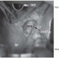
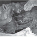
Fractionated radiation therapy may be delivered with stereotactic radiation therapy, intensity-modulated radiation therapy (IMRT), or a wedge pair technique using three-dimensional (3D) conformal treatment planning (Figs. 27-23 and 27-24).2 The lesion is treated with a 1-cm margin of normal tissue to a dose of 45 Gy in 25 fractions over 5 weeks. The disadvantage of the wedge pair technique is that a larger volume of normal tissue is treated. However, even very large tumors can be treated with an ipsilateral technique that will result in a very low dose to the contralateral parotid gland and minimize the risk of xerostomia.
Glomus vagale and carotid tumors are not suitable for stereotactic radiosurgery and are better treated with IMRT or three-dimensional treatment planning using a wedge pair technique.
The technique used for patients with multiple tumors is variable and depends on the location and extent of the lesions. Very large bilateral tumors may require parallel-opposed fields. Alternatively, resection of one lesion and irradiation of the contralateral
tumor may minimize the long-term morbidity of treatment if one of the lesions can be resected with acceptable morbidity. Relatively small bilateral paragangliomas may be treated with IMRT with adequate sparing of one or both parotid glands (Fig. 27-25).
tumor may minimize the long-term morbidity of treatment if one of the lesions can be resected with acceptable morbidity. Relatively small bilateral paragangliomas may be treated with IMRT with adequate sparing of one or both parotid glands (Fig. 27-25).
Stay updated, free articles. Join our Telegram channel

Full access? Get Clinical Tree


