Radiation Therapy for Penile Cancer


Radiation Therapy for Penile Cancer


Juanita M. Crook


INTRODUCTION

Carcinoma of the penis is rare, with an estimated incidence of approximately 1 in 100,000 men in North American and Western European countries, and accounts for a total of <1% of male cancers. Extirpative surgery is highly effective, especially for early-stage disease, but can be associated with considerable psychosexual morbidity. Men treated by irradiation tend to have fewer problems with posttreatment sexual activity, compared to those treated by partial penectomy where sexual activity is frequently severely reduced or completely lacking (1,2). Romero et al. (3) reported that despite capacity for erection of the penile stump and the ability to achieve orgasm, only one third of subjects retained preoperative frequency and satisfaction with intercourse. Other series include reports of suicide or attempted suicide following partial penectomy (4,5,6). The European Urology Association guidelines (7) now strongly recommend a penile-conserving approach for Ta-T1, G1-2 lesions and state that a conservative approach is an alternative in lesions that are T1G3 or ≥T2 if the tumor occupies less than half the glans and the patient is reliable for close follow-up.

Radiotherapy, either external or interstitial, is an organ-sparing alternative that preserves penile morphology and function without compromising disease control or survival in selected patients. Unfortunately, many urologists have not routinely offered this penis-conserving option. One survey reported that even for small, distally situated lesions, 43% of urologists suggested amputative surgery, while for larger distal lesions on the glans, 82% preferred surgery (8). Quality of life and sexual health following such surgery is rarely studied or reported. Although some men might, if necessary, sacrifice sexual activity in favor of improved long-term survival, the converse also applies (1). Posttreatment sexuality and expectations should be discussed when deciding on primary management.

Penis-conserving surgical techniques such as laser or Mohs surgery may be suitable for carcinoma in situ (CIS) or very superficial tumors. These techniques are discussed in detail in the chapter on surgical management of penile cancer.

Because of the rarity of carcinoma of the penis, many series span several decades, during which time, both treatment techniques and staging systems have evolved (Table 48.1). However, despite the medley of techniques, doses, and staging systems, all radiotherapy series suffer similarly from the lack of accurate pathologic staging (5,9). Clinical staging of carcinoma of the penis is very subjective and, in many cases, it is impossible to distinguish T1 (invasion of subepithelial connective tissue) from T2 (invasion of corpus spongiosum or cavernosum). Imaging such as ultrasound, or magnetic resonance with prostaglandin-induced erection may be helpful (10,11,12). Unfortunately, biopsies are often too superficial to accurately assess the depth of invasion (13). For this reason, techniques that treat less than the full thickness of the penis must be restricted to very carefully selected cases.


RADIOTHERAPY TECHNIQUES


External

External radiotherapy has several advantages as the radiation modality of choice for carcinoma of the penis. It is widely available, delivers a reliably homogeneous dose, and does not require the specific expertise of brachytherapy. Well-localized CIS (Tis) may be treated effectively to a depth of less than full thickness of the penile shaft using 125 to 250 kV orthovoltage beams, or 13 MeV electrons (14), using fractionation schemes commonly employed for skin cancer such as 35 to 40 Gy in 10 fractions over 2 weeks. However, most penile cancers, because of uncertainty of the depth of invasion, require irradiation to the full thickness of the penis with full dose to the skin surface. Various techniques have been devised for this purpose. Fraction sizes <2 Gy (4) may be associated with higher recurrence rates, whereas fraction sizes >2 Gy (15) may be associated with greater long-term sequelae such as urethral strictures. The most commonly prescribed dose is 60 Gy in 30 fractions over 6 weeks. Treatment interruptions because of acute reactions (edema, pain, desquamation) should be avoided (4). Circumcision is required before definitive radiotherapy to prevent acute radiation balanitis and long-term phimosis. A setup that is easily reproducible over a 6-week period, comfortable for the patient despite increasing local reaction, and easily verified by technologists is desirable.


Wax Block

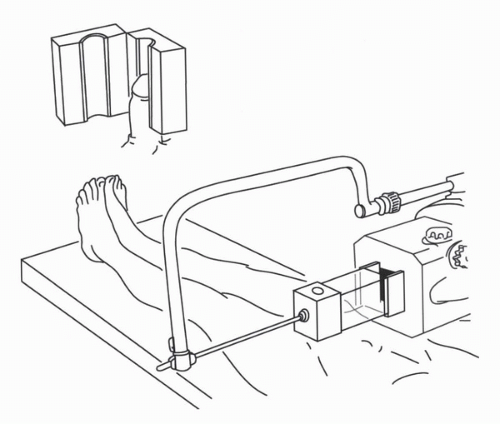
A 10 × 10 cm to 10 × 15 cm wax block is used to encompass the penis for each treatment (16). The block is constructed in two halves, with a central cylindrical chamber fashioned internally. The patient is positioned supine on the treatment couch, and the penis is supported in a vertical position, encased in the wax block. A tissue-equivalent “cork” must be placed in the opening. Parallel opposed beams treat the entire length of the penis. External beam radiation (EBRT) using 60Co, or 4 to 6 MV photons can be used (Fig. 48.1). Daily setup may become increasingly uncomfortable as treatment progresses and penile swelling may require modification of the wax block to enlarge the central chamber. Furthermore, it is not possible to verify penile position within the wax and if the fit is not snug, the penis may “slump” inside the wax, resulting in treatment of a shorter length of the shaft than planned.


Perspex Block

The use of a Perspex block (4) follows the same principles as the wax block technique, providing full buildup to the skin surface and allowing treatment by parallel opposed beams but has several advantages. Blocks can be preconstructed with a range of sizes of the central cylindrical chamber and can be sterilized for reuse. Penile swelling can be accommodated easily by choosing the next larger size, and penile position
within the transparent block can be visually verified before each treatment (Fig. 48.2).


TABLE 48.1 STAGING SYSTEMS FOR CARCINOMA OF THE PENIS COMMONLY SEEN IN THE LITERATURE


A.


Jackson staging system for carcinoma of the penis (19)


1.


Tumor limited to the glans or prepuce


2.


Tumor extending into the shaft or corpora, without node involvement


3.


Tumor confined to the shaft, with malignant but operable lymph nodes


4.


Invasion beyond the shaft, with inoperable lymph nodes or distant metastases


B. TNM staging UICC 1978 (23)


Tis: CIS


N1: metastases in unilateral inguinal lymph nodes


T1: ≤2 cm


N2: metastases in bilateral inguinal lymph nodes


T2: >2 cm and ≤5 cm


N3: fixed inguinal lymph nodes


T3: >5 cm or deep invasion including urethra


T4: tumor invades adjacent structures


C. TNM staging UICC 1987-2002 (24)


Tis: CIS


T1: subepithelial connective tissue


N1: one superficial inguinal lymph node


T2: corpus spongiosum/cavernosum


N2: multiple/bilateral superficial inguinal lymph nodes


T3: urethra/prostate


N3: deep inguinal or pelvic lymph nodes


T4: other adjacent structures


CIS, carcinoma in situ; TNM, tumor, nodes, metastases; UICC, union internationale contre le cancer.


Water Bath

A technique for external beam irradiation of the penis using a water bath in place of wax or Perspex to provide full dosage to the penile surface has been reported (17,18). The patient is positioned prone on the treatment couch, supported under the torso and thighs on Styrofoam slabs, with the water bath positioned to contain the suspended penis (Fig. 48.3). This technique does not require daily manipulation of the penis for positioning but may not be appropriate for all types of body habitus.


FIGURE 48.1. Wax block technique. Wax block with central cylindrical chamber for penis provides full buildup of dose to skin surface for parallel opposed mega voltage beams. (From McLean M, Akl AM, Warde P, et al. The results of primary radiation therapy in the management of squamous cell carcinoma of the penis. Int J Radiat Oncol Biol Phys 1993;25:623-628, with permission.)


FIGURE 48.2. Perspex block technique. Blocks provide full buildup as in the wax block technique but are transparent for verification of penile position. The blocks are bivalved and can be made in a range of sizes to accommodate penile swelling as treatment progresses.


Brachytherapy

The penis is well suited to brachytherapy. For centers with appropriate expertise, either surface molds or interstitial techniques can be used with good effect. Circumcision is required before brachytherapy to ensure complete exposure of the tumor and prevent subsequent phimosis or annular fibrosis of the foreskin.


Molds

Neave et al. (15) have reported experience with iridium molds for Jackson stage 1 or 2 (19) (Table 48.1) penile carcinoma. A hollow cylindrical Perspex tube is fitted over the penis along its long axis and is held in place by a girdle fitting around the patient’s groin and upper thighs. Iridium-192 wire is arranged circumferentially in concentric rings in a second outer sourcecarrying tube, which is positioned daily for periods of approximately 12 hours. Hospitalization is required, and the patient must be cooperative. One advantage of a mold over EBRT is that the acute reaction develops after the treatment is finished. Unlike interstitial brachytherapy, a mold is not invasive and does not require anesthesia. A surface dose of 55 to 60 Gy (16) is prescribed, with a central axis dose of 46 to 50 Gy. The total treatment duration is approximately 1 week.

Akamoto et al. (20) have reported a custom mold technique using silicon monomer. The penis is suspended using traction on a catheter to hold it centrally in a plastic cylinder. Silicon monomer mixed with a catalyst is poured into the cylinder, around the penis, and hardens into a sponge-like material within 10 minutes. The material can be removed easily after it has set, and hollow catheters are inserted longitudinally for after-loading sources. The authors describe using a variety of sources, including 226Ra, 137Cs, and 192Ir. Patients were treated with one to three fractions. Fraction size ranged from 10.7 to 40 Gy (median 20 Gy), with total dosages ranging from 32 to 74 Gy.


Interstitial Brachytherapy

There is a wide experience with interstitial brachytherapy for T1 and T2 penile carcinoma, with reports from many
European countries, from Canada, and from India. Implants are generally performed under general anesthesia, but local or spinal anesthesia is also an option. The Paris system of dosimetry (21) is most commonly employed and is applicable to both manually after-loaded implants and remote after-loading pulse dose rate (PDR), although for the latter, some optimization can be introduced.


FIGURE 48.3. Water bath technique. The patient lies prone on the Styrofoam slabs such that the penis will be suspended in the water bath. The overflow tray catches the runoff. Transparent sides on the water bath permit visual check of penile position.

Penile brachytherapy is generally performed as a volume implant delivering treatment to the full thickness of the glans, although well-selected, very superficial tumors, or Tis disease, may be treated with a single-plane implant. For volume implants, two or three parallel planes of sources are inserted, with intersource and interplane spacing being equal and ranging from 12 to 18 mm.The distribution, spacing, and total number of needles depend on tumor size. Four (2 × 2), six (2 × 3), or nine (3 × 3) needles may be required. Planes are usually oriented with the needles passing from the dorsal to the ventral surface of the glans, but a left-right orientation may be acceptable depending on tumor location. Needle placement requires 30 to 45 minutes in the operating room. Catheterization of the patient during the procedure aids in identification of the urethra so as to avoid transfixing it with implant needles. 19.5-gauge needles are used in association with manually after-loaded 192Ir wire, and 17-gauge needles are compatible with a PDR remote after-loader. The needles are held in place through the duration of the implant with predrilled plexiglass templates. The prescribed dose is generally 60 Gy over 4 to 5 days at a classic low dose rate (LDR) of 50 to 60 cGy/hr (<1 Gy/hr). No dose rate correction is required for PDR as compared with continuous LDR. On the contrary, high dose rate brachytherapy (HDR) requires compensation in total dose because of the elevated dose rate and smaller number of fractions. Suggested fractionation schemes for HDR brachytherapy for penile cancer have not been published.

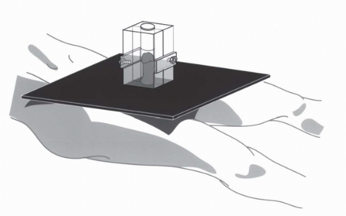
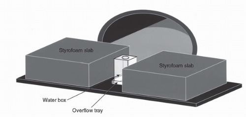
At the completion of the procedure, a Styrofoam collar is positioned around the base of the penis proximal to the needles to support the penis in the upright position and to thereby minimize unnecessary irradiation of the adjacent abdominal wall, thigh, or scrotal skin. If preservation of fertility is a concern, a thin sheet of lead can be inserted into the Styrofoam collar to minimize transmitted dosage to the testicles. Thermoluminescent dosimeters measurements for PDR penile brachytherapy after incorporation of a layer of 2 mm of lead into the Styrofoam collar around the penis indicate a total dose of 53 to 58 cGy to the anterior testicle and of 26 cGy to the posterior testicle (Crook, personal experience).

The brachytherapy needles are remarkably well tolerated. The patient remains catheterized and in bed for the duration of the implant, although with PDR the source cables can be disconnected from the needles to allow the patient to mobilize for a brief period between fractions if desired. Sufficient analgesia is usually provided by acetaminophen, with or without codeine. Leg exercises and subcutaneous heparin (5,000 U q12h) are recommended as prophylaxis against deep vein thrombosis. Removal of the needles occurs at the bedside following premedication with a narcotic analgesic.

Interstitial brachytherapy has the advantage of being completed within 4 to 5 days, with the peak acute reaction occurring 2 to 3 weeks after treatment has finished. Interstitial brachytherapy minimizes unnecessary treatment to the penile shaft, and because the lesion is clearly visible as the needles are being placed, visual verification of coverage can be readily accomplished. Needle placement should be such that the prescription isodose should allow for about 10 mm of coverage beyond visible or palpable tumor. Care must be taken to adhere to the rules of the Paris system (21) and to be aware of the relation of the treated volume to the needles such that the entire tumor and desired margin are contained within the highdose volume (Fig. 48.4). The technical aspects of implanting meatal or unilateral tumors have been described (22).


RESULTS OF TREATMENT

Results for several recent series in the literature are summarized in Table 48.2. Often series span several decades, during which treatment techniques evolve and dose prescription changes. Staging systems also undergo major modifications. The three most commonly seen in the literature [Jackson (19) TNM 1978 (23) and TNM 1992 (24)] are shown in Table 48.1.

From Table 48.2, it can be readily appreciated that radical radiotherapy, as either brachytherapy or EBRT, is effective in achieving local control in a high percentage of patients. Surgery for salvage is highly effective with success rates in excess of 80% (9,16). As in primary management, local excision is rarely appropriate as surgical salvage. The choice between total and partial penectomy (Table 48.3) depends on penile length and the proportion of the shaft irradiated. Brachytherapy, because of the more localized treatment volume, should lend itself to a less radical salvage operation.

Interstitial brachytherapy results in 5-year local tumor-control rates of 70% to 86% and penile preservation rates ranging from 72% to 83%. At 10 years, actuarial LF-free survival is 72% to 80% and penile preservation 67% to 72% (27,28). External radiotherapy does not appear to be quite as effective in local control of the primary tumor (41%-70% at 5 years), with penile preservation rates reflecting the more frequent need for second-line surgery (36%-66%). Careful extended follow-up is recommended, as local failures can occur several years after treatment. Mazeron et al. (29) reported that of the 11 local failures in his series of 50 patients treated by interstitial brachytherapy, only 36% were in the first 2 years, whereas 45% occurred between years 2 and 5, and 18% between years 5 and 8. Similarly, Crook et al. (27) found that although five of eight local failures in an experience of 67 patients occurred in the first 2 years, three occurred between 4.5 and 8 years and de Crevoisier et al. (28) reported that 20% of local recurrences occurred after 8 years. Because of the success of surgical salvage, late local recurrences do not result in a decline in cause-specific survival (CSS). De Crevoisier et al. reported CSS of 92% at 10 years (28) and Crook et al. reported no deaths from penile cancer between 5 and 10 years (27), with CSS being 83.6% at both time points.

Only gold members can continue reading. Log In or Register to continue

Stay updated, free articles. Join our Telegram channel

Jul 15, 2016 | Posted by in ONCOLOGY | Comments Off on Radiation Therapy for Penile Cancer

Full access? Get Clinical Tree

Get Clinical Tree app for offline access