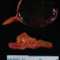
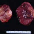
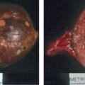
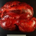
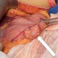
Primary adrenal lymphoma is rare and typically presents in older men with progressive signs and symptoms of adrenal insufficiency, weight loss, and abdominal pain. , Computed cross-sectional imaging usually reveals large (up to 18 cm in diameter) lipid-poor adrenoform bilateral adrenal masses. On magnetic resonance imaging (MRI), the lesions are hypointense in T1-weighted images and hyperintense in T2-weighted images. F-18 fluorodeoxyglucose (FDG) positron emission tomography (PET)-computed tomography (CT) typically shows high FDG avidity and it may reveal extraadrenal locations of disease. Adrenalectomy is not indicated. Chemotherapy is the treatment of choice. Herein we present a case of a patient with primary adrenal lymphoma.
Case Report
The patient was a 57-year-old man who had been previously healthy and running 3 miles per day. He took no regular medications. He had traveled to Europe 3 weeks previously and for 48 hours he had fairly intense abdominal pain. As the abdominal pain resolved, it became more of a pleuritic posterior back and chest discomfort that persisted—leading to a chest CT scan that detected bilateral adrenal masses. The subsequent abdominal CT showed bilateral oblong adrenoform 11-cm adrenal masses ( Fig. 77.1 ). The CT appearance was that of an infiltrating process such as lymphoma or infection. His pleuritic chest pain was 3 out of 10 in severity during the day and 10 out of 10 in severity at night. He had lost 5 pounds because of poor appetite that was associated with the pain. He had no fevers. He had limited laboratory studies before referral and they included: morning serum cortisol concentration of 12.4 mcg/dL (normal, 7–25 mcg/dL), morning serum corticotropin (ACTH) concentration of 92 pg/mL (normal, 10–60 pg/mL), and 1-mg overnight dexamethasone suppression test with a next day cortisol concentration of 1.34 mcg/dL (normal, <1.8 mcg/dL).

INVESTIGATIONS
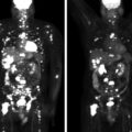
The laboratory studies are listed in Table 77.1 . All adrenal gland–related testing was normal except for a mild elevation in serum ACTH concentration. Hypercalcemia was associated with low blood concentration of parathyroid hormone and elevated 1,25-dihydroxyvitamin D. The imaging findings and hypercalcemia were consistent with lymphoma. CT-guided right adrenal mass biopsy was performed and showed findings consistent with diffuse large B-cell lymphoma. The FDG-PET-CT scan showed the large bilateral adrenal masses to have very intense FDG uptake compatible with malignancy ( Fig. 77.2 ). No other sites of involvement were identified by FDG-PET-CT, including the spleen and lymph nodes.
| Biochemical Test | Result | Reference Range |
Sodium, mmol/L | 140 | 135–145 |
Potassium, mmol/L | 4.2 | 3.6–5.2 |
Creatinine, mg/dL | 1.3 | 0.8–1.3 |
Calcium, mg/dL | 11.3 | 8.9–10.1 |
Phosphorus, mg/dL | 3.2 | 2.5–4.5 |
1,25-Dihydroxy-vitamin D, pg/mL | 84 | 18–64 |
25-Hydroxy-vitamin D, ng/mL | 18 | 20–50 |
Parathyroid hormone, pg/mL | 12 | 15–65 |
8 am serum cortisol, mcg/dL | 12 | 7–25 |
8 am serum ACTH, pg/mL | 10.0 | 10–60 |
Aldosterone, ng/dL | <4 | ≤21 |
Plasma renin activity, ng/mL per hour | 1.2 | ≤0.6–3 |
Metanephrine, nmol/L | <0.2 | <0.5 |
Normetanephrine, nmol/L | 0.35 | <0.9 |
Lactate dehydrogenase, U/L | 668 | 122–222 |
24-Hour urine: | ||
Calcium, mg | 385 | 25–300 |
Cortisol, mcg | 8.9 | 3.5–45 |
Stay updated, free articles. Join our Telegram channel

Full access? Get Clinical Tree


