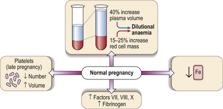
Pregnancy

44


Pregnancy


Haematological changes


Several haematological changes occur in normal pregnancy (Fig 44.1). Beginning in the sixth week there is an increase in plasma volume accompanied by an increase in red cell mass. The plasma volume expansion peaks at around 24 weeks when it is approximately 40% greater than in a non-pregnant woman. As the increase in red cell mass is more modest (15–25%) a dilutional anaemia is inevitable. In practice the haematocrit and haemoglobin level start to fall at 6–8 weeks and reach a trough at around 20 weeks. It is unusual for the haemoglobin level to fall below 100 g/L and if this happens another cause for anaemia should be sought. Negative iron balance can be regarded as routine in pregnancy and as discussed below frank iron deficiency commonly occurs.


The other major changes which may be regarded as a physiological consequence of pregnancy affect the coagulation system. There are increases in the levels of the coagulation factors VII, VIII and X and a marked increase in plasma fibrinogen. The resulting hypercoagulability is helpful in limiting the likelihood of life-threatening bleeding at delivery but it does lead to an increased risk of thromboembolism. The platelet count falls about 10% during an uncomplicated pregnancy. Later in pregnancy there may also be an increase in mean platelet volume (MPV).


Anaemia in pregnancy


There are several causes of anaemia in pregnancy. The most common scenario is an exacerbation of the usual dilutional anaemia by deficiency of iron and/or folate. Erythropoietin levels increase less than in anaemic non-pregnant women, possibly suppressed by hormonal changes.


The identification of iron deficiency relies upon normal laboratory tests (p. 25). However, even in women with no overt clinical deficiency there is a progressive fall in serum iron through pregnancy. Routine dietary supplementation with modest amounts of iron (e.g. ferrous sulphate 200 mg daily) leads to a significant increase in haemoglobin level at term compared with women receiving no supplements. Parenteral iron is contraindicated in the first trimester.


The other major type of anaemia in pregnancy is megaloblastic anaemia. This usually results from deficiency of folate. As for iron, folate requirements are increased during pregnancy and the diet is frequently inadequate to meet this demand. Megaloblastic anaemia most often presents as a macrocytic anaemia in the third trimester or postpartum. It is normal practice to give folate supplements in pregnancy. The amount of folate routinely administered orally should be large enough to avoid megaloblastic anaemia but not so large as to risk masking pernicious anaemia with vitamin B12 deficiency which does occasionally occur in pregnancy. Folate deficiency in pregnancy has been linked with an increased incidence of neural tube defects in the fetus and recommendations for planned pregnancies are the use of folate supplements (400 µg daily) prior to conception and then particularly in the first 12 weeks. Larger doses of folate are recommended where women are at high risk of conceiving a child with a neural tube defect (e.g. previously affected pregnancy). There is no justification for the prescription of multi-ingredient vitamin preparations in pregnancy but a combined iron and folate tablet of adequate dosage may be prescribed.


It should be remembered that not all anaemia in pregnancy is caused by deficiency states. Other blood disorders may present in pregnancy and chronic blood diseases such as sickle cell anaemia can be especially difficult to manage at this time.


Jun 12, 2016 | Posted by in HEMATOLOGY | Comments Off on Pregnancy

Full access? Get Clinical Tree

Get Clinical Tree app for offline access