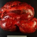
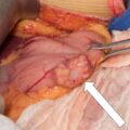
Neurofibromatosis type 1 (NF1) is an autosomal dominant disease caused by mutations in the NF1 gene located on chromosome 17q11.2. Approximately 50% of patients with NF1 present with de novo germline mutations. Clinical diagnosis is based on at least two of the following features: six or more café au lait spots, two or more neurofibromas or one plexiform neurofibroma, freckling in axillae or inguinal areas, optic glioma, two or more Lisch nodules, bony lesions, or a first-degree relative with NF1. Pheochromocytomas can occur in 3% of patients with NF1. ,
Case Report
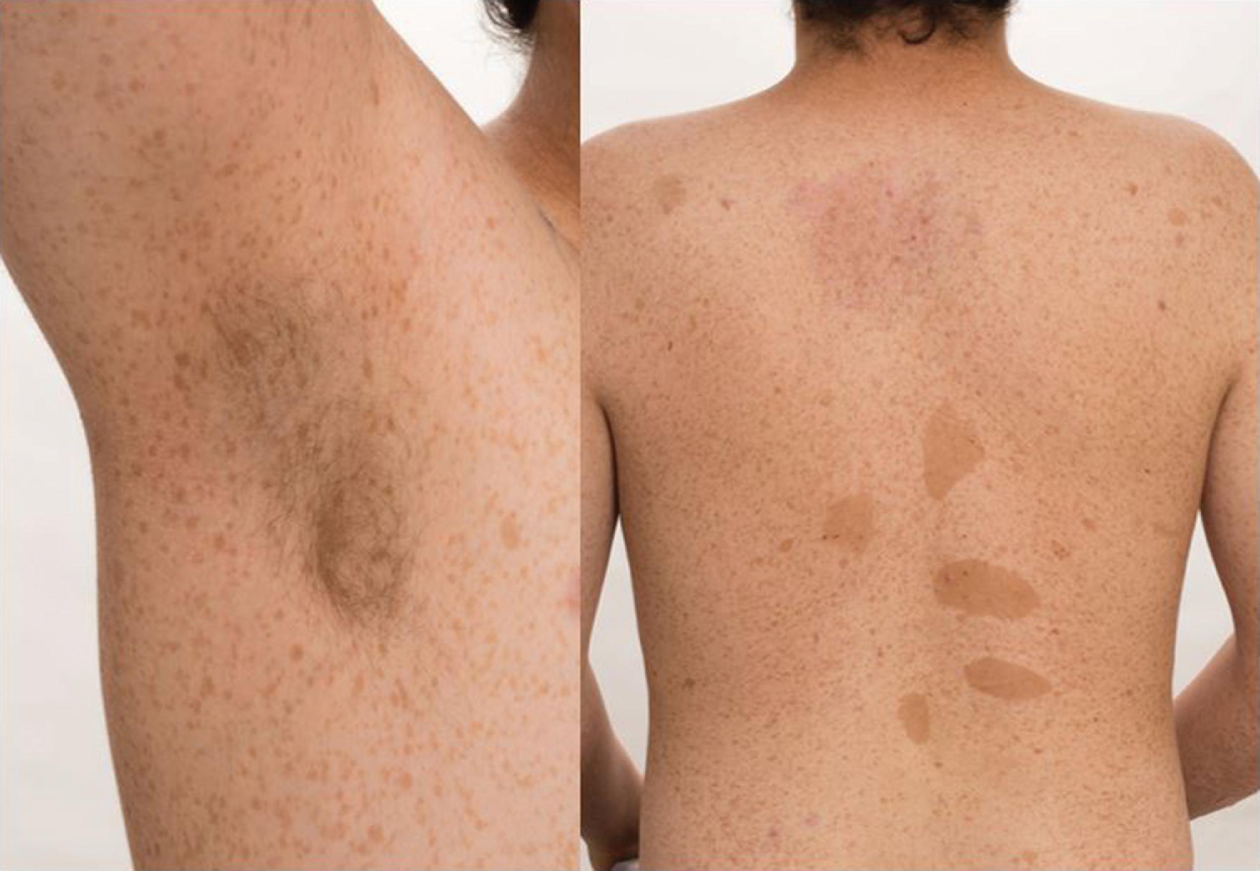
The patient was a 21-year-old man who presented for evaluation of an 8-cm left adrenal mass. He was diagnosed with NF1 at 6 months of age when café-au-lait spots and neurofibromas were detected on physical examination. He described progressive symptoms of palpitations, anxiety, and headaches for the 9 years prior. Over the last year these symptoms were precipitated by straining, and he also developed abdominal pain. Computed tomography (CT) of the abdomen was obtained to investigate the origin of the abdominal pain and led to the incidental discovery of the adrenal mass. The patient’s medical history was positive for attention deficit disorder, and he was not taking any medications. He had no family history of NF1. On physical examination, his blood pressure was 143/92 mmHg, heart rate 80 beats per minute, and body mass index 24.1 kg/m 2 . Café-au lait spots and axillary freckling were visible on examination ( Fig. 39.1 ).
INVESTIGATIONS
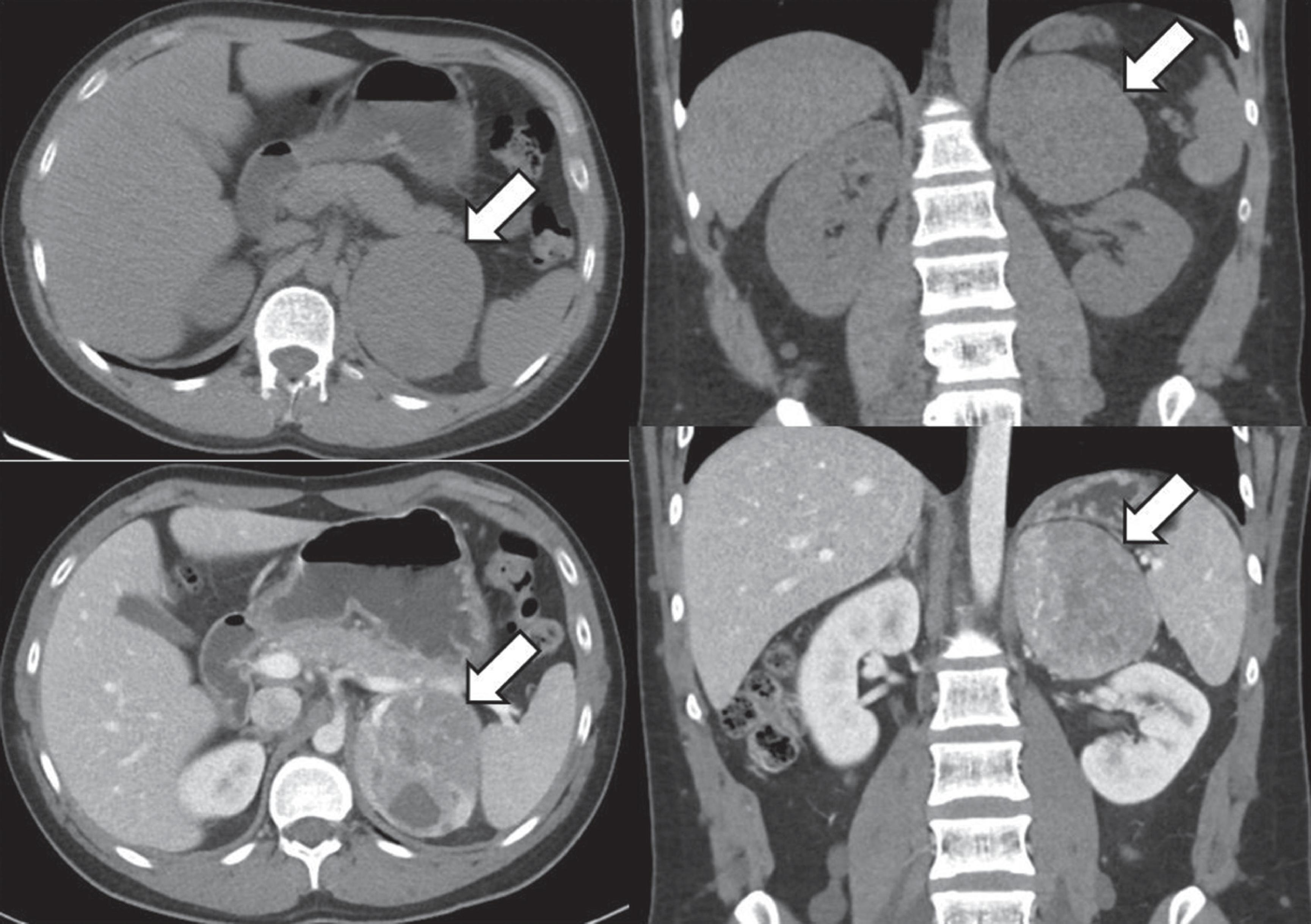
Abdominal CT revealed a heterogeneous left adrenal mass of 6.7 × 7.7 × 7.8 cm causing inferior displacement of the left kidney ( Fig. 39.2 ). The right adrenal gland appeared normal. Subsequent I-123 metaiodobenzylguanidine scintigraphy demonstrated increased radiotracer uptake in the left adrenal mass without any additional abnormal radiotracer uptake in the abdomen and pelvis ( Fig. 39.3 ). Preoperative laboratory studies are shown in Table 39.1 . The levels of metanephrine in the blood and urine were diagnostic of an adrenergic pheochromocytoma.

Stay updated, free articles. Join our Telegram channel

Full access? Get Clinical Tree


