Pathology of Invasive Breast Cancer


Pathology of Invasive Breast Cancer


Deborah Dillon

Anthony J. Guidi

Stuart J. Schnitt


Invasive breast cancers constitute a heterogeneous group of lesions that differ with regard to their clinical presentation, radiographic characteristics, histopathologic and molecular features, and biological potential. The most widely used classification of invasive breast cancers, and that used in this chapter (with minor modifications), is that of the World Health Organization (1). This classification scheme is based on the growth pattern and cytologic features of the invasive tumor cells and does not imply histogenesis or site of origin within the mammary duct system. For example, although the classification system recognizes invasive carcinomas designated “ductal” and “lobular,” this is not meant to indicate that the former originates in extralobular ducts and the latter in lobules. In fact, subgross whole organ sectioning has demonstrated that most invasive breast cancers arise in the terminal duct lobular unit, regardless of histologic type (2). The most common histologic type of invasive breast cancer by far is invasive (infiltrating) ductal carcinoma. In fact, the diagnosis of invasive ductal carcinoma is a diagnosis by default, since this tumor type is defined as a type of cancer not classified into any of the other categories of invasive mammary carcinoma (1). To further emphasize this point, and to distinguish these tumors from invasive breast cancers with specific or special histological features (such as invasive lobular, tubular, mucinous, papillary, and other rare types), some authorities prefer the term invasive or infiltrating ductal carcinoma, not otherwise specified (NOS) or of no special type (NST). In this chapter, the terms invasive ductal carcinoma, infiltrating ductal carcinoma, and infiltrating or invasive carcinoma of no special type are used interchangeably.

The distribution of histologic types of invasive breast cancer has varied among published series (Table 25-1). These differences may be related to a number of factors including the nature of the patient population and variability in the confines of definition for the different histological types. In general, special type cancers comprise approximately 20% to 30% of invasive carcinomas, and at least 90% of a tumor should demonstrate the defining histological characteristics of a special type cancer to be designated as that histological type (6).

The widespread use of screening mammography has had a dramatic impact on the nature of invasive breast cancers encountered in clinical practice. The value of mammography in detecting more cases of ductal carcinoma in situ, smaller invasive breast cancers, and fewer cancers with axillary lymph node involvement is well recognized. However, mammography has also resulted in a change in the distribution of the histological features of the invasive breast cancers detected. In particular, special type cancers (particularly tubular carcinomas) and cancers of lower histological grade are more frequently observed in mammographically screened populations than in patients who present with a palpable mass, particularly in the prevalent round of screening.

Most invasive breast cancers have an associated component of in situ carcinoma, although the extent of the in situ component varies considerably. The prevailing view has long been that the invasive carcinomas derive from the in situ component. This is based not only on the frequent coexistence of the two lesions, but on the histological similarities between the invasive and in situ components within the same lesion. For example, a number of studies have
clearly documented that low-grade invasive cancers are most often associated with low-grade ductal carcinoma in situ, and high-grade invasive cancers with high-grade in situ lesions (7). In addition, studies evaluating profiles of biological markers and genetic abnormalities have shown that coexisting invasive and in situ carcinomas often share the same immunophenotype and genetic alterations. Gene expression profiling studies have confirmed this observation (8).


TABLE 25-1 Histologic Types of Invasive Breast Cancer in Four Large Series before the Widespread Use of Mammographic Screening


Histologic Type (%)


Study


No. of Cancers


Ductalaa


Lobular


Medullary


Mucinous


Tubular


Tubular Mixed


Mixed


Other


Fisher et al. (3)


1,000


53


5


6


2


1


32


Rosen (4)


857


75


10


9


2


2


Ellis et al. (6)


1,547


49


16


3


1


2


14


14


2


Edinburgh (5)


Not stated


70


10


5


2


3


2


8


a In some series, designated “not otherwise specified” (NOS) or “no special type” (NST).


The routine pathologic examination of invasive breast cancers has extended beyond simply determining and reporting the histologic type of the tumor. Although histologic typing provides important prognostic information in and of itself, other morphologic features that are evaluable on routine histologic sections are also of prognostic value. In this chapter, the various histologic types of invasive breast cancer will be discussed as will pathologic features important in the assessment of prognosis (prognostic factors) and response to therapy (predictive factors). Characteristic molecular and immunophenotypic features will also be noted where appropriate.


INVASIVE (INFILTRATING) DUCTAL CARCINOMA

Invasive ductal carcinomas represent the single largest group of invasive breast cancers. Although these tumors are most commonly encountered in pure form, a substantial minority exhibit admixed foci of other histologic types. The classification of tumors composed primarily of invasive ductal carcinoma with a minor component consisting of one or more other histological types is problematic. Some authorities categorize such lesions as invasive ductal carcinomas (or invasive carcinomas of no special type) and simply note the presence of the other types, whereas others classify them as “mixed.”


Clinical Presentation

Invasive ductal carcinomas most often present as a palpable mass and/or mammographic abnormality. There are no clinical or mammographic characteristics that distinguish invasive ductal carcinomas from other histologic types of invasive cancer. Rarely, these lesions present with Paget disease of the nipple.


Gross Pathology

The classic macroscopic appearance of invasive ductal carcinoma is that of a scirrhous carcinoma, characterized by a firm, sometimes rock-hard, mass that on cut section has a gray-white gritty surface (Fig. 25-1). This consistency and appearance is due to the desmoplastic tumor stroma and not the neoplastic cells themselves. Some invasive ductal carcinomas are composed primarily of tumor cells with little desmoplastic stromal reaction, and such lesions are grossly tan and soft. Although most invasive ductal cancers have a stellate or spiculated contour with irregular peripheral margins, some lesions have rounded, pushing margins, and still others are grossly well circumscribed.


Histopathology

The microscopic appearance of invasive ductal carcinomas is highly heterogeneous with regard to growth pattern, cytologic features, mitotic activity, stromal desmoplasia, extent of the associated ductal carcinoma in situ (DCIS) component, and contour. Variability in histologic features may even be seen within a single case. The tumor cells may be arranged as glandular structures; as nests, cords, or trabeculae of various sizes; or as solid sheets. Foci of necrosis are evident in some cases and may be extensive. Cytologically, the tumor cells range from those that show little deviation from normal breast epithelial cells to those exhibiting marked cellular pleomorphism and nuclear atypia. Mitotic activity can range from imperceptible to marked. Stromal desmoplasia is inapparent to minimal in some cases. At the other end of the spectrum, some tumors show such prominent stromal desmoplasia that the tumor cells constitute only a minor component of the lesion. Similarly, some invasive ductal carcinomas have no identifiable component of DCIS, whereas in others, the in situ carcinoma is the predominant component of the tumor. An associated lymphocytic or lymphoplasmacytic infiltrate may or may not be present. Finally, the microscopic margins of the cancer may be infiltrating, pushing, circumscribed, or mixed.


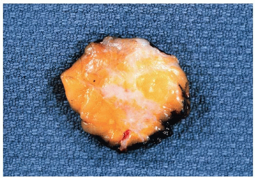
FIGURE 25-1 Cut surface of an excision specimen containing an invasive ductal carcinoma. The tumor appears as an irregular area of whitish tissue.

Recognizing that invasive ductal carcinomas are a histologically diverse group of lesions, many investigators have attempted to stratify them based upon certain microscopic features. The most common method to subclassify invasive ductal carcinomas is grading, which may be based solely on nuclear features (nuclear grading) or on a combination of architectural and nuclear characteristics (histologic grading). Histologic grading is the method of grading most often used in current practice. The histologic grading system currently in most widespread use is that of Elston and Ellis (reviewed in detail in reference 9). This system is a modification of the grading system proposed by Bloom and Richardson in 1957, but provides strictly defined criteria that are lacking in the original description. Tubule formation, nuclear pleomorphism, and mitotic activity are each scored on a 1 to 3 scale. The sum of the scores for these three parameters provides the overall histologic grade, such that tumors in which the sum of the scores is 3 to 5 are designated grade 1 (well differentiated), those with score sums of 6 and 7 are designated grade 2 (moderately differentiated), and those with score sums of 8 and 9 are designated grade 3 (poorly differentiated) (Fig. 25-2; Table 25-2). The prognostic significance of histologic grading is discussed below (see section on prognostic factors).


Biomarkers

The expression of biologic markers, such as estrogen and progesterone receptors, growth factors, oncogene and tumor suppressor gene products, and other markers is
highly variable in invasive ductal carcinomas as might be anticipated from their histologic heterogeneity. Although a large number of biomarkers have been studied in invasive ductal carcinomas, only estrogen receptor, progesterone receptor, and HER2 are reported in routine clinical practice at this time. Overall, 70% to 80% of invasive ductal carcinomas are estrogen receptor positive and approximately 15% are HER2 amplified and overexpressed.


FIGURE 25-2 Invasive ductal carcinoma. (A) Histologic grade 1. (B) Histologic grade 2. (C) Histologic grade 3.


TABLE 25-2 Histologic Grading System for Invasive Breast Cancers (Elston and Ellis Modification of Bloom and Richardson Grading System)


Components of Grade


Score


Tubules


>75% of tumor composed of tubules


1 point


10%-75% of tumor composed of tubules


2 points


<10% of tumor composed of tubules


3 points


Nuclear grade


Nuclei small and uniform


1 point


Moderate variation in nuclear size and shape


2 points


Marked nuclear pleomorphism


3 points


Mitotic rate


Dependent on microscope field area


1-3 points


Histologic Grade


Total points


1 (well differentiated)


3-5


2 (moderately differentiated)


6-7


3 (poorly differentiated)


8-9


Adapted from Ellis IO. Assessment of histologic grade. In: Elston CW, Ellis IO, eds. The breast. Edinburgh, Scotland: Churchill Livingstone, 1998:365-384.


Invasive ductal carcinomas also display a wide variety of genetic and genomic alterations. In gene expression profiling studies, invasive ductal carcinomas may be found within all major molecular subtypes (24).


Clinical Course and Prognosis

The prognosis of invasive ductal cancer varies according to tumor size, histologic grade, lymph node status, and presence of lymphovascular invasion as well as expression of hormone receptors and HER2 (see section on prognostic factors). However, even within this group prognostically favorable specialized tumor types can be identified, as discussed below.


INVASIVE (INFILTRATING) LOBULAR CARCINOMA

Invasive lobular carcinomas constitute the second most frequent type of invasive breast cancer, in most series accounting for approximately 5% to 15% of cases. Although some of this variability may be related to differences in patient populations, much of it appears to be due to differences in diagnostic
criteria. In particular, since the “classical” form of invasive lobular carcinoma was first described by Foote and Stewart (11), a variety of authors have described invasive breast cancers that they consider variants of invasive lobular carcinoma, thereby expanding the spectrum of this histologic type and accounting for a higher incidence of invasive lobular carcinoma in more recent series than in the past. In addition, recent studies have suggested that the increase in the frequency of infiltrating lobular carcinoma may be in part related to the use of postmenopausal hormone replacement therapy (14).

Invasive lobular carcinomas are characterized by multifocality in the ipsilateral breast and appear to be more often bilateral than other types of invasive breast cancer. Lobular carcinoma in situ coexists with invasive lobular carcinoma in the majority of cases, with 70% to 80% of cases of invasive lobular carcinoma associated with foci of lobular carcinoma in situ.


Clinical Presentation

Invasive lobular carcinoma may present as a palpable mass or a mammographic abnormality with characteristics similar to those of invasive ductal carcinomas (i.e., discrete, firm mass on palpation; spiculated mass on mammogram). However, both the findings on physical examination and the mammographic appearance of invasive lobular carcinomas may be quite subtle. On physical examination, there may be only a vague area of thickening or induration, without definable margins. Mammographic findings may be equally subtle, with many invasive lobular carcinomas appearing as poorly defined areas of asymmetric density with architectural distortion and others revealing no mammographic abnormalities, even in the presence of a palpable mass. In fact, the extent of the tumor may be substantially underestimated by both physical examination and mammography.


Gross Pathology

Some invasive lobular carcinomas appear as firm, gritty, gray-white masses, indistinguishable from invasive ductal carcinomas. However, in other cases, no mass is grossly evident and the breast tissue may have only a rubbery consistency. In still other cases, no abnormality is evident on visual inspection or upon palpation of the involved breast tissue and the presence of carcinoma is revealed only upon microscopic examination.


Histopathology

Invasive lobular carcinomas as a group show distinctive cytologic features and patterns of tumor cell infiltration of the stroma. The classical form is characterized by small, relatively uniform neoplastic cells that invade the stroma singly and in a single-file pattern which results in the formation of linear strands (Fig. 25-3). These cells frequently encircle mammary ducts in a targetoid manner. Furthermore, the tumor cells may infiltrate the breast stroma and adipose tissue in an insidious fashion, invoking little or no desmoplastic stromal reaction. This feature accounts for the difficulty in detecting some invasive lobular carcinomas on physical examination, mammography and gross pathologic examination. The nuclei of the neoplastic cells are usually small, show little variation in size, and are often eccentric. Mitotic figures are infrequent. The cells may contain intracytoplasmic lumina which, in some, may be large enough to impart a signet ring cell appearance. However, in the classical form of invasive lobular carcinoma, cells with a signet ring configuration comprise only a small proportion of the tumor cell population.

Many examples of invasive lobular carcinoma (as well as lobular carcinoma in situ) are characterized histologically by tumor cells that are loosely cohesive. This phenotype may be, at least in part, related to the fact that both in situ and invasive lobular carcinomas typically show loss of expression of the adhesion molecule E-cadherin. This is associated, in many cases, with mutations in the gene encoding this protein (17) or to loss of heterozygosity on chromosome 16q22.1, the region of the E-cadherin gene (18). Although loss of E-cadherin expression characterizes lobular carcinomas and distinguishes them from ductal-type carcinomas, a subset of lobular carcinomas are reported to be E-cadherin positive (19). Thus, membrane expression of E-cadherin in an invasive carcinoma with morphologic characteristics of invasive lobular carcinoma is not, by itself, sufficient for classification as invasive ductal carcinoma.

Variant forms of invasive lobular carcinoma differ from the classical form with regard to architectural and/or cytologic features. In the solid and alveolar variants, the cells comprising the tumor have features characteristic of the classical form of invasive lobular carcinoma, but differ from the classical form with regard to the growth pattern of the tumor cells (10). In the solid form, the neoplastic cells grow in large confluent sheets with little intervening
stroma (Fig. 25-4). The alveolar form is characterized by tumor cells that grow in groups of 20 or more cells. These cellular aggregates are separated from one another by a delicate fibrovascular stroma (Fig. 25-5). Although a trabecular variant has also been described (10), there is considerable overlap between this pattern and that seen in the classical form of invasive lobular carcinoma. In the pleomorphic variant, the neoplastic cells are larger, exhibit more nuclear variation than that seen in the classical form, and may show apocrine features (13) (Fig. 25-6). Although signet ring cells can be seen in the classical type of invasive lobular carcinoma as well as in some examples of invasive ductal carcinoma, tumors that are composed of a prominent component of signet ring cells that otherwise have the characteristic features of invasive lobular carcinoma are considered to represent the signet ring cell variant of invasive lobular carcinoma (12). Histiocytoid carcinoma is an apocrine variant of invasive lobular carcinoma in which the tumor cells have a histiocyte-like appearance with abundant foamy pale eosinophilic cytoplasm and mild nuclear atypia (20). Some authors have recognized a “mixed” category of invasive lobular carcinoma. This term is generally used to designate lesions in which no single pattern comprises more than 80% to 85% of the lesion (21).


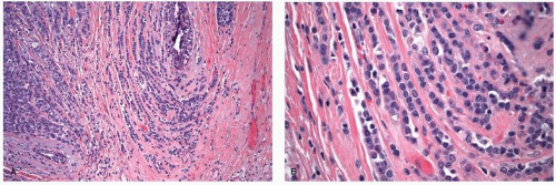
FIGURE 25-3 Invasive lobular carcinoma, classic type. (A) Linear strands of tumor cells infiltrate the stroma. (B) Higher-power view to demonstrate cytologic detail. The tumor cells have small, relatively uniform nuclei.


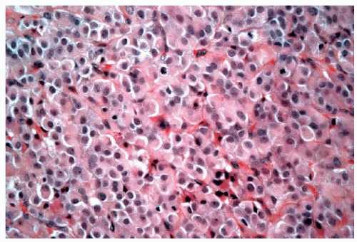
FIGURE 25-4 Invasive lobular carcinoma, solid type. The tumor cells grow in a confluent sheet with little intervening stroma.


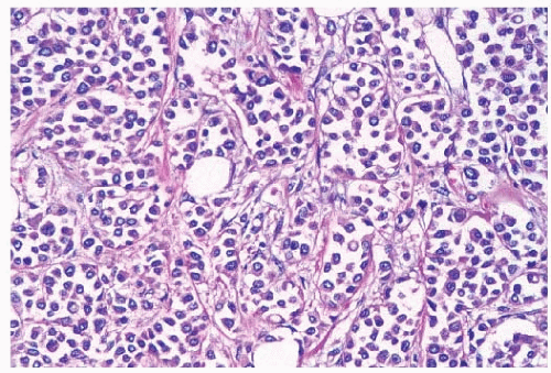
FIGURE 25-5 Invasive lobular carcinoma, alveolar type. Loosely cohesive tumor cell aggregates are separated by delicate fibrous septa.


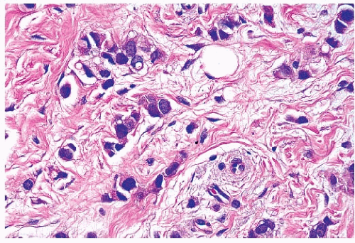
FIGURE 25-6 Invasive lobular carcinoma, pleomorphic type. The tumor cells infiltrate the stroma in linear strands, similar to those seen in the classic type of invasive lobular carcinoma. However, the cells in this lobular variant show considerable nuclear pleomorphism, in contrast to the small, monomorphic nuclei characteristic of the classic type of invasive lobular carcinoma (compare with Fig. 25-3B).

The relative frequency of the various lobular subtypes is difficult to discern since not all subtypes have been recognized in all series. In addition, patient selection criteria have varied among these studies. In the series of Dixon et al., among 103 invasive lobular carcinomas, 30% were of the classical type, 22% were solid, 19% were alveolar, and 29% were mixed lesions (15). In the experience of Ellis et al., 40% of invasive lobular carcinomas were of the classical type, 10% were solid type, 4% were alveolar, and 40% were mixed (6). In contrast, in a study from Memorial Sloan-Kettering Cancer Center, 176 of 230 invasive lobular carcinomas (77%) were of the classical type and the remainder were variants: 4% solid, 6% alveolar, and 13% mixed (16) (Table 25-3).


Biomarkers

Classical invasive lobular carcinomas typically show expression of estrogen and progesterone receptors and rarely show HER2 overexpression or amplification. Although pleomorphic lobular carcinomas are also frequently estrogen receptor and progesterone receptor positive, they may also show overexpression and amplification of HER2 (23). Gross cystic disease fluid protein 15 is seen in about one-third of all invasive lobular carcinomas, but is present in the vast majority of lesions that show prominent signet ring cell features (22).

In gene expression profiling studies, most invasive lobular carcinomas are classified as luminal A subtype; however, some cases fall within the luminal B, HER2 and basal-like groups (24). Chromosomal analysis shows characteristic loss of 16q and gain of material on 1q and 16p (18).


Clinical Course and Prognosis

There are several aspects of the clinical course of invasive lobular carcinomas that merit consideration. First, a number of studies have noted differences in the pattern of metastatic spread between invasive lobular and invasive ductal carcinomas. In particular, metastases to the lungs, liver, and brain parenchyma appear to be less common in patients with lobular than ductal cancers (25, 26). In contrast, lobular carcinomas have a greater propensity to metastasize to the
leptomeninges, peritoneal surfaces, retroperitoneum, gastrointestinal tract, and reproductive organs and bone (25). In fact, the majority of cases of carcinomatous meningitis in patients with metastatic breast cancer occur in patients with lobular cancers (98, 26). Peritoneal metastases may appear as numerous small nodules studding the peritoneal surfaces in a manner similar to that seen in ovarian carcinoma (25, 26). Metastases to the stomach can produce an appearance that simulates an infiltrative (linitis plastica) type of primary gastric carcinoma (27). Involvement of the uterus may result in vaginal bleeding (28), whereas metastatic tumor in the ovary may produce ovarian enlargement and the appearance of a Krukenberg tumor.


TABLE 25-3 Frequency of Invasive Lobular Carcinoma Subtypes in Series with More Than 100 Patients


Subtypes


Study


No. Invasive Lobular Carcinomas


Classic


Solid


Alveolar


Tubulolobular


Mixed


Dixon et al. (15)


103


30


22


19


Not included


29


Ellis et al. (6)


243


40


10


4


6


40


DiCostanzo et al. (16)


230


77


4


6


Not included


13


Whether or not invasive lobular carcinomas differ in overall prognosis from invasive ductal carcinomas is difficult to determine due in large part to variations in the application of histologic criteria for the diagnosis of invasive lobular carcinoma. However, the prognosis of patients with invasive lobular carcinoma as a group has not consistently been shown to differ from that of patients with invasive ductal carcinoma. Several studies have suggested that the prognosis for the classical form of invasive lobular carcinoma is better than variant types and than invasive ductal carcinomas (6, 15, 16). Available evidence suggests that the pleomorphic variant and the signet ring cell variant (when defined as lesions in which greater than 10% of the neoplastic cells are of the signet ring cell type) appear to be associated with a particularly poor clinical outcome (13).

Numerous clinical follow-up studies have indicated that patients with invasive lobular carcinoma can be adequately treated with conservative surgery and radiation therapy following complete gross excision of the tumor, with local recurrence rates comparable to those seen in patients with invasive ductal carcinoma (reviewed in reference 29).


INVASIVE CARCINOMAS WITH DUCTAL AND LOBULAR FEATURES

A small proportion of invasive breast cancers, up to 5% in most studies, are not readily classifiable as either ductal or lobular (6). Invasive cancers may be difficult to classify definitively as either ductal or lobular either because they show distinct features of both or have features that are indeterminate. Cancers that show distinct areas of invasive ductal carcinoma and invasive lobular carcinoma are best classified as mixed invasive ductal and invasive lobular carcinoma. Tubulolobular carcinoma is a distinctive type of low-grade invasive breast cancer that is E-cadherin positive but shows both ductal and lobular morphologic features. In this variant, some of the tumor cells invade the stroma in linear strands characteristic of the classical form of invasive lobular carcinoma whereas others form small tubules with round to ovoid contours. These tubules are smaller and less angulated than those seen in tubular carcinoma (see below). Some invasive cancers have both cytologic and architectural features that are intermediate between those of invasive ductal and invasive lobular carcinomas. Immunohistochemical staining for E-cadherin and cyto-keratin 8 has been proposed as a useful adjunct in making the distinction between ductal and lobular carcinomas in histologically problematic or indeterminate cases (30). The fact that it may be difficult for the pathologist to categorize a given lesion as ductal or lobular in some cases should not be surprising in view of reports suggesting that some invasive ductal carcinomas exhibit cytogenetic alterations that are similar to those seen in invasive lobular carcinomas (18).

Given the heterogeneous nature of the lesions included in this group, data on clinical features and outcome of patients with invasive carcinomas with ductal and lobular features are difficult to interpret. However, these lesions do not appear to be distinctive in their rate of local recurrence or distant failure when compared with patients with invasive ductal or invasive lobular carcinomas.


TUBULAR CARCINOMA

Tubular carcinoma is a special type cancer that is associated with limited metastatic potential and an excellent prognosis. Prior to the widespread use of screening mammography, tubular carcinomas accounted for less than 4% of all breast cancers (31). However, these tumors account for a much higher proportion of cancers detected in mammographically screened populations.


Clinical Presentation

The mean age at presentation for patients with tubular carcinoma is in the early sixth decade (31, 32). Historically, the majority of tubular carcinomas were detected as palpable lesions; however, the majority (60% to 70%) now present as nonpalpable mammographic abnormalities. Not infrequently, tubular carcinomas are discovered incidentally in biopsies performed for unrelated reasons. Rare examples of tubular carcinoma have been reported in men.

Mammographic abnormalities, in the absence of a palpable mass, have been reported in the majority (80%) of patients with tubular carcinomas; however, mammographically occult tubular carcinomas are not infrequent. When a mammographic abnormality is present, it is usually a mass lesion, occasionally associated with microcalcifications. The mass may be irregular, round, oval, or lobulated. The majority of tubular carcinomas have spiculated margins, and cannot be distinguished radiologically from infiltrating ductal carcinomas.


Gross Pathology

Tubular carcinomas are typically small, with an average diameter less than 1.0 cm in most series (32). Tubular carcinomas detected by screening mammography are typically smaller than palpable lesions. Grossly tubular carcinomas are firm, spiculated lesions that are indistinguishable from infiltrating ductal carcinomas.


Histopathology

Tubular carcinomas are characterized by a proliferation of well-formed glands or tubules formed by a single layer of epithelial cells without surrounding myoepithelial cells. These tubules tend to be ovoid in shape and have sharply angular contours with tapering ends, and open lumens. The cells comprising these tubules are characterized by low-grade nuclear features and are usually polarized toward the lumen, often exhibiting apical cytoplasmic “snouts” (Fig. 25-7). The stroma of tubular carcinomas usually has desmoplastic features, and prominent elastosis may be present. There is now general agreement that more than 90% of the tumor should exhibit this characteristic morphology to be categorized as a “pure” tubular carcinoma (1); however, the proportion required for this diagnosis in published studies has varied from 75% to 100%.

The majority of tubular carcinomas have an associated intraductal component. The DCIS seen in association with tubular carcinoma is usually of low nuclear grade, with cribriform and micropapillary patterns, and does not typically comprise a large proportion of the tumor mass. In addition, flat epithelial atypia may be found in the vicinity of tubular carcinomas (36). Lobular carcinoma in situ may also be observed in association with tubular carcinoma. The frequency of multifocality and multicentricity in tubular carcinoma is difficult to determine due to varying definitions and methods of specimen sampling employed by different investigators. In one report in which 17 mastectomy specimens with tubular carcinomas were examined using the Egan serial subgross method (37), Lagios et al. (33) found a 56% incidence of multicentricity, defined in that study as carcinoma of any type present 5 cm from the index lesion. This incidence was significantly greater than a control group comprised of mastectomy specimens containing breast cancers of other types (33).

Because these lesions are extremely well differentiated, several benign entities such as sclerosing adenosis, radial scars, complex sclerosing lesions, and microglandular adenosis may enter into the differential diagnosis. In such cases, the use of immunohistochemical stains may be necessary in order to arrive at the correct diagnosis.


Biomarkers

The expression of various biologic markers in tubular carcinomas generally reflects the well-differentiated nature and favorable prognosis associated with these lesions. Tubular carcinomas are strongly positive for estrogen receptor and usually positive for progesterone receptor. In addition, these lesions are almost always diploid, have a low proliferative rate and no HER2 overexpression or amplification (38). When compared to invasive carcinomas of no special type, tubular carcinomas exhibit fewer overall chromosomal changes, more often show losses of 16q and less often show losses of 17p (39). On gene expression profiling studies, tubular carcinomas fall into the luminal A category (24).


Clinical Course and Prognosis

The reported incidence of axillary lymph node metastases in patients with tubular carcinomas is up to 29% (31, 32); however, there is considerable variation in the histologic definitions employed in these studies. A number of studies have shown an inverse relationship between the degree of tubular differentiation and the incidence of lymph node metastases (34). Nevertheless, even patients with “pure” tubular carcinomas (over 90% tubules) have nodal metastases in up to 15% of cases (35). However, as with other types of breast cancer, the size of the tumor strongly influences the likelihood of axillary metastases. Winchester reported that 67% of tubular carcinomas associated with nodal metastases were greater than the median size of 1.0 cm (35). The relative infrequency of nodal disease in patients with small tubular carcinomas has led some investigators to advocate abandoning axillary lymph node dissection in these patients. With regard to survival, patients with tubular carcinoma have an excellent prognosis, equivalent in some series to age-matched women without breast cancer (40). In the randomized prospective NSABP-B06 trial, 1090 node-negative and 651 node-positive patients were classified with regard to histologic type, and the “favorable” category included 120 patients with tubular carcinoma (43). Both node-negative and node-positive patients in the “favorable” category
experienced significantly greater overall survival at 10 years compared to other patients in a univariate analysis, and “favorable” histology proved to be an independent predictor of survival in node-negative patients by multivariate analysis (43). Similar improved survival rates in patients with tubular carcinoma were reported in a series of 1,621 patients, although these patients were not stratified by node status (6). In this latter study, even patients with “tubular mixed” tumors (which were defined as stellate cancers composed of cells typical of invasive ductal carcinoma but with central tubules identical to tubular carcinoma) experienced significantly better overall survival compared to patients with invasive ductal carcinoma (6). In addition, two series, one examining node-negative early stage breast cancer patients treated with mastectomy, and the other examining early stage patients treated with breast-conserving therapy, both reported that patients with tubular carcinoma had significantly lower rates of distant recurrences compared to patients with invasive ductal carcinoma (41, 42).


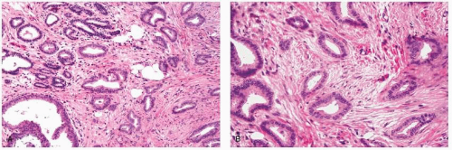
FIGURE 25-7 Tubular carcinoma. (A) This tumor is composed of well-formed glandular structures in a desmoplastic stroma. (B) The glands, or tubules, are elongated, and some have tapering ends. Numerous cytoplasmic “snouts” are evident at the luminal aspect of the tumor cells.

Other investigators have suggested that even patients with node-positive tubular carcinomas have a good prognosis. When tubular carcinoma does metastasize to axillary lymph nodes, usually one and seldom more than three nodes are involved. Several investigators have concluded that the presence of nodal disease in patients with tubular carcinoma does not affect disease-free or overall survival in these patients (35).

Reports examining the use of conservative surgery and radiation therapy in patients with tubular carcinoma show no significant differences in local recurrence rates when patients with tubular carcinomas are compared to patients with invasive ductal carcinoma (42). Although it is tempting to speculate that at least some patients with tubular carcinoma may be adequately treated with local excision alone (i.e., without radiation therapy), there are currently insufficient data to consider this a standard treatment option.


MUCINOUS CARCINOMA

Mucinous carcinoma (also known as colloid carcinoma) is another special type cancer that is associated with a relatively favorable prognosis. The reported incidence of mucinous carcinoma varies depending on the histologic criteria. Most studies have indicated that less than 5% of invasive breast carcinomas have a mucinous component and of these, less than half represent pure mucinous carcinomas (44).


Clinical Presentation

The mean age at presentation for patients with mucinous carcinoma is in the seventh or early eighth decade in most studies, and is greater than that for patients with breast cancers of no special type. Many patients with mucinous carcinoma present with palpable tumors. However, with widespread screening mammography, a substantial proportion (30% to 70%) present with nonpalpable mammographic abnormalities, most often poorly defined or lobulated mass lesions that are rarely associated with calcification (46). Wilson and coworkers reported that pure mucinous carcinomas were more often associated with a circumscribed, lobulated contour than the irregular borders characteristic of tumors with a mixture of mucinous and nonmucinous components (mixed mucinous tumors) (46). In addition, mammographically occult mucinous carcinomas are not infrequent.


Gross Pathology

Mucinous carcinomas average approximately 3 cm in size, with a wide range reported in the literature (47). Mucinous carcinomas have a distinctive gross appearance. These lesions are typically circumscribed and have a variably soft, gelatinous consistency, and a glistening cut surface. However, lesions with a greater amount of fibrous stroma may have a firmer consistency.


Histopathology

The hallmark of mucinous carcinomas is extracellular mucin production. However, the extent of extracellular mucin varies from tumor to tumor. Typically, tumor cells in small clusters are dispersed within pools of extracellular mucin (Fig. 25-8). This characteristic histology should comprise at least 90% of the tumor (or 100% according to some) (6) to qualify for the diagnosis of mucinous carcinoma. The cells comprising mucinous carcinomas are usually of low or intermediate nuclear grade. Mucinous neoplasms intermixed with other non-mucinous histologic features are classified as “mixed” mucinous tumors. The cellularity of mucinous carcinomas is variable, with some tumors being highly cellular (type B) and others relatively paucicellular (type A). For paucicellular type A mucinous carcinomas, the differential diagnosis may
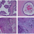
include mucocele-like lesions, benign lesions characterized by cystically dilated ducts associated with rupture and extravasation of mucin into the stroma. Type B mucinous carcinomas may show endocrine differentiation, including immunoreactivity for chromogranin or synaptophysin (48). Mucinous carcinomas are often accompanied by a DCIS component which may have a papillary, micropapillary, cribriform, or solid pattern. In some cases, the DCIS may also exhibit prominent extracellular mucin production (47).


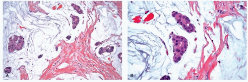
FIGURE 25-8 Mucinous carcinoma. (A) The tumor is composed of clusters of neoplastic cells dispersed in mucous pools. (B) In this specimen, the neoplastic cells have intermediate-grade nuclei.


Biomarkers

The expression of various biological markers in mucinous carcinomas reflects the good prognosis associated with these lesions. Mucinous carcinomas are generally estrogen receptor positive. The majority of cases (about 70%) are also progesterone receptor positive. In addition, mucinous carcinomas usually do not overexpress the HER2 protein or show HER2 amplification (38). Mucinous carcinomas show relatively little genomic instability, with substantially fewer chromosomal gains and losses than invasive carcinomas of no special type (49). In gene expression studies, mucinous carcinomas generally cluster within the luminal A subtype. Type B mucinous carcinomas are distinct from type A mucinous carcinomas and cluster with other breast carcinomas showing neuroendocrine differentiation (50).


Clinical Course and Prognosis

Only 12% of patients presented with axillary lymph node metastasis in the SEER database review of over 11,000 patients with pure mucinous carcinoma (45). This is significantly less than the incidence of node positivity seen in mixed mucinous tumors or invasive breast cancers of no special type. Lymph node involvement is related to tumor size and is extremely rare in mucinous carcinomas measuring less than 1 cm (38).

With regard to survival, 38 patients with mucinous carcinoma were enrolled in the NSABP-B06 trial, and they experienced the same significantly increased survival as patients with tubular carcinoma, particularly in the nodenegative group (43). Similar results were reported by Ellis and coworkers in their retrospective series; however, these patients were not stratified by nodal status (6). A report utilizing the SEER database compared 20-year survival data from 11,422 patients with mucinous carcinoma and patients with invasive ductal carcinoma diagnosed between 1973 and 2002 (45). Similar to the studies cited above, this report indicated that the patients with mucinous carcinoma present most often with localized disease (86%), with only 12% having regional lymph node involvement and 2% with distant metastases at the time of diagnosis. Although there were no significant differences in overall survival, survival at 10, 15, and 20 years for mucinous carcinoma was 89%, 85%, and 81%, respectively, compared with 72%, 66%, and 62% for invasive ductal carcinoma. The most significant prognostic factors in multivariate analyses were nodal status, then age, tumor size, progesterone receptor status, and nuclear grade (45). In addition, two series, one examining node-negative early stage breast cancer patients treated with mastectomy (with 20-year follow-up), and the other examining early stage patients treated with breast-conserving therapy (with 10-year follow-up), both reported that patients with mucinous carcinoma had significantly lower rates of distant recurrences compared to patients with invasive ductal carcinoma (41, 42). Several studies have noted that a significant number of late recurrences are seen in patients with mucinous carcinoma, with one report documenting a recurrence 30 years after initial treatment (51).

Several studies have examined the use of conservative surgery and radiation therapy in patients with mucinous carcinoma, and report no significant differences in local recurrence rates compared to patients with invasive ductal carcinoma (42). Given the relatively good prognosis in patients with mucinous carcinoma, some authors have raised the question of whether radiation therapy can be safely omitted after breast-conserving surgery in patients with this tumor type; however, at this time, there are insufficient data on which to base such a recommendation.

Mucinous carcinomas have rarely been associated with unusual metastatic manifestations, including mucin embolism resulting in fatal cerebral infarcts and pseudomyxoma peritonei (52, 53).


CARCINOMAS WITH MEDULLARY FEATURES

Classic medullary carcinomas are rare, accounting for less than 1% of all invasive breast cancers. Initial reports indicated that this type of breast cancer had a favorable prognosis despite its aggressive histologic appearance (54, 55). However, there is considerable controversy regarding the appropriate histologic definition of medullary carcinoma, as well as the reproducibility of this diagnosis among pathologists. Carcinomas with some but not all of the features of medullary carcinoma have been called “atypical medullary carcinomas,” “invasive carcinomas with medullary features,” and “invasive ductal carcinomas with medullary features.” Given the difficulties in applying the criteria for medullary carcinoma reproducibly, the current WHO classification combines medullary carcinomas, atypical medullary carcinomas and invasive ductal carcinomas with medullary features into a single group designated “carcinomas with medullary features” (1).


Clinical Presentation

Patients with carcinomas with medullary features usually present at a younger age than patients with other breast cancers, owing, at least in part, to the inclusion in this group of patients with inherited BRCA1 mutations. The majority of patients present with a palpable mass. Of interest, some patients with this tumor type exhibit axillary lymphadenopathy at the time of presentation with histologic examination of the lymph nodes showing only benign reactive changes (59). Rare examples of carcinoma with medullary features have been reported in males.

Most carcinomas with medullary features are associated with a moderately well-defined mass without calcifications (59); however, a significant proportion are associated with an ill-defined margin. Moreover, the majority of mammographically well-circumscribed cancers are infiltrating ductal carcinomas rather than medullary carcinomas (60). On ultrasound examination, medullary carcinomas are generally well-circumscribed, frequently lobulated, and hypoechoic (60).


Gross Pathology

The mean size of medullary carcinomas is similar to that of breast cancers of no special type (47). Grossly, these lesions are well circumscribed, soft, tan-brown to gray tumors that bulge above the cut surface of the specimen. A multinodular appearance may be appreciated in some cases. Areas of hemorrhage, necrosis, or cystic degeneration may be present in tumors of any size, but prominent necrosis is often seen in larger tumors.


Histopathology

Several similar but distinct classification systems for the histologic diagnosis of medullary carcinomas have been proposed (56, 57 and 58). All three classification schemes recognize the following attributes, but the relative importance and the mandatory nature of each are stressed to a different degree: (i) syncytial growth pattern of the tumor cells in more than 75% of the tumor, (ii) admixed lymphoplasmacytic infiltrate, (iii) microscopic circumscription,(iv) grade 2 or 3 nuclei, and (v) absence of glandular differentiation (Fig. 25-9). Tumors that lack a variable number of these characteristics (depending on the system used) have been classified either as “atypical medullary carcinoma,” or invasive ductal carcinoma.

Carcinomas with medullary features may show hemorrhage, tumor necrosis, cystic degeneration, and various types of metaplasia of the tumor cells, most often squamous metaplasia (47). Some tumors exhibit bizarre cytologic features with marked nuclear atypia and multinucleated tumor giant cells. There is usually little or no associated in situ carcinoma.


Biomarkers

Carcinomas with medullary features are typically estrogen receptor negative, progesterone receptor negative, and lack HER2 overexpression/amplification (“triple negative”) and show a high proliferation rate. They often show a basal-like phenotype, with expression of basal cytokeratins (CK5/6, 14, and 17) and epidermal growth factor receptor (EGFR) and may have TP53 mutations. Genomic instability is a characteristic feature of these lesions. Most breast cancers in women with germline BRCA1 mutations show medullary features; however, only about 13% of cancers with medullary features are associated with germline BRCA1 mutations (61). Many of the sporadic carcinomas show inactivation of BRCA1 by somatic mutation or promoter hypermethylation (62).

Gene expression profiling studies have compared patterns of gene expression in 22 cancers diagnosed as medullary carcinoma with 44 high-grade invasive ductal carcinomas. Results of these studies showed that 95% of medullary carcinomas displayed a basal-like profile, similar to that seen in the basal group of invasive ductal carcinomas. Compared with the basal group of invasive ductal carcinomas, however, medullary carcinomas showed lower expression of genes involved in smooth muscle differentiation and greater expression of genes on 12p13 and 6p21, regions known to contain genes involved in pluripotency. Together, these findings suggest that carcinomas with medullary features represent a subset of basal-like breast cancers (63). Studies using array comparative genomic hybridization (CGH) have further clarified the similarities and differences between cancers diagnosed as medullary and other basallike carcinomas. In one study, medullary carcinomas and other basal-like carcinomas were both characterized by 1q and 8q gains and X losses, with medullary carcinomas typically showing greater chromosomal instability and a wider spectrum of chromosomal gains and losses (64).


Clinical Course and Prognosis

Although studies have differed in the histologic criteria employed, most studies indicate that the incidence of axillary lymph node metastases is lower in patients with medullary carcinomas (19% to 46%) than in those with atypical medullary carcinomas (30% to 52%) or invasive ductal carcinomas with medullary features (29% to 65%) (56, 57).

Data regarding survival rates in patients with medullary carcinoma and carcinomas with medullary features are difficult to interpret given the different classification systems employed and general lack of reproducibility. Although a number of earlier studies reported a more favorable prognosis for medullary carcinomas, this finding has not been confirmed in all studies. In addition, a number of studies have also questioned the practical applicability of the diagnostic criteria. Thus, although there may be patients with medullary carcinoma who have improved survival compared to patients with breast cancers of no special type, the ability of pathologists to reliably and reproducibly identify this subset of patients is limited at the current time. It is essential that clinicians be aware of these limitations when confronted with a pathology report suggesting the diagnosis of medullary carcinoma or carcinoma with medullary features. Recently, some studies have suggested that features of the associated inflammatory infiltrate may be important in the prognosis of both high-grade medullary-like and non-medullary-like carcinomas (65, 66).

The results of the use of breast-conserving therapy in patients with medullary carcinoma have been reported in several studies (42) with no significant differences in local recurrence rates among patients with medullary carcinoma compared to patients with invasive ductal carcinoma. Thus,
the available data suggest that conservative surgery and radiation therapy is appropriate local treatment for patients with medullary carcinoma and carcinoma with medullary features.


FIGURE 25-9 Medullary carcinoma. (A) Low-power photomicrograph demonstrating the well-circumscribed border of the tumor. (B) The tumor cells show high-grade nuclear features, and a prominent admixture of lymphocytes and plasma cells is seen.


INVASIVE CRIBRIFORM CARCINOMA

Invasive cribriform carcinoma is a well-differentiated cancer that shares some morphologic features with tubular carcinoma, and is also associated with a favorable prognosis. Invasive cribriform carcinoma accounts for 1% to 4% of invasive breast cancers (67).


Clinical Presentation

The majority of patients with invasive cribriform carcinoma present in the sixth decade (range 19 to 86 years) (67). Tumors may present as a palpable mass, but are often clinically occult and detected by mammography as spiculated masses with or without associated calcifications.


Gross Pathology

No distinctive gross features of invasive cribriform carcinoma have been described.


Histopathology

Invasive cribriform carcinomas are characterized by low- to intermediate-grade tumor cells that invade the stroma in a cribriform or fenestrated growth pattern similar to that seen in the cribriform pattern of DCIS (Fig. 25-10). These tumors often show admixtures of other histologic patterns of invasive breast cancer, particularly tubular carcinoma, which is seen in approximately 20% of cases. The “classic” variant of invasive cribriform carcinoma, described by Page et al. (67), is defined as a tumor composed of an exclusively invasive cribriform pattern, or a tumor with more than 50% invasive cribriform features in which the remainder of the tumor exhibits features of tubular carcinoma. Tumors with any component of nontubular carcinoma were described as “mixed” in that study. Most invasive cribriform carcinomas are associated with DCIS, usually of the cribriform type. The average size of these tumors is relatively large, 3.1 cm (range 1 to 14 cm) for the classical variant of cribriform carcinoma, to 4.2 cm (range 2 to 9 cm) for tumors of mixed histology (67).

Only gold members can continue reading. Log In or Register to continue

Stay updated, free articles. Join our Telegram channel

Jul 9, 2016 | Posted by in ONCOLOGY | Comments Off on Pathology of Invasive Breast Cancer

Full access? Get Clinical Tree

Get Clinical Tree app for offline access