Pathogenesis of Atherosclerosis and the Unstable Plaque


Pathogenesis of Atherosclerosis and the Unstable Plaque


Frank D. Kolodgie

Gaku Nakazawa

Aya Nakazawa

David R. Fowler

Robert Kutys

Renu Virmani


The earliest descriptions of atherosclerotic lesions focused on morphologies of fatty streaks to fibroatheromas (FA) and advanced plaques complicated by hemorrhage, calcification, ulceration, and thrombosis. This long-standing terminology, however, lacked an understanding of how plaques progressed to entities responsible for acute coronary syndromes and was later refined in the mid-1990s by the American Heart Association (AHA) consensus group headed by Dr. Stary (1,2). The consensus classification consists of six different numerical categories to include early lesions of initial type I, adaptive intimal thickening; type II, fatty streak; and type III, transitional or intermediate lesion and advanced plaques characterized as type IV, atheroma; type V, fibroatheroma or atheroma with thick fibrous cap; and type VI, complicated plaques with surface defects, and/or hematoma-hemorrhage, and/or thrombosis.

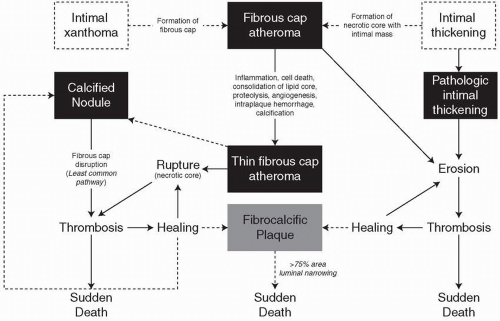
Our laboratory, however, found the numeric nomenclature challenging, and the lesion categories were essentially incomplete since luminal thrombi were thought to develop exclusively from plaque rupture (PR), excluding alternative existence of plaque erosion, and nodular calcification (3). These limitations prompted us to develop a modified version of the AHA classification. In our modified classification, numeric AHA lesion types I to IV are now replaced by descriptive terminology to include adaptive intimal thickening, intimal xanthoma, pathologic intimal thickening (PIT), and fibroatheroma, respectively (Figs. 3.1 and 3.2). Lesion reference to AHA types V and VI was discarded since it failed to account for the three different morphologies (rupture, erosion, and calcified nodule) that give rise to acute coronary thrombosis.


FATAL CORONARY PLAQUES

Fatal coronary plaques are depicted in (Fig. 3.1).


  • Plaque rupture is defined by fibrous cap disruption or fracture whereby the overlying thrombus is in continuity with the underlying necrotic core (4).


  • Plaque erosion is identified when serial sectioning through a thrombus fails to show communication with a necrotic core or deep intima; the endothelium is absent and the thrombus is superimposed on a plaque substrate primarily composed of smooth muscle cells (SMCs) and proteoglycans (5).


  • Calcified nodule is characterized by eruptive, dense calcified bodies protruding into the luminal space and represents the least frequent morphology associated with luminal thrombosis (3).

Another lesion of emphasis, the presumed precursor to PR, is the thin-cap fibroatheroma (TCFA). This lesion contains a fibrous cap measuring < 65 µm in thickness, which is heavily infiltrated by macrophages and lymphocytes accompanied by a paucity of α-actin-positive SMCs (6).

In approximately 50% to 60% of sudden coronary deaths examined at autopsy, the culprit lesion (fatal plaque) exhibits an acute coronary thrombus, whereas the remainder include stable coronary plaques with >75% cross-sectional area luminal narrowing (5). More than half of patients without acute coronary thrombi have healed myocardial infarcts, and in 15% to 20% of cases, there is no underlying myocardial pathology implicating a terminal arrhythmia (3).


EARLY LESIONS OF ATHEROSCLEROSIS


Fatty Streaks and PIT

Although the earliest lesion of atherosclerosis (Table 3.1) ascribed by the AHA classification is considered the fatty streak, in our experience this lesion is not representative of a progressive disease process in the majority of cases. Moreover, fatty streak lesions in coronary arteries examined at autopsy are uncommon relative to other morphologic subtypes. The intimal fatty streak is primarily composed of macrophages-rich foam cells interspersed with SMCs and proteoglycan matrix. In man, this lesion is known to completely regress as demonstrated in young individuals 15 to 30 years of age (7). In these studies, young individuals had fatty streak lesions in the thoracic aorta and in the mid-right coronary artery, which were not observed in older young adults 25 to 30 years of age.

PIT is the earliest of progressive plaques considered by our laboratory. This lesion is primarily composed of SMCs near the lumen with chiefly proteoglycan and type III collagen matrix (3). Focal areas of accumulated lipid (“lipid pools”) are found localized toward the abluminal medial wall as areas devoid of SMCs, but rich in proteoglycans, such as versican, and in glycosaminoglycans such as hyaluronan. The confirmation of lipids by oil red O staining is highly suggestive of a lipid retention process facilitated by select proteoglycans. Accumulated free cholesterol represented by fine spicules may also be present in varying degrees. Another important lesion hallmark is the finding of macrophages on the adluminal aspect of the plaque, although not observed in all cases. It is thought that lesions of PIT that show presence of macrophages are at a more advanced stage as demonstrated by Nakashima et al. in their study of early coronary lesions progression near branch points (8). Although the precise origin of the “lipid pool” is
debatable, studies suggest that the loss of SMCs (death by apoptosis) may be involved as their remnant basement membranes can be visualized by periodic acid-Schiff (PAS) staining. Another feature of these lesions is microcalcification as prominently seen with anionic stains such as von Kossa’s method. These cell remnants and calcium apatite crystals can be observed by transmission electron microscopy as well. Calcium deposits may also be visualized on routine hematoxylin and eosin stains, provided the arteries have not undergone decalcification processes.
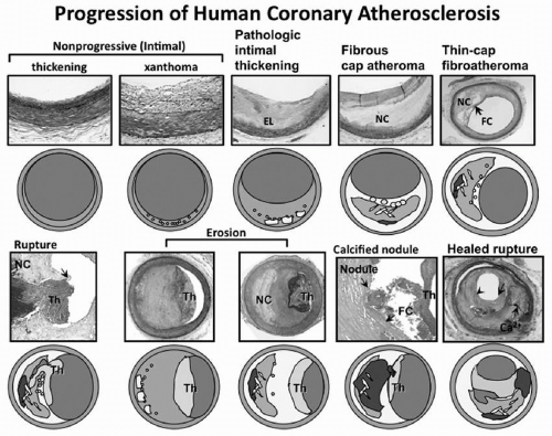


FIGURE 3.1 Spectrum of representative coronary lesion morphologies seen in our sudden coronary death population forming the basis for our modified AHA descriptive classification. The two nonprogressive lesions are intimal thickening and intimal xanthomas (foam cell collections known as fatty streaks, AHA type II). PIT (AHA type III transitional lesions) marks the first of the progressive plaques since they are the assumed precursors to more advanced FA. TCFAs are considered precursors to plaque rupture. Essentially missing from the AHA consensus classification are two alternative entities that give rise to coronary thrombosis, namely erosion and the calcified nodule. Erosions can occur on a substrate of PIT or fibroatheroma while calcified nodules (a minor but viable mechanisms of thrombosis) depict eruptive fragments of calcium that protrude into the lumen causing a thrombotic event. Lastly, healed plaque ruptures are lesions with generally smaller necrotic cores and focal areas of calcification where the surface generally shows areas of healing rich in proteoglycans. Multiple healed plaque ruptures are thought responsible for progressive luminal narrowing. AHA, American Heart Association; PIT, pathologic intimal thickening; FA, fibroatheromas; TCFAs, thin-cap fibroatheromas. (Reproduced with permission from Virmani R, Kolodgie FD, Burke AP, et al. Lessons from sudden coronary death: a comprehensive morphological classification scheme for atherosclerotic lesions. Arterioscler Thromb Vasc Biol. 2000;20:1262-1275.) (see color insert.)


ADVANCED LESIONS WITH NECROSIS


Fibroatheroma

FA are lesions with necrotic cores, which are well distinguished from lipid pool lesions of PIT since they confer a further progressive stage of atherosclerotic disease (3) (Table 3.1). Our laboratory defines fibroatheroma into those with “early” and “late” necrosis, as this distinction may provide mechanistic insight into how necrotic cores evolve. Recognition of early necrosis is identified by macrophage infiltration within lipid pools coinciding with a substantial increase in free cholesterol and breakdown of extracellular matrix, presumably by proteases. Early necrotic cores characteristically exhibit hyaluronan and versican matrix, which are typically absent in more advanced plaques with necrotic cores exhibiting late necrosis. Notably, the majority of macrophages within areas of necrosis display features consistent with apoptotic cell death. Moreover, it is widely held that the size of the necrotic core is a strong predictor of lesion vulnerability (3,9).

Accumulated free cholesterol visualized as empty clefts by routine histologic sectioning is another distinguishing feature of the late necrotic core. The production of free cholesterol in macrophages is in part regulated by a re-esterification process involving acyl coenzyme A:cholesterol acyltransferase, or ACAT1 (10). Manipulating the activity or expression of ACAT1 in culture or animal models favors necrotic core
formation. This together with the death of macrophages in the setting of defective phagocytic clearance of apoptosis cells is thought to contribute to the development of late plaque necrosis.


FIGURE 3.2 Simplified scheme for classifying atherosclerotic lesions modified from the current AHA recommendations. The boxed areas represent the seven categories of lesion. Dashed lines are used for two categories because there is controversy over the role that each plays in the initial phase of lesion formation, and both “lesions” can exist without progressing to a fibrous cap atheroma (i.e., AHA type IV lesion). The processes responsible for progression are listed between categories. Lines (solid and dotted, the latter representing the least-established processes) depict current concepts of how one category may progress to another, with the thickness of the line representing the strength of supportive evidence that the events occur. AHA, American Heart Association. (Reproduced with permission from Virmani R, Kolodgie FD, Burke AP, et al. Lessons from sudden coronary death: a comprehensive morphological classification scheme for atherosclerotic lesions. Arterioscler Thromb Vasc Biol. 2000;20:1262-1275.)


FREE CHOLESTEROL AS A MECHANISM OF LESION INSTABILITY

The few biochemical studies available regarding lipid composition in plaques report significant changes in various forms of cholesterol as lesion progresses (11,12). In the study by Katz et al., fatty streaks contained mostly esterified cholesterol, with relatively little free cholesterol content in contrast to intermediate or PIT, fibrous, and gruel plaques (12). The precise origin of free cholesterol in PIT is speculative since the bulk of lesional macrophages are confined to the more luminal aspect of the plaque outside lipid pools. Thus, it is suspected that the free cholesterol typically observed in the deeper intima may be derived from the membranes of apoptotic SMCs (13). In the study by Felton et al., the burden of free cholesterol was associated with lesion instability where disrupted plaques showed a correlation with increased free cholesterol, cholesterol esters, and free-to-esterified cholesterol ratio while the triglyceride content was essentially unchanged (11). The relationship of lipid composition and plaque instability in coronary sudden death is also apparent from our own studies where the percentage of cholesterol clefts was greater in lesions with rupture compared to eroded or stable plaques (3).


PUTATIVE SIGNALING PATHWAYS INVOLVED IN NECROTIC CORE EXPANSION

The presenting inflammatory stimuli for macrophage recruitment into lipid pools are poorly understood along with the respective signaling pathways for subsequent apoptotic cell death and necrosis (Fig. 3.3). Recent reports from the laboratories of Tabas (14) and notably others (15,16) point toward the involvement of the endoplasmic reticulum (ER) stress pathway or so-called unfolded protein response (UPR) as the primary mechanism of macrophage cell death in plaques. The regulation of this pathway is critical as the accumulation of dead macrophages coupled with defective phagocytic clearance (efferocytosis) is perhaps one of the principal factors causing necrotic core expansion.

Emerging data are beginning to unravel candidate molecules involved in the phagocytic clearance of apoptotic cells by professional scavengers, thereby suppressing proinflammatory signaling while activating anti-inflammatory pathways. In the laboratory of Mallat, milk fat globule-EGF factor 8 (Mfge8 or lactadherin) was identified as a potential bridging factor between apoptosis, phagocytic clearance, and secondary necrosis (17). In this study, low-density lipoprotein receptor-deficient
mice subjected to medullar aplasia by radiation were given bone marrow-derived cells from Mfge8-deficient mice leading to the substantial accumulation of apoptotic debris within the developing lesion. Accumulated apoptotic material was associated with a marked acceleration of atherosclerosis accompanied by decreased anti-inflammatory (IL-10) production by protective T cells. In addition to lactadherin, other putative molecules in plaques potentially linked to efferocytosis and defective clearance of apoptotic debris include Fas ligand (18) and transglutaminase-2 (TG2) (19). Taken together, these correlative studies provide potential evidence for the involvement of key regulatory components in necrotic core expansion and further lesion development.


TABLE 3.1 MODIFIED AHA CONSENSUS CLASSIFICATION BASED ON MORPHOLOGIC DESCRIPTION


Description


Thrombosis


Nonatherosclerotic intimal lesions


Intimal thickening


Normal accumulation of SMCs in the intima in the absence of lipid or macrophage foam cells


Absent


Intimal xanthoma


Superficial accumulation of foam cells without a necrotic core or fibrous cap. Based on animal and human data, such lesions usually regress


Absent


Progressive atherosclerotic lesions


Pathologic intimal thickening


SMC-rich plaque with proteoglycan matrix and focal accumulation of extracellular lipid


Absent


Fibrous cap atheroma


Early necrosis: focal macrophage infiltration into areas of lipid pools with an overlying fibrous cap


Absent


Late necrosis: loss of matrix and extensive cellular debris with an overlying fibrous cap


Absent


Thin-cap fibroatheroma


A thin fibrous cap (<65 µm) infiltrated by macrophages and lymphocytes with rare or absence of SMCs and relatively large underlying necrotic core. Intraplaque hemorrhage/fibrin may be present


Absent


Lesions with acute thrombi


Plaque rupture


Fibroatheroma with fibrous cap disruption; the luminal thrombus communicates with the underlying necrotic core


Occlusive or nonocclusive


Plaque erosion


Plaque composition, as above; no communication of the thrombus with the necrotic core. Can occur on a plaque substrate of pathologic intimal thickening or fibroatheroma


Usually nonocclusive


Calcified nodule


Eruptive (shedding) of calcified nodules with an underlying fibrocalcific plaque with minimal or absence of necrosis


Usually nonocclusive


Lesions with healed thrombi


Fibrotic (without calcification) Fibrocalcific (±necrotic core)


Collagen-rich plaque with significant luminal stenosis. Lesions may contain large areas of calcification with few inflammatory cells and minimal or absence of necrosis. These lesions may represent healed erosions or ruptures


Absent


Modified from Virmani R, Kolodgie FD, Burke AP, et al. Lessons from sudden coronary death: a comprehensive morphological classification scheme for atherosclerotic lesions. Arterioscler Thromb Vasc Biol. 2000;20:1262-1275.

Only gold members can continue reading. Log In or Register to continue

Stay updated, free articles. Join our Telegram channel

Oct 7, 2016 | Posted by in ENDOCRINOLOGY | Comments Off on Pathogenesis of Atherosclerosis and the Unstable Plaque

Full access? Get Clinical Tree
Get Clinical Tree app for offline access