Dyslipidemia in Children and Adolescents


Dyslipidemia in Children and Adolescents


Peter O. Kwiterovich Jr.

Sarah B. Clauss

Brian W. McCrindle


This chapter presents a theoretical and practical approach to the diagnosis and treatment of dyslipidemia in infants, children, and adolescents. The major clinical complication of dyslipidemia is a predilection to atherosclerosis starting in childhood and leading to cardiovascular disease (CVD) in adulthood. At the extremes of dyslipidemia, where inherited disorders of lipid and lipoprotein metabolism are more likely to occur, premature CVD is more frequent and can be accompanied by deposition of lipid (xanthomas) in various tissues. Children with profound hypertriglyceridemia are at high risk of pancreatitis.

While dyslipidemia can result from the expression of a mutation in a single gene that plays a paramount role in lipoprotein metabolism, more often dyslipidemia reflects the influence of multiple genes. Environmental influences such as excessive dietary intake of fat and calories and limited physical activity, particularly when associated with overweight or obesity, can also contribute significantly to dyslipidemia. Oligogenic and environmentally driven dyslipidemia constitute the overwhelming majority of dyslipidemia in youth. Such dyslipidemia also predicts atherosclerosis later in life and warrants early identification and treatment.

Data from a number of pathologic, epidemiologic, and genetic studies and from randomized clinical trials support strongly the tenet that the origins of atherosclerosis and CVD risk factors begin in childhood and adolescence, and that treatment should begin early in life (1).


THEORETICAL CONSIDERATIONS


Atherosclerosis in Infants, Children, and Adolescents

Almost 100 years ago, Klotz and Manning described the presence of fatty streaks in the large arteries of children (2). Over 50 years ago, it was reported that 77% of young (average age 22 years) soldiers killed in the Korean War had significant atherosclerosis; 15% of these subjects had coronary plaques that narrowed the vessel lumen 50% or greater (2). Similar observations were made in deceased soldiers killed in the Vietnam War. Strong, McGill, and Holman demonstrated that some childhood fatty streaks progressed to clinically significant fibrous plaques during young adulthood (2). Using modern methods of experimental pathology, the evolution and progression of atherosclerosis from a fatty streak to an intermediate lesion to a fibrous plaque was unequivocally shown (2). Further, when lesions in the coronary arteries were studied among different racial and ethnic groups in an international study, the conversion of fatty streaks in childhood into fibrous plaques and complicated lesions during the third and fourth decades of life was accelerated in subjects from countries and groups where adult coronary disease was a major problem. Fibrous plaques then underwent a variety of changes (hemorrhage, rupture, thrombosis) that led to obstruction and clinically manifest coronary disease (see Chapter 3).


Lipids and Lipoproteins

The plasma lipoproteins and the lipids that they transport play a central role in the development of, or protection from, atherosclerosis starting in childhood. The lipoproteins can be usefully divided into the apolipoprotein B (apoB)-containing lipoproteins and the apoA-I-containing lipoproteins. The apoB-containing lipoproteins include chylomicrons and very low density lipoproteins (VLDL) and their remnants, intermediate-density lipoproteins (IDL), low-density lipoproteins (LDL), and Lp(a) [lipoprotein(a)]. For each molecule of an apoB-containing lipoprotein, there is one molecule of apoB. The apoA-I-containing lipoproteins include high-density lipoproteins (HDL) and their subfractions HDL2 and HDL3. ApoA-I constitutes about 70% of the protein on HDL. There are usually three to four molecules of apoA-I for each HDL particle.

Most of the cholesterol in plasma is transported by LDL (60% to 70%) with the remainder carried on HDL (20% to 30%) and VLDL (10% to 15%). Triglycerides (TG) are transported primarily by chylomicrons, VLDL, and their remnants. Increased levels of the apoB-containing lipoproteins promote atherosclerosis and CVD, while low levels of these lipoproteins are associated with reduced CVD and longevity. In distinct contrast, high levels of HDL are usually associated with reduced CVD, while low levels of HDL often promote atherosclerosis and CVD.


Lipoprotein Structure and Function and Transport Pathways

A detailed summary of lipoprotein structure and function may be found in Chapters 1 and 2. As well, each of the three lipoprotein pathways, exogenous lipid transport, endogenous lipid transport, and reverse cholesterol transport (RCT), are covered in fine detail in Chapters 7, 8, and 9, respectively. Only a brief overview is provided here to provide a background to discuss the observation studies, the randomized controlled clinical trials, and the disorders of lipid and lipoprotein metabolism and their relevance to the diagnosis and treatment of dyslipidemia germane to pediatrics.


FIGURE 12.1 Overview of lipoprotein metabolism. Three major pathways of plasma lipoprotein metabolism are shown: (i) transport of dietary (exogenous) fat (left); (ii) transport of hepatic (endogenous) fat (center); (iii) reverse cholesterol transport (right and center). A detailed description may be found in the text. Sites of action of the six major lipid-altering drugs on exogenous and endogenous pathways of lipoprotein metabolism: (1) inhibition of HMG-CoA reductase by statins; (2) binding of bile acids by sequestrants, interfering with their reabsorption by IBAT; (3) binding of a CAI to the NPC1L1, decreasing the absorption of dietary and biliary cholesterol; (4) decreased mobilization of FFA by nicotinic acid, leading to decreased uptake of FFA by liver and reduced VLDL, IDL, and LDL production; (5) inhibition of TG synthesis by omega-3 fatty acids; (6) upregulation of LPL and decreased production of apoC-III, an inhibitor of LPL, by fibric acid derivatives, leading to decreased VLDL-TG. The hepatic cholesterol pool is decreased by the agents at steps 1, 2, and 3, each leading to an upregulation of the LDLR. HMG-CoA, hydroxymethylglutaryl; IBAT, intestinal bile acid transporter; CAI, cholesterol absorption inhibitor; FFA, free fatty acids; LPL, lipoprotein lipase. (Reproduced from Kwiterovich PO Jr. Clinical and laboratory assessment of cardiovascular risk in children: guidelines for screening, evaluation and treatment. J Clin Lipidol. 2008;2:248-266, with permission.)

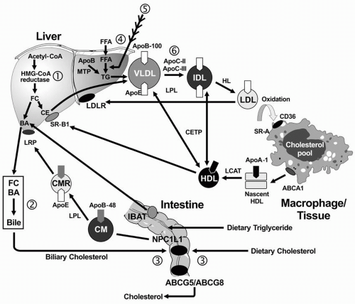
Exogenous Lipid Transport. In the small intestine, lipids are emulsified by bile salts and hydrolyzed by pancreatic lipases. The bile acids are then reabsorbed by the intestinal bile acid transporter (IBAT) for return to the liver through the entero-hepatic pathway (Fig. 12.1). TG are broken down into FFA acids and monoglycerides; cholesteryl esters (CE) are hydrolyzed into FFA and unesterified cholesterol. These components are then absorbed by the intestinal cells. The absorption of cholesterol occurs in the jejunum, through the high-affinity uptake of dietary and biliary cholesterol by the Niemann-Pick C1 Like 1 (NPC1L1) protein (Fig. 12.1). Normally, about half the dietary and biliary cholesterol is absorbed daily. Excessive cholesterol absorption is prevented by the ATP binding casette (ABC) transporters, ABCG5/ABCG8, which act together to pump excess cholesterol and plant sterols from the intestine back into the lumen for excretion into the stool (see Fig. 1.4) In intestinal cells, monoglyceride is re-esterified into TG and cholesterol is esterified by acyl cholesterol acyltransferase (ACAT), and both lipids and apoB-48 and other apolipoproteins are packaged into chylomicrons.

After absorption of dietary fat, the CE and TG are packaged with apoB-48 to form chylomicrons, which are then secreted from intestinal cells. The enzyme lipoprotein lipase (LPL) on the surface of endothelial cells (with apoC-II required as a cofactor) hydrolyzes the TG into free fatty acids (FFA) and monoglycerol. The released FFA are primarily taken up by adipose tissue (for storage) and muscle cells (for energy). The components that remain constitute the “chylomicron remnants,” which are removed from the blood in the liver by interaction of apoE with the chylomicron remnant receptor, also called the low density lipoprotein receptor-related protein (LRP).

Endogenous Lipid Transport. TG-rich VLDL is secreted by the liver into plasma, a process requiring apoB-100 (Fig. 12.1). The VLDL-TG are subsequently hydrolyzed by LPL and apoC-II
into FFA and monoglycerols, resulting in the formation of VLDL remnants and subsequently IDL. ApoE is the ligand for the hepatic uptake of some of the IDL particles by the LDL receptor (LDLR), while the remaining IDL are hydrolyzed by LPL and hepatic lipase (HL), producing the final product of VLDL catabolism, namely LDL. LDL are normally removed from plasma following the binding of apoB-100 to the hepatic LDLR (1).

Reverse Cholesterol Transport. RCT refers to the pathway by which cholesterol is transported away from peripheral cells, such as macrophages and foam cells in the walls of blood vessels to the liver for uptake and excretion into bile. Free cholesterol is removed from peripheral cells by the interaction of apoA-I on the nascent HDL particle with the ATP-binding casette (ABC) transporter, ABCA1. The cholesterol in the nascent HDL is esterified by lecithin-cholesterol acyltransferase (LCAT), with apoA-I as a cofactor, producing a more mature and larger HDL particle with cholesteryl ester in its core. The mature HDL can deliver the cholesteryl ester directly to the liver through the interaction of apoA-I with the HDL receptor (also called scavenger receptor, class B, type I [SR-BI]; Fig. 12.1). The cholesterol derived from this process may be excreted into bile either as cholesterol or by conversion of cholesterol into bile acids. Less than half of the cholesterol from peripheral cells is delivered to the liver through this pathway. The remaining CE is transferred from HDL to the apoB-containing lipoproteins in exchange for TG by the cholesteryl ester transfer protein (CETP).


Relationship between Atherosclerosis and CVD Risk Factors

Pathologic Studies. Several longitudinal pathologic studies from the general population found that early atherosclerotic lesions of fatty streaks and fibrous plaques in children, adolescents, and young adults, who died from accidental deaths, are significantly related to higher antecedent levels of total cholesterol (TC) and LDL cholesterol (LDL-C), lower levels of HDL cholesterol (HDL-C), and other CVD risk factors, such as obesity, higher blood pressure, and cigarette smoking (1,2). These effects of risk factors on coronary lesion severity are multiplicative rather than additive.

Prospective Epidemiologic Studies. Five major prospective population studies—two from Muscatine and Bogalusa, and the Coronary Artery Risk Development in Young Adults (CARDIA), the Special Turku Coronary Risk Factor Intervention Project (STRIP), and the Cardiovascular Risk in Young Finns Study—showed that CVD risk factors in children and adolescents, particularly LDL-C and obesity, predicted clinical manifestations of atherosclerosis in young adults, as judged by coronary artery calcium, carotid intima-media thickness (IMT), or brachial flow-mediated dilatation (FMD) (1,2,3,4). In contrast, HDL-C levels in childhood and early adulthood have been related to decreased IMT and increased FMD (3). There are little data on prediction of CVD events; however, medical students at Johns Hopkins who had a TC level >207 mg/dL had five times the risk of developing CVD 40 years later than those who had a TC level <172 mg/dL (1).

Human Genetic Studies. Studies have also been performed in high-risk youth selected by virtue of CVD in one parent or because they have inherited a known metabolic disorder of lipoprotein metabolism that produces premature CVD. Half of the young progeny of men with premature CVD before 50 years of age had one of seven dyslipidemic profiles: elevated LDL-C alone (type IIa) or combined with high TG (type IIb); elevated TG alone (type IV); low HDL-C alone (hypoalpha); and type IIa, type IIb, or type IV also accompanied by low HDL-C (1). Elevated levels of apoB, in the presence of normal LDL-C (hyperapobetalipoproteinemia or hyperapoB), were prevalent in young offspring of adults with premature CVD and hyperapoB (1). The levels of apoB and apoA-I, the major apolipoproteins of LDL and HDL, respectively, and the ratio of apoB to apoA-I in young offspring from Bogalusa, were stronger predictors of premature coronary artery disease (CAD) in their parents than LDL-C and HDL-C levels (1). Related findings were recently reported from the Cardiovascular Risk in Young Finns Study, where childhood levels of serum apoB and apoA-I predicted carotid IMT and brachial FMD in adulthood better than LDL-C and HDL-C (3).

Inherited Disorders of Lipoprotein Metabolism. Examples of inherited lipoprotein disorders that often present in youth at high risk of future CVD include familial hypercholesterolemia (FH), due to a defect in the LDLR, and familial combined hyperlipidemia (FCHL), and its metabolic cousin, hyperapoB, the prototypes for hepatic overproduction of VLDL, which are often accompanied by insulin resistance and the dyslipidemic triad of hypertriglyceridemia, increased small, dense LDL particles (LDL-P), and low HDL-C (see also the following text). Recently, the presence of a type IIb phenotype (elevated LDL-C and TG) in childhood predicted increased carotid IMT, elasticity, and FMD in early adulthood (4).


SCREENING FOR DYSLIPIDEMIA IN YOUTH

Two major approaches have been considered to detect dyslipidemia in youth, namely screening in the general population or in a selected population. The extensive literature related to these two screening approaches has been reviewed in detail (5).

Traditionally, screening for dyslipidemias in high-risk children is recommended because they have multiple CVD risk factors, or a family history of premature CVD and/or hypercholesterolemia. LDL-C has been the main focus of diagnosis and treatment. Less attention has been paid to HDL-C and TG. Now with obesity and the metabolic syndrome evident in our youth (1,6), the focus of screening is likely to be expanded to include other factors, such as obesity, low HDL-C, non-HDL-C (TC minus HDL-C), elevated TG, elevated apoB (reflecting increased small, dense LDL-P), glucose intolerance and insulin resistance, and higher blood pressure levels. Both the current and evolving concepts in screening for dyslipidemia in youth will now be discussed.


Who to Screen


Selective Screening

The National Cholesterol Education Program (NCEP) Expert Panel on Blood Cholesterol Levels in Children and Adolescents
recommended in 1992 that selective, not general, screening be performed (7). We have expanded some of these recommendations for selective screening:


  • A lipoprotein profile in youth whose parents and/or grandparents required coronary artery bypass surgery or balloon angioplasty prior to age 55.


  • A lipoprotein profile in those with a family history of myocardial infarction, angina pectoris, peripheral or cerebral vascular disease, or sudden death prior to age 55.


  • A TC in those whose parents have high TC levels (>240 mg/dL who have any dyslipidemia, involving elevated LDL-C, non-HDL-C, apoB, TG, or low HDL-C).


  • A lipoprotein profile if the parental/grandparental family history is not known, and the patient has two or more other risk factors for CAD including obesity (BMI > 30), hypertension, cigarette smoking, low HDL-C, physical inactivity and diabetes mellitus.


  • A lipoprotein profile if either obesity (BMI > 95th percentile) or overweight (BMI 85th to 94th percentile) is detected per se, regardless of the presence of other nonlipid CVD risk factors.


Universal Screening

Universal lipid screening of all children is controversial (5,7,8). The advantages and disadvantages of universal screening will now be briefly discussed.

What are some of the arguments in favor of universal screening? First, current screening recommendations based on family history of CVD or hypercholesterolemia will fail to detect substantial numbers (from 17% to 90%) of children who have elevated lipid levels (5).

Universal screening might be performed to detect those with undiagnosed heterozygous FH or more marked FCHL, who will require more intensive treatment including the possibility of drug therapy. In a recent meta-analysis of screening for FH in a primary care setting, use of TC detected 88%, 94%, and 96% of cases, with false positive rates of 0.1%, 0.5%, and 1%, respectively (9). This approach might be combined with a case finding strategy in relatives of patients with FH (9).

Identification of children and adolescents affected with hypercholesterolemia through universal screening may bring to attention their adult relatives who will have greater coronary mortality than relatives of children with normal cholesterol levels (1). If universal lipid screening is combined with an assessment of obesity and high blood pressure, this can also lead to the detection of additional relatives from families at high risk for CVD (1).

It is clear that CVD risk factors cluster in childhood and persist into adulthood (1,5,7,8). While it is known that offspring of parents with CVD generally have higher LDL and TG and lower HDL-C both in childhood and young adulthood, the majority of children with dyslipidemia and multiple risk factors will be missed by selective screening (5).

That each child and adolescent should ideally have an assessment of their plasma lipids and lipoproteins makes sense. While there are practical problems (see following text), and no longitudinal studies are available to show that treatment starting in childhood decreases adult CVD (5), one might argue that universal screening seems all the more urgent, given the epidemic of obesity and the metabolic syndrome in American youth.

What are some of the concerns about universal lipid screening in childhood? The use of TC in childhood to predict TC or LDL-C in young adults, sufficiently high to warrant treatment, is often associated with less than optimal sensitivity, specificity, and predictive power of a positive test. A number of longitudinal studies (5) have found that when the 75th percentile for TC in children is used as a screening cut point, about half the individuals who will require treatment as adults are identified by universal lipid screening. In one report, the sensitivity was much lower when screening occurred during adolescence, presumably reflecting the temporary shift of LDL-C to lower values during this period of rapid growth and development (1). Another unresolved issue is whether the detection of elevated TC or LDL-C in children and young adults will predict those who are destined to manifest premature CVD. Few data are available to address this question.


What to Measure


Lipoprotein Profile

For selective screening, a lipoprotein profile after an overnight fast is measured for screening youth with a positive family history of premature CVD or dyslipidemia, with obesity, with multiple CVD risk factors, and for those suspected of having secondary dyslipidemia. Such a profile includes TC, TG, LDL-C, HDL-C, and non-HDL-C. Levels of lipoproteins are typically measured and expressed in terms of their cholesterol content. LDL-C is calculated from the Friedewald equation: LDL-C = TC − (HDL-C + TG/5). Total TG in the fasting state divided by 5 is used to estimate the levels of VLDL-C. If the TG is >400 mg/dL, this formula cannot be used and a direct LDL-C may be measured. If the patient is nonfasting, TC, HDL-C, and non-HDL-C levels can be measured.


Apolipoproteins A-I and B

ApoB and apoA-I might also be determined using wellstandardized immunochemical methods (10,11). Such measurements might provide additional useful information, particularly in youth with premature CAD in parents (1). Age-, gender-, and race-specific cut points for apoB and apoA-I, empirically derived from the National Health and Nutrition Education Survey (NHANES) sample, are available, providing cut points that might be used to define elevated apoB and low apoA-I (10) (Table 12.1). ApoB provides an assessment of the total number of apoB-containing lipoprotein particles (11).


Non-HDL-C

Non-HDL-C is determined by subtracting HDL-C from TC and can be measured in plasma from nonfasting patients. Non-HDL-C reflects the amount of cholesterol carried by the “atherogenic” apoB-containing lipoproteins [VLDL, IDL, LDL, and Lp(a)]. In adults, non-HDL-C appears to be a better independent predictor of CVD than LDL-C (11). In children, non-HDL-C is at least as good a predictor as LDL of future dyslipidemia in adulthood (1). Percentiles for non-HDL-C in children are available from Bogalusa (12) (Table 12.1).


Advanced Lipoprotein Testing

The plasma levels of VLDL, LDL, and HDL subclasses have been determined in children and adolescents by nuclear magnetic resonance (NMR) spectroscopy (1) or by vertical-spin
density-gradient ultracentrifugation (1) in research studies (see also the following text), but cut points derived from these methods for the diagnosis and treatment of dyslipidemia in youth are not currently available.


TABLE 12.1 ACCEPTABLE, BORDERLINE, AND HIGH PLASMA LIPID, LIPOPROTEIN, AND APOLIPOPROTEIN CONCENTRATIONS FOR CHILDREN AND ADOLESCENTSa


Category


Acceptable


Borderline


Highb


TC


<170


170-199


≥200


LDL-C


<110


110-129


≥130


Non-HDL-C


<123


123-143


≥144


ApoB


<90


90-109


≥110


TG


0-9 years


<75


75-99


≥100


10-19 years


<90


90-129


≥130


HDL-C


>45


35-45


<35


ApoA-I


>120


110-120


<110


aValues for plasma lipid and lipoprotein levels are from the National Cholesterol Education Program (NCEP) Expert Panel on Cholesterol Levels in Children (7). Non-HDL-C values from Bogalusa are equivalent to NCEP Pediatric Panel cut points for LDL-C (12). Values for plasma apoB and apoA-I are from the National Health and Nutrition Examination Survey III (NHANES III) (10).

b The cut points for a high or low value represent approximately the 95th and 5th percentiles, respectively.


TC, total cholesterol; LDL-C, low-density lipoprotein cholesterol; Non-HDL-C, non-high-density lipoprotein cholesetrol; apoB, apolipoprotein B; TG, triglycerides; HDL-C, high-density lipoprotein cholesterol; apoA-I, apolipoprotein A-I.


Data from National Cholesterol Education Program. Report of the Expert Panel on Blood Cholesterol Levels in Children and Adolescents. Pediatrics. 1992;89:525-584, (suppl); Bachorik PS, Lovejoy KL, Carroll MD, et al. Apolipoprotein B and AI distributions in the United States, 1988-1991: results of the National Health and Nutrition Examination Survey III (NHANES III). Clin Chem. 1997;43:2364-2378; and Srinivasan SR, Myers L, Berenson GS. Distribution and correlates of non-high-density lipoprotein cholesterol in children: the Bogalusa Heart Study. Pediatrics. 2002;110:e29.


Summary

For universal screening, the simplest approach appears to be the measurement of TC, HDL-C, and non-HDL-C in nonfasting specimens. However, treatment algorithms in pediatrics are usually focused on fasting LDL-C. Hypertriglyceridemia is usually assessed as part of the dyslipidemic triad and is often elevated in obesity and the metabolic syndrome (1,13). Thus, in an ideal screening program, TC, TG, LDL-C, HDL-C, and non-HDL-C would be assessed by performing a lipoprotein profile in the fasting state.


When to Sample for Dyslipidemia

Human plasma cholesterol levels are lowest during intrauterine life (1). At birth, the mean (1 SD) plasma levels (mg/dL) are: TC, 74 (11); LDL-C, 31 (6); HDL-C, 37 (8); and TG, 37 (1). TC and LDL-C increase rapidly in the first weeks of life. The lipids and lipoproteins continue to increase gradually until 2 years of age, during which time the kind and source of the milk in the infant’s diet can markedly influence these levels. Screening for dyslipidemia is therefore not generally recommended before 2 years of age. After 2 years of age the levels of the lipids and lipoproteins become quite constant up to adolescence (5,7,8)

Ten years of age has been proposed as a good time to obtain a lipoprotein profile (5,7,8). The children are older, able to fast easier, the values are predictive of future adult lipoprotein profiles, and adolescence has not yet set in. Since TC and LDL-C may fall from 10% to 20%, or more, during adolescence (1), it is preferable to screen children at risk for familial dyslipidemias before adolescence, between 2 and 10 years of age. Even in FH heterozygotes, there is a significant fall in the 1:1 ratio of affected to normal in adolescence (1). If sampling occurs during adolescence, and the results are abnormal, they are likely to be even higher after adolescence. If the results during adolescence are normal, sampling will need to be repeated toward the end of adolescence (for girls, 16 years of age; for boys, 18 years of age).

The complete phenotypic expression of some disorders, such as FCHL, can be delayed until adulthood, so the continued evaluation of such subjects from high-risk families with FCHL should occur well into adulthood. However, elevated apoB is the first expression of FCHL in adolescents and young adults (1). Age-related factors, such as increased BMI, contribute to the degree of dyslipidemia in such youth.


Definition of Dyslipidemia

Cut points (7,10,12) to define elevated TC, LDL-C, apoB, non-HDL-C and TG, and low HDL-C and apoA-I in children and adolescents are given in Table 12.1. Dyslipidemia is present if one or more of these lipid, lipoprotein, or apolipoprotein factors are abnormal. In offspring of young progeny of men with premature CVD before 50 years of age, seven different dyslipidemic profiles were present (1). Such results emphasize the importance of evaluating a lipoprotein profile in the fasting state.


Single versus Multiple Cut Points

Using data from three major population-based prospective cohort studies, TC, LDL-C, HDL-C, and TG variables in adolescence were classified according to NCEP cut points (7) (Table 12.1) and to age and gender (not race specific) NHANES cut points and compared for their ability to predict abnormal levels in adulthood (14). NCEP cut points (compared with NHANES cut points) were more strongly predictive of high TC, LDL-C, and TG levels in adults but less predictive of low HDL-C (14). The continued use of the current NCEP cut points for TC, LDL-C, and TG levels in adolescents appears indicated. The cut point for HDL-C might be revised upward, perhaps to 40 mg/dL, to improve the sensitivity of this measurement to predict low HDL-C in adults.


PRIMARY VERSUS SECONDARY DYSLIPOPROTEINEMIA

Before considering a dyslipoproteinemia to be primary, secondary causes must be excluded (Table 12.2). Each child with dyslipidemia should have routine blood tests to help rule out
secondary causes of dyslipidemia. These include fasting blood sugar, and tests of kidney, liver, and thyroid function. In secondary dyslipidemia, the associated disorder producing the dyslipidemia should be treated first in an attempt to normalize lipoprotein levels; however, if the dyslipidemia persists, for example, as it often does in type I diabetes and the nephrotic syndrome, the patient will require dietary treatment, and if indicated, drug therapy using the same guidelines as in primary dyslipidemias.


TABLE 12.2 CAUSES OF SECONDARY DYSLIPIDEMIA IN CHILDREN AND ADOLESCENTS


Exogenous


Storage disease


Alcohol


Cystine storage disease


Oral contraceptives


Gaucher disease


Prednisone


Glycogen storage disease


Anabolic steroids


Juvenile Tay-Sachs disease


13-cis-Retinoic acid


Niemann-Pick disease


Tay-Sachs disease


Endocrine and metabolic


Acute intermittent porphyria


Acute and transient


Type I and type II diabetes


Burns


Hypopituitarism


Hepatitis


Hypothyroidism


Lipodystrophy


Pregnancy


Others


Anorexia nervosa


Renal


Cancer survivor


Chronic renal failure


Heart transplantation


Hemolytic-uremic syndrome


Idiopathic hypercalcemia


Nephrotic syndrome


Kawasaki disease


Klinefelter syndrome


Progeria (Hutchinson-Gilford syndrome)


Rheumatoid arthritis


Hepatic


Systemic lupus erythematosis


Benign recurrent intrahepatic cholestasis


Werner syndrome


Congenital biliary atresia


Alagille syndrome


INHERITED METABOLIC DISORDERS OF DYSLIPIDEMIA IN YOUTH

The dyslipidemia in most children who are screened will not reflect the presence of an inherited metabolic disorder. Most will have a combination of the influence of at least several genes (oligogenic) and of environmental factors such as obesity, diet, and lack of exercise. There will be, however, those who are more severely affected and who will carry one or more mutant alleles for a specific metabolic disorder of dyslipidemia. Further, there are some inherited disorders of dyslipidemia that will be relatively prevalent, such as FH and FCHL.


Disorders Affecting LDL Receptor Activity

There are five disorders expressed in pediatrics that result from mutations either in the LDLR per se, or from mutations in other genes that impact LDLR activity (see Fig. 1.4 on page 10). Elevated LDL-C can vary considerably in these five conditions (see also the following text) but each disorder manifests early atherosclerosis and premature CVD (15,16). These disorders include FH (15,16), familial ligand defective apoB-100 (FDB) (15,16), autosomal recessive hypercholesterolemia (ARH) (15,16), sitosterolemia (15,16), and mutations in proprotein convertase subtilisin-like kexin type 9 (PCSK9) (16,17). Each disorder warrants diet and drug therapy in childhood in an attempt to decrease atherosclerosis and subsequent CVD.

Only gold members can continue reading. Log In or Register to continue

Stay updated, free articles. Join our Telegram channel

Sep 7, 2016 | Posted by in ENDOCRINOLOGY | Comments Off on Dyslipidemia in Children and Adolescents

Full access? Get Clinical Tree

Get Clinical Tree app for offline access