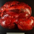
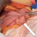
A higher incidence of paraganglioma or pheochromocytoma (PPGL) has been reported in people living at high altitudes, indicating that environmental hypoxia could be a risk factor for development of PPGL. Limited literature suggests that patients with cyanotic congenital heart disease also demonstrate an increased risk for development of PPGL. Herein we share such a case.
Case Report
The patient was a 51-year-old woman with a history of complex cyanotic congenital heart disease who was recently discovered to have a left carotid body tumor after noticing a lump on self-palpation. She was asymptomatic, without signs of catecholamine excess. Family history was negative for PPGL syndromes.
COMPLEX CYANOTIC CONGENITAL HEART DISEASE HISTORY
The patient had been noted to be “blue” on birth. The details of the initial evaluation were not available. At 5 months of age, she underwent a classical Glenn anastomosis. Between age 5 and 6 years, she was treated with the Potts anastomosis (descending aorta to left pulmonary artery anastomosis). Between ages 5 and 34 years, the patient had a gradual reduction in her exercise tolerance and progressive fatigue. At age 35, she had another surgical intervention consisting of atrial septectomy, attempted establishment of pulmonary artery confluence with a homograft right central shunt and patch augmentation of right superior vena cava, and ligation of the left superior vena cava. At age 38, she underwent insertion of a left Blalock-Taussig shunt (left subclavian artery-to-distal left pulmonary artery graft) with a 7-mm interposition GORE-TEX graft. The shunt resulted in improvement in oxygenation and cardiovascular status.
INVESTIGATIONS
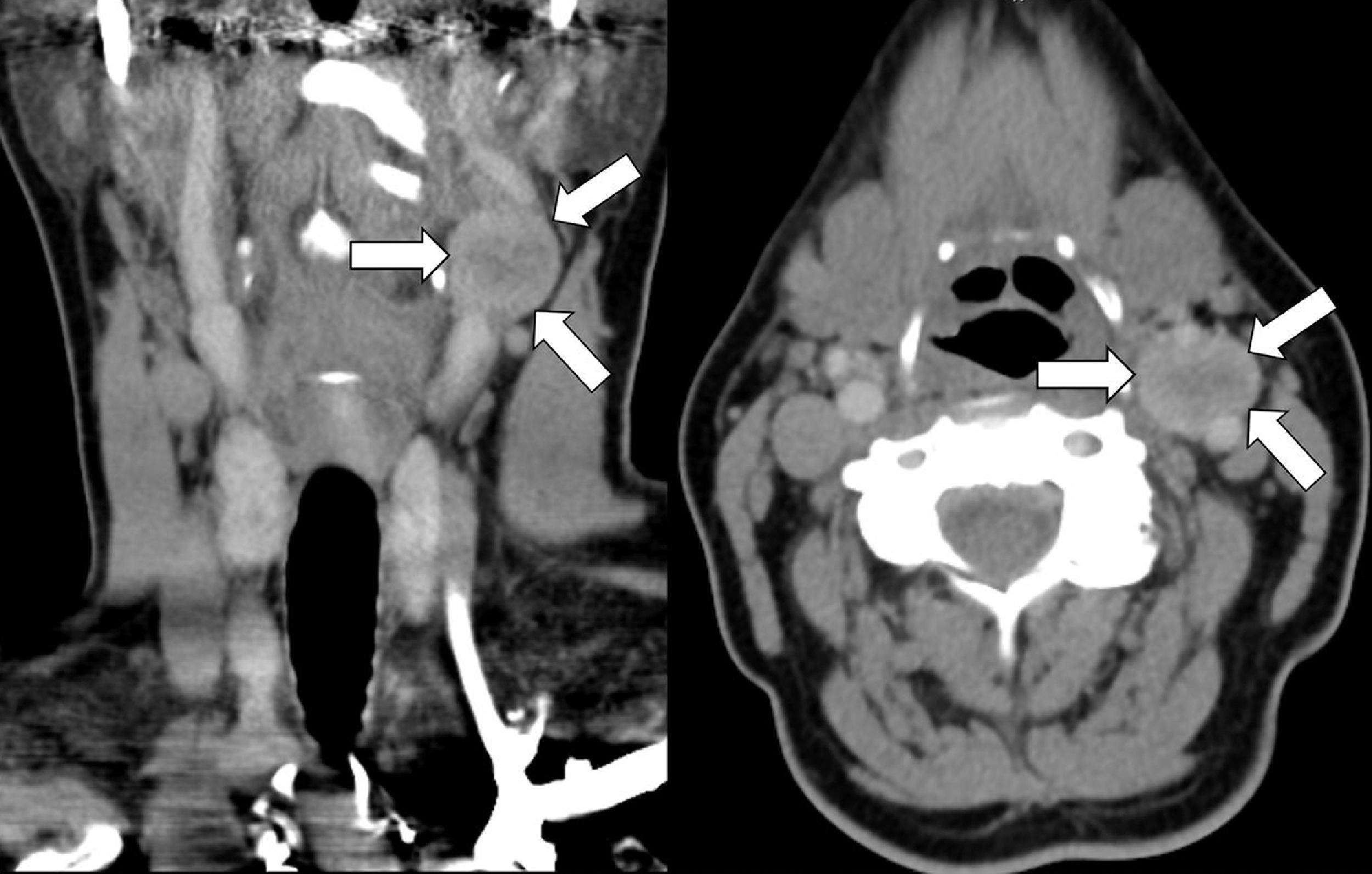
The initial computed tomography (CT) scan of neck demonstrated a 2.0 × 1.8 × 1.9–cm mild to moderately enhancing circumscribed mass within the left carotid bifurcation, consistent with paraganglioma ( Fig. 52.1 ). The laboratory studies were unremarkable, with fractionated catecholamines and metanephrines within the normal range in the 24-hour urine collection ( Table 52.1 ). CT of the abdomen and pelvis did not detect additional PPGLs.
| Biochemical Test | Result | Reference Range |
| 24-Hour urine: | ||
Metanephrine, mcg | 101 | <400 |
Normetanephrine, mcg | 288 | <900 |
Norepinephrine, mcg | 58 | <80 |
Epinephrine, mcg | 6.2 | <20 |
Dopamine, mcg | 152 | <400 |
Stay updated, free articles. Join our Telegram channel

Full access? Get Clinical Tree


