Pancreas Cancer


Pancreas Cancer


Scott E. Kern

Ralph H. Hruban


Pancreatic ductal adenocarcinoma is a genetic disease. This perspective is supported by reproducible patterns of genetic mutations that accumulate during tumorigenesis. These patterns indicate the operation of a selective process favoring the emergence of specific constellations of genetic changes. Individuals who inherit a mutant form of certain genes have an increased risk of developing pancreatic cancer. According to this genetic theory, most pancreatic cancers share a common foundation of genetic mutations disrupting specific cellular regulatory controls. These shared abnormalities are responsible for the processes of growth, invasion, and metastasis in individual patients.

Four categories of mutated genes play a role in the pancreatic tumorigenesis: oncogenes, tumor-suppressor genes, genome-maintenance genes, and tissue-maintenance genes (summarized in Table 17.1). Some of these mutations are germ line, for example, they are transmitted within a family. Genetic mutations acquired during life, termed somatic mutations, contribute to tumorigenesis within a tissue but are not passed to off-spring.

Very recently, techniques were developed to sequence all of the genes of individual cancers. Whole-exomic sequencing of pancreatic ductal cancers revealed an average of 63 somatic mutations per tumor.1 Most of these mutations undoubtedly were nonfunctional “passenger” mutations, each mutated at a low frequency and not contributing to tumorigenesis. Indeed, most passenger mutations might arise as a normal aspect of tissue aging before tumorigenesis begins.2 Smoking is associated with a doubling of the risk for pancreatic cancer, and remarkably is also associated with a 40% increase of the prevalence of low-frequency mutations in the cancers.3 A subset of the mutations, however, is responsible for “driving” the neoplastic process in the ducts and is the focus here.

Telomere abnormalities and signs of chromosome instability are the most common alterations in pancreatic neoplasia. Four genes are mutated in most pancreatic cancers: the KRAS, p16/CDKN2A, TP53, and SMAD4 genes. Other genetic abnormalities are seen at a much lower frequency, including mutations in the genes BRCA2, PALB2, FANCC, FANCG, FBXW7, BAX, RB1, the TGFβ (transforming growth factorbeta) receptors TGFBR1 and TGFBR2, the activin receptors ACVR1B and ACVR2, MKK4, STK11, GUCY2F, NTRK3, EGFR, and cationic trypsinogen, alterations in the mitochondrial genome, amplifications, various chromosomal deletions, inactivation of DNA mismatch-repair genes, and rarely the presence of the Epstein-Barr virus genome as an episome.

Knowing the genes mutated in a cancer can have direct clinical impact. For example, many cancers occur from an inherited mutation, and these patients and their families could benefit from genetic counseling.4,5,6,7 A distinct morphologic subtype of pancreatic cancer, the medullary cancer, can suggest such an inherited mutation.8,9 Another example includes the analysis of the genetic alterations in precursors to invasive pancreatic neoplasia, which has indicated that most carcinomas arise by a process of progressive intraductal tumorigenesis.10 Epigenetic changes in DNA methylation and in gene expression are also highly specific for the cancerous cells and can serve as diagnostic markers.


COMMON MOLECULAR CHANGES

Telomere shortening is the earliest and most prevalent genetic change identified in the precursor lesions.11 Telomere shortening is thought to predispose to chromosome fusion (translocations) and the mis-segregation of genetic material during mitosis.12 Later during tumorigenesis, telomerase is reactivated,13,14 moderating the telomere erosive process while permitting continued chromosomal instability.15

The KRAS gene mediates signals from growth factor receptors and other signaling inputs (Fig. 17.1). The mutations convert the normal Kras protein (a protooncogene) to an oncogene, causing the protein to become overactive in transmitting the growth factor-initiated signals.16 KRAS is mutated in over 90% of conventional pancreatic
ductal carcinomas.17 The first genetic change in the ducts is probably not (or not always) a KRAS gene mutation, for the prevalence of this mutation is highest in the more advanced lesions (Table 17.1).18,19 KRAS is one of a family of RAS genes that can harbor mutations in human cancers. The other RAS genes include NRAS and HRAS, although it is possible that only KRAS is mutated in pancreatic carcinomas.


TABLE 17.1 GENETIC PROFILE OF PANCREATIC CARCINOMA


Gene


Gene Locations


Frequency in Cancers (%)


Timing During Tumorigenesisa


Mutation Origin


Oncogenes


KRAS


12p


95


Early-mid


Som.


BRAF


7q


4


Som.


AKT2


19q


10-20


Som.


GUCY2F


3


Som.


NTRK3


1


Som.


EGFR


1


Som.


EBV genome


<1


Tumor Suppressors/Genome-Maintenance Genes


p16


9p


>90


Mid-late


Som. > Germ.


TP53


17p


75


Late


Som.


SMAD4


18q


55


Late


Som.


BRCA2/PALB2


13q/16p


8


Late


Germ. > Som.


FANCC/FANCG


9q/9p


3


Germ. or Som.


MAP2K4


17p


4


Som.


LKB1/STK11


19p


4


Som. > Germ.


ACVR1B


12q


2


Som.


TGFBR1


9q


1


Som.b


MSI/TGFBR2


3p


1


Som.b


MSI+/TGFBR2


3p


4


Som. > Germ.c


ACVR2


2q


4


Som. > Germ.c


BAX


19p


4


Som. > Germ.c


MLH1


3q


4


Som. > Germ.c


FBXW7/Cyclin E dereg.


4q


6


Som.d


Tissue-Maintenance Genes


PRSS1


7q


<1


Prior


Germ.


Som., (prevalence of) somatic mutation or methylation; Germ., (prevalence of) germ line mutation.


a Stage of appearance of the genetic changes during the intraductal precursor phase of the neoplasm, where known. For BRCA2, most mutations are inherited, but the loss of the second allele is reported only in a single advanced pancreatic intraepithelial neoplasm.

b Single examples of homozygous deletion of the TGFBR1 gene and TGFBR2 gene have been identified in MSI-negative pancreatic cancer.

c In MSI-positive tumors, the mismatch repair defect is usually somatic in origin; the TGFBR2, ACVR2, and BAX alterations are somatic.

d dA single example of homozygous mutation of the FBXW7 gene is reported in a series having a 6% prevalence of cyclin E overexpression. Cyclin E amplification is reported to date only in cell lines.


As one of the most commonly mutated genes in pancreatic cancer, Ras is an attractive target for the development of gene-specific therapies, and an understanding of the normal biology of the Ras protein should help in the development of these Ras targeted therapies. The Ras proteins require an attachment to the plasma membrane for activity. For many proteins, including Ras, a hydrophobic prenyl group is essential for the attachment. Either farnesyl (15-carbon) or geranylgeranyl (20-carbon) makes a covalent thioether linkage at a cysteine residue located near the C-terminal end of Ras proteins, termed the CAAX motif. Working mostly in artificial legacy models of the HRAS oncogene (rather than the more widely available but experimentally less tractable natural KRAS-mutant cancer cell lines), the farnesylation reaction was readily inhibited by various means; in these models, the Ras protein was rendered inactive and often accompanied by cytotoxicity limited to the mutant cells.

Although many types of compounds capable of blocking the farnesyltransferase enzyme were
developed as drugs, they have not been successful anticancer agents. There are many reasons for this. Although Hras protein is linked predominantly through farnesyl groups, the Kras protein can be alternately prenylated by geranylgeranyl linkages. Unfortunately, the latter type of linkage is thought to be critical for a wider number of cellular proteins, and for fear of excessive toxicity, geranylgeranyl linkages have not usually been considered as an attractive drug target. Kras protein also appears to bind more tightly than Hras to the farnesyltransferase enzyme, requiring higher drug concentrations.16 Additionally, the artificial models usually employed the engineered overexpression of the Ras protein, a situation in which the unattached Ras proteins would serve as a dominant-negative inhibitor, binding the necessary interacting proteins and sequestering them in the cytoplasm to ensure the inactivation of all three Ras pathways. Such a concentrationdriven mechanism would presumably not occur under the normal levels of Ras proteins present in human cancers.20 Indeed, it is proposed that the limited efficacy of farnesyltransferase inhibitors observed in some experimental models and in clinical trials may be attributable to a cellular target not yet identified.21 Attention has turned to compounds that target the downstream mediators, such as Raf and Mek protein kinase inhibitors.


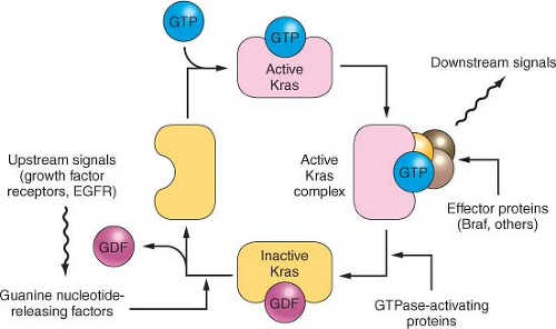
FIGURE 17.1 The KRAS pathway. KRAS normally integrates and regulates signals arising in the growth factor receptors that are passed to KRAS using the Grb2 and the Sos1 nucleotide exchange factor. The active GTP-bound form of KRAS recruits effector proteins such as Raf1 and Braf, in turn stimulating the downstream mitogen-activated protein kinases such as MEK and ERK and activating certain transcription factors. The EGF receptor can be overexpressed and occasionally mutated to provide inappropriately strong upstream signals, and the BRAF protein can be activated by point mutation, but more often in pancreatic cancer the Kras protein is mutated. These latter mutations impair the GAP (GTPase-activating protein)-stimulated reaction that normally returns Kras to the inactive state.

The Smad pathway mediates signals initiated on the binding of the extracellular proteins TGFβ and activin to their receptors (Fig. 17.2). These signals are transmitted to the nucleus by the Smad family of related genes, including SMAD4 (DPC4).22 Smad protein complexes bind specific recognition sites on DNA and cause the transcription of certain genes.23 Mutations in the SMAD4 gene are found in nearly half of pancreatic carcinomas, including both homozygous deletions and intragenic mutations combined with loss of heterozygosity (LOH).24 Other Smad genes are also mutated occasionally.1

Homozygous deletions and mutation/LOH affecting the TGFβ receptor genes are seen in a few pancreatic cancers.25 A more common abnormality, in pancreatic as well as in other tumor types, is the underexpression of TGFβ receptors, which results in cellular resistance to the usual suppressive effects of the TGFβ ligand.26

The p16/RB1 pathway is a key control of the cell division cycle (Fig. 17.3). The retinoblastoma protein (Rb1) is a transcriptional regulator and regulates the entry of cells into S phase. A complex of cyclin D and a cyclin-dependent kinase (Cdk4 and Cdk6) phosphorylates and thereby regulates Rb1. The p16 protein is a Cdk-inhibitor that binds Cdk4 and Cdk6.27,28,29 Virtually all pancreatic carcinomas suffer a loss of p16 function, through homozygous deletions, mutation/LOH, or promoter methylation of the p16/CDKN2A gene associated with a lack of gene expression.30,31 In addition, inherited mutations of the p16/CDKN2A gene cause a familial melanoma/pancreatic cancer syndrome known as familial atypical multiple mole melanoma.32,33,34,35,36 Occasional pancreatic cancers have inactivating mutations of the RB1 gene.37


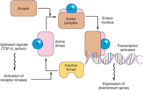
FIGURE 17.2 The transforming growth factor-beta (TGFβ)/Activin/Smad pathway. Dimeric kinase receptors of the TGFβ superfamily respond to extracellular ligands, causing phosphorylation of one or more of the receptor-associated Smad proteins and leading them to complex with the unphosphorylated common Smad, Smad4. This complex binds to specific DNA sequences and works with other transcription factors to stimulate gene expression. Mutations in pancreatic cancer can inactivate either partner of the dimeric receptors that respond to extracellular TGFβ or activin. More commonly, however, mutations and large deletions in the SMAD4 gene destabilize its protein product or ablate gene expression.


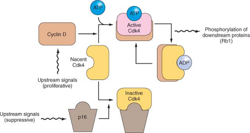
FIGURE 17.3 The p16/RB1 pathway. p16 binds to, inhibits, and thereby controls the availability of the cyclin-dependent kinases Cdk4 and Cdk6 (not shown). When activated by binding to cyclin D, these kinases phosphorylate and thereby inactivate the Rb1 tumor suppressor protein. The activity of p16 is controlled in a complex manner, through changes in gene expression and by displacement reactions involving other similar kinase inhibitor proteins. p16 mutations and deletions are nearly ubiquitous in pancreatic cancer, resulting in dysregulation of these cyclin-dependent kinases that regulate the cell division cycle.


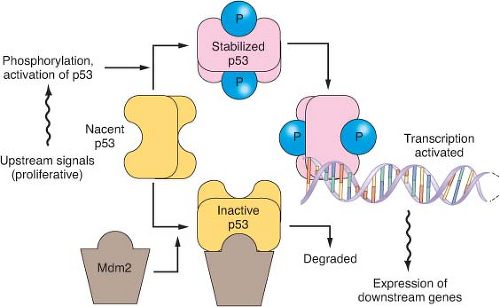
FIGURE 17.4 The p53 pathway. Many modes of control affect p53 activity, one of which is shown in the diagram. Stresses such as DNA damage result in phosphorylation of p53, preventing its degradation by an Mdm2-directed pathway. When stabilized, p53 binds to specific DNA sequences and activates the transcription of many genes, including Mdm2 as part of a negative feedback loop. When p53 is mutated, it fails to bind effectively to DNA to activate transcription. Because Mdm2 then lacks its transcriptional stimulus from p53, mutant but inactive p53 proteins are usually expressed at very high levels.

The protein product of the TP53 gene binds to specific sites of DNA and activates the transcription of certain genes that control the cell division cycle and apoptosis.38

Only gold members can continue reading. Log In or Register to continue

Stay updated, free articles. Join our Telegram channel

May 27, 2016 | Posted by in ONCOLOGY | Comments Off on Pancreas Cancer

Full access? Get Clinical Tree

Get Clinical Tree app for offline access