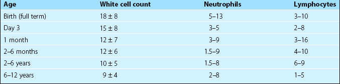
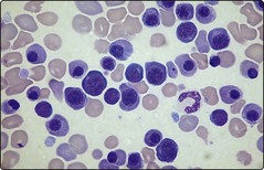
Paediatric haematology

45


Paediatric haematology


Many of the blood disorders encountered in children have been discussed in the preceding pages. For instance, acute lymphoblastic leukaemia is the most common leukaemia of childhood, haemophilia is usually diagnosed in infancy and the haemoglobinopathies are a significant cause of ill health in children worldwide. Chronic and severe diseases of the blood pose particular problems in childhood and usually are best managed by a paediatrician with a special interest in haematology or in a combined paediatric/haematology clinic. The child’s growth and development, and educational needs often require special attention. In this section we discuss some haematological disorders encountered in paediatric practice which are not addressed elsewhere.


Normal values


It is important to appreciate that the normal ranges for many haematological tests vary with age. Table 45.1 illustrates reference values for the total white cell count (WCC) and the differential count in children. More detailed listings of normal ranges of laboratory tests in childhood can be found in specialised paediatric haematology texts.


Neonatal disorders


Haemolytic disease of the newborn


Haemolytic disease of the newborn (HDN) is a disease of the fetus and newborn child. The haemolysis is caused by maternal IgG antibodies traversing the placenta and attaching to fetal red cells which are destroyed in the child’s reticuloendothelial system. The antibodies are directed against a fetal red cell antigen not shared by the mother. Incompatibility for one of a large number of different red cell blood group systems can cause HDN but most cases of clinically significant disease affect a Rhesus (Rh)D-positive child where the mother is RhD negative. Sensitisation of the mother (i.e. the formation of anti-D) occurs following the haemorrhage of fetal red cells into the maternal circulation. This usually occurs at parturition following a normal pregnancy but may also arise earlier in pregnancy or following abortion. ABO incompatibility between mother and fetus gives some protection against sensitisation to RhD as fetal red cells are quickly destroyed by the mother’s naturally occurring anti-A or anti-B antibodies. Unfortunately, in most cases baby and mother are ABO compatible. With the considerable success of prophylaxis against HDN due to RhD incompatibility (see below), the most common cause of the disorder is the formation of immune antibodies against ABO; most cases are associated with only mild haemolysis.


Management


Management of HDN is complex, requiring close liaison between the haematology laboratory and obstetrician. In RhD alloimmunisation, if maternal anti-D levels are high and paternal testing indicates RhD heterozygosity, the fetal Rh genotype can be determined non-invasively by applying PCR technology to a maternal blood sample. Another advance is velocimetry of the fetal middle cerebral artery during an affected pregnancy. High peak systolic velocities predict severe fetal anaemia and allow the selective use of more invasive techniques such as fetal blood sampling and intrauterine transfusion. Newborns may experience ongoing anaemia and require exchange transfusion. Later anaemia may respond to erythropoietin therapy. With optimal management, a healthy child is the outcome in more than 90% of cases.


Jun 12, 2016 | Posted by in HEMATOLOGY | Comments Off on Paediatric haematology

Full access? Get Clinical Tree

Get Clinical Tree app for offline access