Fig. 4.1
An 18-French Foley catheter is placed transurethrally, and an extraperitoneal incision is made in the lower midline, approximately 8 cm in length. Reprinted with permission, Cleveland Clinic Center for Medical Art & Photography © 1996–2013. All Rights Reserved
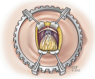
Fig. 4.2
A self-retaining, table-fixed ring retractor is placed. The prostatic apex is to the top. Reprinted with permission, Cleveland Clinic Center for Medical Art & Photography © 1996–2013. All Rights Reserved
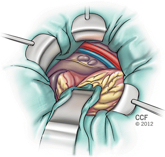
Fig. 4.3
A malleable blade is used for exposure of the obturator fossa when pelvic lymphadenectomy is performed. Reprinted with permission, Cleveland Clinic Center for Medical Art & Photography © 1996–2013. All Rights Reserved
Pelvic Lymphadenectomy
Based on published nomograms and our own experience, pelvic lymphadenectomy is omitted in selected patients at low risk for lymph node metastases based on preoperative serum prostate-specific antigen (PSA), tumor grade, and palpable tumor extent. Generally, lymphadenectomy is omitted in patients with AUA or D’Amico low-risk criteria. Such patients have a minuscule risk of positive nodes and omission of lymphadenectomy in patients with these characteristics does not increase the likelihood of biochemical failure [4]. For prognostic and potential therapeutic purposes, an extended lymphadenectomy is performed in all patients not meeting the low risk criteria. The dissection includes the tissue medial to the external iliac artery, all tissue surrounding the external iliac, the internal iliac artery superiorally, the bifurcation of the external and internal iliac veins cephalad, the origin of the superficial circumflex iliac vein caudally, and the pelvic sidewall in the obturator fossa deeply. In addition, the nodes immediately medial to the common iliac artery are excised. The extent of the dissection is guided by the study by Mattei et al. showing that the nodes within these boundaries constitute 75 % of the primary lymphatic drainage of the prostate [5]. Frozen section analysis is not routinely performed unless the nodes are grossly suspicious, and only if a finding of positive nodes would result in aborting the prostatectomy.
Endopelvic and Lateral Pelvic Fascia, Santorini’s Plexus, and Dorsal Vein Complex
The apical dissection begins with vertical incisions of the endopelvic fascia at the apex bilaterally (Fig. 4.4). The attachments of the levator muscles to the lateral surface of the prostate are taken down sharply with scissors. Blunt dissection of these attachments should be avoided to prevent shearing of small blood vessels, which may be difficult to control. The puboprostatic ligaments are divided.
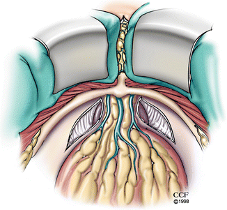

Fig. 4.4
The endopelvic fascia is incised bilaterally just lateral to the prostatic apex. The attachments of the levator muscles to the lateral surface of the prostate are taken down sharply with scissors. The puboprostatic ligaments are left intact. The apex is to the top. Reprinted with permission, Cleveland Clinic Center for Medical Art & Photography © 1996–2013. All Rights Reserved
Next, the lateral pelvic fascia (the visceral portion of the endopelvic fascia) covering the prostate is incised bilaterally beginning from the initial incision in the apical endopelvic fascia and extending to the base of the prostate (Fig. 4.5). The incision is performed high on the lateral surface of the prostate to avoid injuring the neurovascular bundles (NVBs). When completed, this maneuver allows clear visualization of the prostatourethral junction and location of the NVBs and facilitates bunching of the ramifications of the dorsal vein over the prostate. The cut edges of the lateral fascia are then grasped with Turner-Babcock clamps, incorporating the branches of the venous plexus on the dorsolateral surface of the prostate (Fig. 4.6a). The bunched tissue is suture-ligated with two individual figure-of-8 0-chromic ligatures (Fig. 4.6b, c). This technique is a modification of the dorsal venous plexus bunching technique originally described by Myers [6]. It prevents back-bleeding when the dorsal vein is divided and helps identify the plane between the dorsal vein and urethra. The prostate is next retracted superiorly with a sponge stick, and the fat between the puboprostatic ligaments is gently removed to expose the superficial dorsal vein which is then divided between clips.
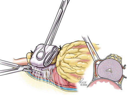
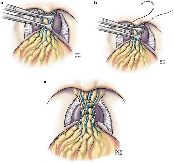

Fig. 4.5
The lateral pelvic fascia (the visceral layer of the endopelvic fascia covering the prostate) is elevated with a right-angled clamp and incised sharply with a knife (along the dotted line) from the apex to base of the prostate. This maneuver exposes the anterior prostatourethral junction and the position of the neurovascular bundles and facilitates control of the ramifications of the dorsal vein over the prostate. The maneuver is then repeated on the opposite side (not shown). The apex is to the left. Reprinted with permission, Cleveland Clinic Center for Medical Art & Photography © 1996–2013. All Rights Reserved

Fig. 4.6
Bunching technique for control of the dorsal venous complex. (a) Turner-Babcock clamps are used to bunch together the branches of the dorsal vein covering the dorsal surface of the prostate. (b) Two figure-of-8 sutures are used to ligate these branches, incorporating the cut edges of the endopelvic fascia. (c) Appearance after both sutures have been placed. The apex is to the top. Reprinted with permission, Cleveland Clinic Center for Medical Art & Photography © 1996–2013. All Rights Reserved
The dorsal vein and urethra are closely approximated and passage of an instrument between them carries the risk of damaging the anterior-striated sphincter. Therefore, the dorsal vein is next divided with scissors, exposing the anterior surface of the urethra (Fig. 4.7a). The distal portion of the incised dorsal vein is then oversewn with a figure-of-8 suture using a 3-0 absorbable monofilament suture on a 26 mm 5/8 circle needle (Fig. 4.7b). When correctly performed this technique does not compromise the anterior prostatic margin and results in excellent visualization of the urethra (Fig. 4.7c).
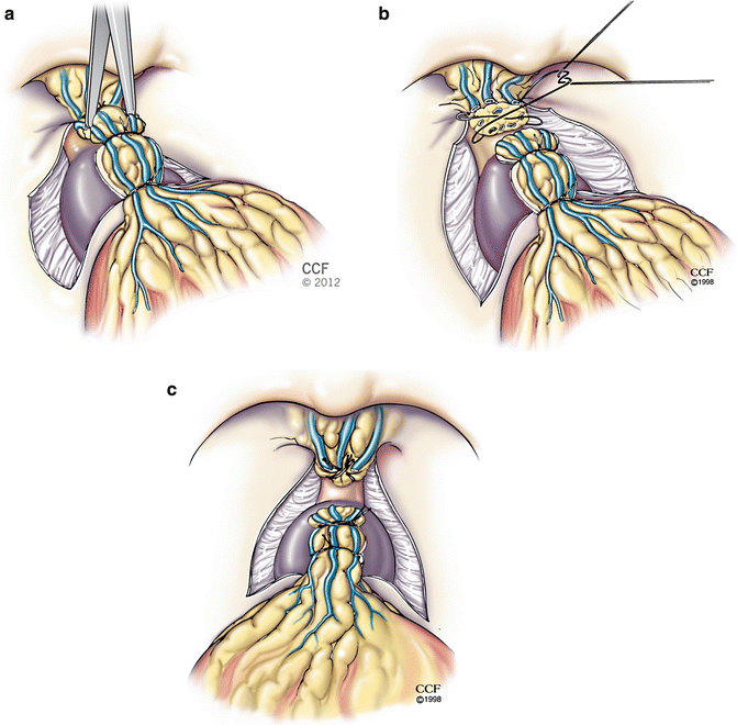

Fig. 4.7


Division and control of the dorsal vein. The prostatic apex is to the left. (a) The dorsal vein is divided with scissors, exposing the anterior surface of the urethra. (b) The cut surface of the dorsal vein is suture-ligated for hemostasis. (c) Appearance of the urethra after division and ligation of the dorsal vein. Reprinted with permission, Cleveland Clinic Center for Medical Art & Photography © 1996–2013. All Rights Reserved
Stay updated, free articles. Join our Telegram channel

Full access? Get Clinical Tree


