Normal newborn peripheral blood morphology

23 Normal newborn peripheral blood morphology


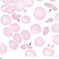
In the healthy, full-term newborn, peripheral blood collected within the first 12 hours of birth has distinctive morphology. Some morphological changes persist for up to 3 to 5 days after birth. These changes should be recognized as physiological and not pathological. For a fuller discussion of hematology in the newborn, refer to a hematology textbook such as Hematology: Clinical Principles and Applications* or a pediatric hematology text such as Nathan and Oski’s Hematology of Infancy and Childhood.


Entire books have been written to address abnormal hematology in neonates and especially in the premature infant. This chapter does not attempt to address those disorders but rather depicts morphological changes commonly seen in the healthy newborn.


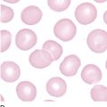
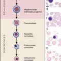
Erythrocyte morphology demonstrates macrocytes, with a mean cell volume of 110 ± 15 fL, which declines dramatically after the first 12 hours. Up to 3 to 10 orthochromic normoblasts (nucleated red blood cells) may be seen per 100 white blood cells and should disappear by day 5. Polychromasia reflects the erythropoietic activity of the newborn. Anisocytosis is reflected in the red blood cell distribution width index, which ranges from 15.2% to 18.0%.

Stay updated, free articles. Join our Telegram channel

Jun 12, 2016 | Posted by in HEMATOLOGY | Comments Off on Normal newborn peripheral blood morphology

Full access? Get Clinical Tree
Get Clinical Tree app for offline access