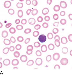
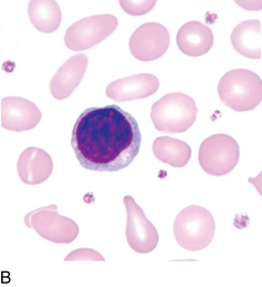
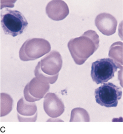
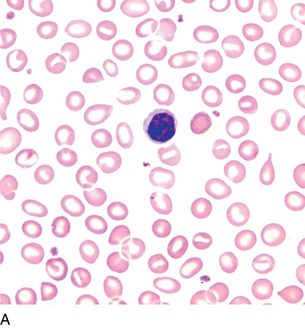
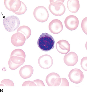
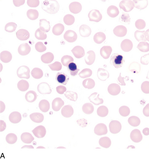
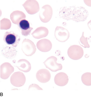
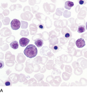
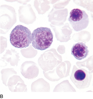
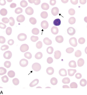
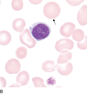
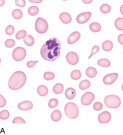
13 Diseases affecting erythrocytes Microcytic/hypochromic anemia Iron deficiency anemia FIGURE 13–1A Severe iron deficiency anemia (peripheral blood [PB] ×500). FIGURE 13–1B Iron deficiency anemia (PB ×1000). FIGURE 13–1C Iron deficiency anemia (bone marrow [BM] ×1000; showing shaggy cytoplasm). Peripheral blood: Erythrocytes are hypochromic and microcytic; large variation in size; possible thrombocytosis Note: Small lymphocyte depicted for size comparison. Bone marrow: Erythrocyte precursors are smaller and more numerous than normal and have shaggy cytoplasm. There is nuclear cytoplasmic asynchrony, with cytoplasmic maturation lagging behind that of the nucleus. β-thalassemia minor β/β+ β/β0 β/δβ0 β/δβlepore FIGURE 13–2A β-Thalassemia minor (PB ×500). FIGURE 13–2B β-Thalassemia minor (PB ×1000). The presence of basophilic stippling (arrow) is common in thalassemia minor but not in iron deficiency anemia. Peripheral blood: Microcytosis, slight hypochromia, target cells, basophilic stippling β-thalassemia major β0β0 β+β+ β0β+ δβlepore/δβlepore FIGURE 13–3A β-Thalassemia major (PB ×500). FIGURE 13–3B β-Thalassemia major (PB ×1000). Peripheral blood: Marked variation in size and shape, numerous nucleated erythrocytes, microcytes, hypochromia, target cells, basophilic stippling, tear drop cells, schistocytes, polychromasia α-thalassemia* Hemoglobin h −−/−α Peripheral blood: Microcytes, hypochromia, marked poikilocytosis, target cells, polychromasia (see Figure 12-5, C). Hemoglobin bart hydrops fetalis syndrome −−/−− Peripheral blood: Numerous nucleated erythrocytes, marked variation in size, hypochromia, variable polychromasia, macrocytes FIGURE 13–4A Bart hemoglobin (PB ×500). FIGURE 13–4B Bart hemoglobin (PB ×1000). Macrocytosis Nonmegaloblastic FIGURE 13–5A Macrocytic (nonmegaloblastic) (PB ×500). FIGURE 13–5B Macrocytic (nonmegaloblastic) (PB ×1000). Peripheral blood: Round macrocytes, leukocyte and platelet counts usually normal Bone marrow: No megaloblastic changes Associated with: Normal newborn, liver disease, chronic alcoholism Megaloblastic anemia FIGURE 13–6A Megaloblastic anemia (PB ×500). Only gold members can continue reading. Log In or Register to continue Share this:Click to share on Twitter (Opens in new window)Click to share on Facebook (Opens in new window) Related Related posts: Hematopoiesis Basophil maturation Megakaryocyte maturation Inclusions in erythrocytes Neutrophil maturation Myelodysplastic syndromes Stay updated, free articles. Join our Telegram channel Join Tags: Clinical Hematology Atlas Jun 12, 2016 | Posted by admin in HEMATOLOGY | Comments Off on Diseases affecting erythrocytes Full access? Get Clinical Tree
13 Diseases affecting erythrocytes Microcytic/hypochromic anemia Iron deficiency anemia FIGURE 13–1A Severe iron deficiency anemia (peripheral blood [PB] ×500). FIGURE 13–1B Iron deficiency anemia (PB ×1000). FIGURE 13–1C Iron deficiency anemia (bone marrow [BM] ×1000; showing shaggy cytoplasm). Peripheral blood: Erythrocytes are hypochromic and microcytic; large variation in size; possible thrombocytosis Note: Small lymphocyte depicted for size comparison. Bone marrow: Erythrocyte precursors are smaller and more numerous than normal and have shaggy cytoplasm. There is nuclear cytoplasmic asynchrony, with cytoplasmic maturation lagging behind that of the nucleus. β-thalassemia minor β/β+ β/β0 β/δβ0 β/δβlepore FIGURE 13–2A β-Thalassemia minor (PB ×500). FIGURE 13–2B β-Thalassemia minor (PB ×1000). The presence of basophilic stippling (arrow) is common in thalassemia minor but not in iron deficiency anemia. Peripheral blood: Microcytosis, slight hypochromia, target cells, basophilic stippling β-thalassemia major β0β0 β+β+ β0β+ δβlepore/δβlepore FIGURE 13–3A β-Thalassemia major (PB ×500). FIGURE 13–3B β-Thalassemia major (PB ×1000). Peripheral blood: Marked variation in size and shape, numerous nucleated erythrocytes, microcytes, hypochromia, target cells, basophilic stippling, tear drop cells, schistocytes, polychromasia α-thalassemia* Hemoglobin h −−/−α Peripheral blood: Microcytes, hypochromia, marked poikilocytosis, target cells, polychromasia (see Figure 12-5, C). Hemoglobin bart hydrops fetalis syndrome −−/−− Peripheral blood: Numerous nucleated erythrocytes, marked variation in size, hypochromia, variable polychromasia, macrocytes FIGURE 13–4A Bart hemoglobin (PB ×500). FIGURE 13–4B Bart hemoglobin (PB ×1000). Macrocytosis Nonmegaloblastic FIGURE 13–5A Macrocytic (nonmegaloblastic) (PB ×500). FIGURE 13–5B Macrocytic (nonmegaloblastic) (PB ×1000). Peripheral blood: Round macrocytes, leukocyte and platelet counts usually normal Bone marrow: No megaloblastic changes Associated with: Normal newborn, liver disease, chronic alcoholism Megaloblastic anemia FIGURE 13–6A Megaloblastic anemia (PB ×500). Only gold members can continue reading. Log In or Register to continue Share this:Click to share on Twitter (Opens in new window)Click to share on Facebook (Opens in new window) Related Related posts: Hematopoiesis Basophil maturation Megakaryocyte maturation Inclusions in erythrocytes Neutrophil maturation Myelodysplastic syndromes Stay updated, free articles. Join our Telegram channel Join Tags: Clinical Hematology Atlas Jun 12, 2016 | Posted by admin in HEMATOLOGY | Comments Off on Diseases affecting erythrocytes Full access? Get Clinical Tree