Chapter 75 Non-Hodgkin’s Lymphoma
Etiology and Epidemiology
The incidence of NHL has been increasing worldwide,1 including in North America.2,3 In 2011, the American Cancer Society estimated that 66,360 new cases were diagnosed and 19,320 deaths occurred in the United States.4 In many developed countries there was evidence for a 2% to 4% per year rise in the incidence of NHL from the 1970s to 1990s, and this stabilized in the past decade. This increase is more marked for older persons. In the United States the annual rise in incidence rate had slowed to 1% to 2% in the 1990s.2,3 Some of the increase has been due to increase in immunosuppression (HIV infection and iatrogenic), spread of infectious agents such as human T-cell lymphotropic virus type 1 (HTLV-1) and Epstein-Barr virus (EBV), increased use of chemicals, and increased case finding due to improved pathologic diagnosis. However, these factors do not explain the magnitude of the rise in incidence.
The epidemiology of certain histologic subtypes of lymphomas follows distinct patterns related to the epidemiology of the putative causative agent and any environmental cofactors. The classic example is Burkitt’s lymphoma in Africa, with the endemic role of EBV and the contribution of immunosuppression because of malaria infection.5 Similarly, EBV-related T-cell/natural killer (NK) cell lymphoma of the nasal region is more frequent in the Asian population in south Asia.6,7 Adult T-cell leukemia/lymphoma caused by HTLV-1 is endemic in the Caribbean and southern Japan.8 The frequency of HIV-related lymphomas is proportional to the HIV infection rate in the population, explaining the observation of high incidence rates in large urban cities and a role of Kaposi sarcoma–associated herpesvirus.9 MALT lymphomas of the stomach are seen more frequently in regions where Helicobacter pylori infection is endemic.
Despite an increasing body of knowledge on the genetic and phenotypic events underlying the development and progression of NHL, the causative agent has not been identified in the majority of cases. The putative causative agents and associated conditions with a predisposition for the development of NHL are listed in Table 75-1. There are some genetic predispositions to the development of NHL, including specific human leukocyte antigen (HLA) allele associations.10,11 Changes in the immune state, either immunosuppression or autoimmune disease with immune stimulation, can predispose to NHL. The immunosuppression can be primary (i.e., inherited as in severe combined immunodeficiency syndrome), secondary to retrovirus infection (HIV), or iatrogenic (after solid organ or bone marrow transplantation). The incidence of NHL in HIV-positive individuals has declined since the late 1990s, owing to highly effective antiretroviral therapy.12,13 Autoimmune diseases such as Sjögren’s syndrome, rheumatoid arthritis, and celiac disease are also associated with an increased risk of NHL.14
TABLE 75-1 Causative Agents and Associated Conditions with a Predisposition to the Development of Non-Hodgkin’s Lymphoma
| Immunodeficiency |
| Congenital: Severe combined immunodefiencies, ataxia-telangiectasia |
| Acquired: Organ or hematopoietic stem cell transplantation, HIV infection |
| Genetic Predisposition |
| Variants within 6q21 (HLA) |
| TNF G308A polymorphisms |
| Autoimmune Disorders |
| Sjögren’s syndrome |
| Hashimoto’s thyroiditis |
| Celiac disease |
| Rheumatoid arthritis |
| Viral Agents |
| Epstein-Barr virus |
| Human T-cell lymphotropic virus 1 (HTLV-1) |
| Herpesvirus type 8 |
| Hepatitis C |
| Bacteria |
| Helicobacter pylori (gastric lymphoma) |
| Chlamydia psittaci (orbital lymphoma) |
| Borrelia burgdorferi (cutaneous lymphoma) |
| Campylobacter jejuni (alpha heavy chain disease) |
| Drugs |
| Alkylating agents |
| Other immunosuppressive drugs |
| Pesticides |
| Phenoxyl herbicides |
| Organophosphate insecticides |
| Fungicides |
| Solvents and Other Chemicals |
| Benzene |
| Trichloroethylene |
| Hair dyes |
| Diet |
| N-nitroso compounds |
| Fat |
Infectious agents have been shown to be etiologic factors in NHL, such as Helicobacter pylori in gastric MALT lymphomas and Chlamydia psittaci in ocular adnexal lymphomas.15 There is a large variation in the Chlamydia positivity rate in orbital lymphomas depending on geographic location.16,17 The viruses that have been implicated include EBV,5 HTLV-1, human herpesvirus 8 (HHV-8), and hepatitis C virus (HCV). HTLV-1 carriers have a 2% cumulative risk of developing adult T-cell leukemia/lymphoma, and this is a major public health problem in some parts of the world where the seropositivity rate is high.8 HHV-8 virus sequences have been implicated in primary effusion lymphomas and multicentric Castleman’s disease. Hepatitis C infection is also a predisposing factor for some B-cell lymphomas, often in association with essential mixed cryoglobinemia.18
Epidemiologic studies suggest an association with environmental factors, including industrial chemicals, rural residence possibly linking to use of pesticides or other agricultural chemicals, hair dyes,19 diet,20 and obesity.21 A small excess risk was also found in atomic bomb survivors for males but not for females,22 whereas minimal or no risk was found for low-dose x-ray exposures.
Prevention and Early Detection
With a better definition of environmental causative factors for NHL, it is expected that preventive strategies will be developed. For adult T-cell leukemia/lymphoma, initiatives are being implemented to reduce the disease burden in susceptible populations, the most important being the control of breast feeding to prevent maternal-fetal transmission of the HTLV-1 virus. Other promising areas of prevention currently include heightened awareness of potentially toxic agents and the development of improved products (chemicals and hair dyes). Genetic tests that aim to identify persons at higher risk of developing lymphoma are under investigation.11,23 For viral agents, the development of vaccines is under active study.24 For organ transplantation, avoidance of transplanting an EBV-positive donor organ into a EBV-negative recipient should reduce the risk of post-transplant NHL. For persons infected with HIV, effective combination antiviral treatments have contributed to a decreased incidence of lymphomas.13 The detection and eradication of Helicobacter pylori infection in the stomach is an important strategy with potential to control or prevent MALT lymphoma25,26 and its transformation. As additional microorganisms and viruses other than the ones mentioned here are being discovered to play critical roles in the pathogenesis of some lymphomas,9,27 prevention and early treatment strategies will become increasingly important in the future.
Biologic Characteristics and Pathology
The present classification of NHL emphasizes lineage, function, and distinct clinicopathologic disease entities, with a liberal use of phenotypic and molecular techniques. The World Health Organization (WHO) classification, fourth edition,28 is shown in Table 75-2, (B-cell) and Table 75-3 (T-cell). The WHO classification recognized three major categories of lymphoid malignancies: B-cell, T-cell, and Hodgkin’s lymphoma.
TABLE 75-2 World Health Organization Classification for Mature B-Cell Neoplasms
• B-cell lymphoma, unclassifiable, with features intermediate between DLBCL and classical Hodgkin lymphoma |
NOTE: The italicized histologic types are provisional entities.
TABLE 75-3 World Health Organization Classification of Mature T-Cell and NK-Cell Neoplasms
NOTE: The italicized histologic types are provisional entities.
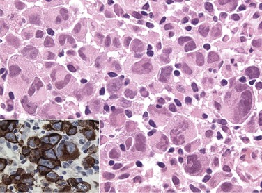
The diagnosis of the new pathologic and clinical entities continues to be based on morphology but is assisted by immunophenotyping and genotyping. The most common lymphoma entities were diffuse, large B-cell lymphoma (DLBCL) (31%) and follicular lymphoma (22%). MALT/marginal zone lymphoma constituted 7.6% of cases, peripheral T-cell lymphomas, 7%, small B-lymphocytic, 6.7%, mantle cell, 6%, anaplastic large T/null cell, 2.4%, primary mediastinal large B-cell, 2.4%, and high-grade non-Burkitt’s and Burkitt’s, less than 3% of cases.29 Surface marker studies provide an objective basis for resolving difficult morphologic problems (Table 75-4). The lineage of the B-cell lymphomas is confirmed by pan–B-cell markers (CD22, CD19, CD20). T-cell lineage is evident from the presence of T-cell markers (CD3, CD2, CD7), and T-cell subset is determined from CD4 and CD8 analysis. Among indolent lymphomas, the CD5+, CD10−, CD23+ phenotype is characteristic of small lymphocytic lymphoma (chronic lymphocytic leukemia), the CD5−, CD10+, CD23± phenotype of follicular lymphoma, and the CD5−, CD10−, CD23− phenotype of MALT lymphoma (see Table 75-4). The CD5+, CD10−, CD23− phenotype is characteristic of mantle cell lymphoma. Among T-cell lymphomas the CD30+ phenotype is characteristic of anaplastic large cell lymphoma (Fig. 75-1), whereas CD56+ is associated with extranodal T-cell/NK-cell lymphoma of nasal type. Although surface marker analysis enhances the accuracy of the diagnosis, few surface marker characteristics are totally lineage specific. Ancillary cytologic and histologic techniques used to establish proliferative activity include labeling index, S-phase fraction, and Ki-67 antigen staining,30,31 which has been documented as a strong prognostic factor.
TABLE 75-4 Phenotypic Characteristics of Non-Hodgkin’s Lymphoma
| Lymphoma Type | Characteristic CD Antigen Profile |
|---|---|
| B-cell markers | CD19, CD20, CD22 |
| T-cell markers | CD2, CD3, CD4, CD7, CD8 |
| Anaplastic large cell lymphoma | CD30+ (Ki-1 antigen) |
| Small lymphocytic lymphoma (B-CLL) | CD5+, CD10–, CD23+, B-cell markers |
| Follicular lymphoma | CD5–, CD10+, CD23±, CD43–, B-cell markers |
| Marginal-zone (MALT) lymphoma | CD5–, CD10–, CD23–, B-cell markers |
| Mantle cell lymphoma | CD5+, CD10±, CD23–, CD43+, B-cell markers |
Mantle Cell Lymphoma
Mantle cell lymphoma occurs in older adults and commonly presents as generalized disease with spleen, bone marrow, and gastrointestinal tract involvement. Circulating lymphoma cells are frequently found in the blood. This type of lymphoma is associated with a characteristic immunophenotype (CD5+, CD10±, and CD23−) and genotype with t(11;14) translocation and BCL1 (CCND1/PRAD1) gene overexpression.28 Currently, advanced disease is not curable with chemotherapy. The median survival for patients with generalized disease is 4 to 6 years, and early use of high-dose therapy is promising.30,32,33 Expression of a microarray gene profile characterized by high proliferation,34 or a high Ki-67 growth fraction,35 confers a worse prognosis. Localized stage I and II mantle cell lymphoma is uncommon, and there are little data on curability. Existing datasets suggest that the survival of patients with localized mantle cell lymphoma is better than those with advanced disease and that RT has an important role in localized disease.36,37 Stage I and II mantle cell lymphomas may be curable.
T-Cell Lymphoma
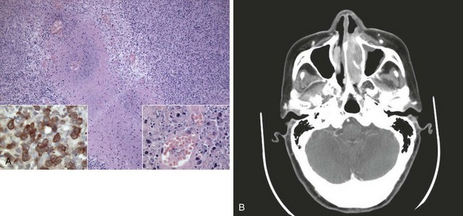
Peripheral T-cell lymphomas are a heterogeneous group of T-cell neoplasms that are more common in Asia38,39 and usually affect adults and are commonly generalized at presentation. An aggressive clinical course is typical; and although potentially curable, many T-cell lymphomas are resistant to current chemotherapy regimens. TCR gene rearrangements may be identified but this is not mandatory for diagnosis. Specific subtypes of T-cell lymphoma to consider include extranodal natural killer (NK)-cell/T-cell lymphoma of nasal type and enteropathy-associated T-cell lymphoma (EATL), previously called malignant histiocytosis of the intestine. This latter disease (EATL) usually involves the jejunum and in approximately 50% of cases is associated with a history of celiac disease.40 Most EATLs express the human mucosal lymphocyte-1 (HML-1) antigen, supporting an origin from the intraepithelial T cells of MALT. EATLs are high-grade pleomorphic large cell lymphomas associated with a very poor prognosis. The entity of NK-cell/T-cell lymphoma of nasal type includes disorders previously known as lethal midline granuloma and nasal T-cell lymphoma. It is characterized by an angiocentric and angioinvasive infiltrate28,41 (Fig. 75-2), with CD56-positive (T-cell/NK-cell) immunophenotype and is characteristically EBV positive. This disease responds poorly to chemotherapy and usually follows an aggressive course.42,43,44 Other rare presenting sites may include skin, lung, testis, and central nervous system (CNS).
The WHO classification, although aimed to classify distinct disease entities and acknowledging the uniqueness of certain presentations (e.g., the T cell lymphomas just cited), does not always consider the presenting site of the lymphoma. Recent evidence strongly suggests that the presenting site is an important factor for primary extranodal lymphomas. Examples include different causes for MALT lymphoma arising in different sites that are associated with differences in relapse rate.45 Similar histology lymphomas may have distinct outcomes (e.g., diffuse large B-cell lymphoma involving the brain and thus with a poor prognosis) that are now clearly identified in the fourth edition of the WHO classification versus stomach or Waldeyer’s ring structures, which have a good prognosis.46 With further understanding of etiology and pathogenesis of lymphomas, future modifications to classification of these diseases are expected.
Molecular Biology
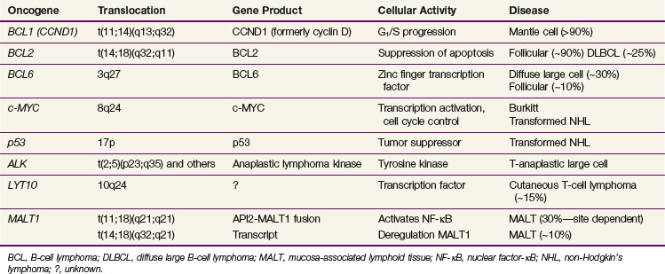
The various pathologic subtypes of NHL are characterized by distinctive nonrandom genetic alterations. The categories of molecular lesions include chromosomal translocations with activation of oncogene as the most commonly observed abnormality (e.g., BCL2 in follicular lymphomas), oncogenic viruses (e.g., EBV and HTLV-1), and inactivation of tumor suppression gene by chromosome mutation or deletions (e.g., TP53). Some of the well-characterized genetic lesions with their corresponding pathologic subtype are presented in Table 75-5. Many of the genetic lesions involve the translocation of an oncogene to a juxtaposition of the immunoglobulin gene (in B-cell lymphomas) or T-cell receptor gene (in T-cell lymphomas). Locations of breakpoints usually occur at the joining (J) or switch (S) sequences involved in antigen receptor gene rearrangement as part of normal lymphoid development to produce antigenic variation. The causes of these translocations are largely unknown, but once established these lesions appear to be highly specific for the type of malignant lymphoma that they characterize.
BCL2
The t(14;18)(q32;q21) translocation involving BCL2 is the most common chromosome translocation in NHL. BCL2 encodes a 26-kd membrane protein that controls and prevents cellular apoptosis. The gene is translocated to a J segment of the IGH gene on chromosome 14, giving rise to deregulation of BCL2, with the result being the inhibition of apoptosis of B cells. This lesion is present in more than 90% of follicular lymphomas and in transformed follicular lymphomas.47 In the latter situation, additional genetic lesions are acquired (TP53 mutation, deletion of chromosome 6q27, MYC) in parallel with more rapid growth and an aggressive clinical course. The TP53 mutation is associated with a poor survival rate in aggressive B-cell lymphomas, independent of the predictive effects of the International Prognostic Index (IPI).48 The t(14;18) translocation is also found in 20% of de novo DLBCL and appears to be an unfavorable prognostic factor.
BCL6
The BCL6 gene located on 3q27 is translocated in 40% of diffuse large B-cell lymphomas.49,50 Reciprocal translocations can involve a number of other chromosomal locations, including 14q32 (IGH), 2p11 (IGK), and so on, all juxtaposing to 3q27.51 BCL6 is thought to mediate the DNA-binding activity of a number of zinc-finger transcription factors and characterizes the germinal center B-cell gene signature in microarray studies.52 The presence of the BCL6 translocation in DLBCL carries a favorable prognosis, in comparison with those carrying BCL2 who have a worse outcome and those with neither translocation who have an intermediate prognosis.49,53
MYC
The MYC oncogene, located on chromosome 8q24, regulates cellular proliferation and differentiation. Its translocation is seen in 100% of Burkitt’s lymphomas typically to chromosome 14q32 (IGH), less commonly to 2p11 (IGK) and 22q11 (IGG). EBV infection is responsible for and can be documented in almost all cases of endemic Burkitt’s lymphoma and about 30% of the sporadic cases.51,54 Aberrations of 8q24 can also be seen in follicular lymphoma, DLBCL, and mantle cell lymphoma and is characterized by transformation or progression of the disease, generally denoting a poor prognosis.55,56
Other Chromosome Translocations
The t(2;5)(p23;q35) translocation characterizes anaplastic large cell lymphomas of T-cell origin. This translocation involves the fusion of the NPM1 (nucleophosmin) gene on 5q35 and the ALK (anaplastic lymphoma kinase) gene on 2p23.28 Others involving ALK-2p23 include t(1;2)(q25;p23), Inv2 (p23q35), and many others.28 T-cell lymphomas may have 14q11 abnormalities involving the T-cell receptor-alpha gene. In MALT lymphomas, characteristic genetic abnormalities can include trisomy 357 and chromosomal translocations t(11;18)(q21;q21),58 t(1;14)(p22; q32),59 and t(14;18)(q32;q21)60 (see Table 75-5).
Microarray Gene Profiling Studies
Gene-expression studies have been increasingly utilized to characterize various lymphomas. Microarray analysis of cDNA (the “lymphochip”) using hierarchical clustering techniques initially classified DLBCL into two groups.61 One expressed “germinal center B-cell–like” genes (GCB signature), such as MYBL1, LMO2, JNK3, CD10, BCL6, CREL, and BCL2, whereas the other expressed “activated B-cell–like” genes (ABC signature), such as CCND2, IRF4, FLIP, NF-κB, and CD44. For patients treated with anthracycline-based chemotherapy in the pre-rituximab era, the 5-year survival for the GCB group was 60%, compared with the ABC group of 35%, whereas that for a type 3 group was 39%.52 Based on a selection of the most prognostic genes, either a larger series of 17 genes52 or condensing further to three immunostains for CD10, BCL6, and MUM1,62 studies have shown that gene expression profiling provides additional prognostic information in addition to the clinical IPI scores.52,62 Additional studies using oligonucleotide microarrays and a different analysis technique have also successfully identified two categories of patients with very different survival rates (70% vs. 12%) in DLBCL.63 Gene profiling of the nonmalignant infiltrating cells in the tumor has also been found to be prognostic in DLBCL, whether treatment included rituximab or not.64 The “stromal-1” group reflected extracellular matrix deposition, and histiocytic infiltration had a better prognosis than the “stromal-2” group, which reflected tumor angiogenesis activity.64 Presently, the microarray assays are still not widely available. Other examples of microarray studies discovering significant findings correlating with clinical outcome include primary mediastinal B-cell lymphomas,65 Burkitt’s lymphoma,66,67 mantle cell lymphoma,34,68 and follicular lymphoma.69 In the case of follicular lymphoma it is of interest that it was the gene signature of the nonmalignant infiltrating immune cells, rather than the malignant cells, that had the prognostic effect.69 It is likely that within the next decade most lymphomas will have their distinct genetic signatures identified. In addition to its usefulness in obtaining an accurate diagnosis (e.g., between DLBCL and Burkitt’s lymphoma),66 and enhancing prognostication, these techniques could also be helpful in the study of minimal residual disease, that is, the detection of a small number of morphologically normal but genetically monoclonal population of malignant cells. The most important aspect of this evolving field is the insight provided into the molecular events underlying malignant transformation, cell cycle regulation, signal transduction, and cell death. Characterization of these mechanisms opens up the potential for novel therapeutic strategies, for example, targeting small molecules such as NF-κB.70
Clinical Manifestations, Patient Evaluation, and Staging Classification
Patient Evaluation
The goal of evaluation is to determine disease extent and bulk and its anatomic distribution and to ascertain normal organ and immune function relevant to the choice of therapy. With the diverse presentation of lymphomas, the recommended investigations may vary,71 but the following procedures serve as minimum recommended investigations (Table 75-5). Additional tests may be necessary based on such factors as the presenting complaint, the subtype of lymphoma, and predilection of organ involvement (e.g., brain and cerebrospinal fluid).
Imaging Studies
Computed tomography (CT) of the neck, thorax, abdomen, and pelvis is the minimum standard today. The radiographic assessment of the extent of extranodal involvement is of particular importance in the planning of RT, since 50% of localized lymphomas occur in extranodal sites. Other radiographs should be obtained based on the actual or suspected organ involved. Magnetic resonance imaging (MRI) is indicated in delineating areas of suspected involvement of bone or CNS, such as spinal cord and epidural space, brainstem, base of skull and cavernous sinuses, and leptomeninges. The role of routine MRI for the thorax, abdomen, and pelvis has not been established. Bone scintigraphy is not recommended as a routine test except in patients with bone pain or those presenting with localized bone lymphoma. Total-body scan with 18F-fluorodeoxyglucose positron emission tomography (FDG-PET) is now a standard staging procedure in many countries and is used also to document response to therapy. FDG-PET has replaced gallium-67 scans in clinical practice, and nonrandomized studies comparing the two imaging modalities in terms of sensitivity concluded that FDG-PET is more sensitive than gallium-67 (100% vs. 71.5%).72–74 Whole-body FDG-PET has been shown to have a high predictive value for differentiating active from necrotic residual masses in lymphomas75,76 and has been incorporated in the revised International Working Group response criteria.77 The category of “complete response, unconfirmed” has been eliminated because PET-negative patients are now classified as complete responders and those with PET-positive disease will be partial responders. Consensus guidelines regarding standardization of PET reporting has also been published by the International Harmonization Project in lymphoma.78 Mediastinal blood pool activity was recommended as the reference background activity to define PET positivity for a residual mass 2 cm or larger in maximum diameter. A smaller residual mass or a normal-sized lymph node is considered positive if its activity is above that of the surrounding background.78 Increasing evidence also suggest that persistent FDG-PET activity after one to four cycles of chemotherapy confers a worse prognosis.79 In a systematic review of seven studies, interim FDG-PET in DLBCL has a sensitivity of 0.78 and specificity of 0.87.79
Staging Classification
The American Joint Committee on Cancer (AJCC) and Union Internationale Contre le Cancer (UICC) have endorsed the use of the Ann Arbor classification for staging of NHL. The Ann Arbor staging classification has been used for more than 40 years.80,81 In the Ann Arbor system, Waldeyer’s ring, thymus, spleen, appendix, and Peyer’s patches of the small intestine are considered as lymphatic tissues and involvement of these areas does not constitute an “E” lesion, originally defined as extralymphatic involvement. However, because of the unique pathologic and clinical characteristics of primary lymphoma affecting these organs, most clinicians consider them as separate clinical entities and report their involvement as extranodal presentation. The Ann Arbor classification differentiates locoregional from widespread lymphoma and documents anatomic extent of disease and B symptoms, but it is not optimal for describing the extent of local disease, invasion of adjacent organs, tumor bulk, or multiple sites of involvement within one organ (e.g., skin, gastrointestinal tract). Several modifications to the Ann Arbor classification have been proposed in the past. In gastric lymphoma, substaging of stage I to reflect the depth of the stomach wall penetration has been suggested. In stage II disease, distinction of involvement of the immediate nodal region (II1) versus more extensive regional lymph node involvement (II2) has been found to be of prognostic significance in primary gastrointestinal lymphomas.82,83 Currently, the Ann Arbor staging classification supplemented by description of prognostic factors, including tumor bulk, hematologic and biochemical parameters, sites of involvement, and pathology, remains the basis for patient assessment.
TABLE 75-6 Diagnostic Workup for Non-Hodgkin’s Lymphoma
| General |
• History, including systemic symptoms (unexplained fever, night sweats, weight loss > 10% of body weight), risk factors for HIV infection |
| Special Tests |
| Standard |
| Imaging Studies |
Standard
Stay updated, free articles. Join our Telegram channel
Full access? Get Clinical Tree
 Get Clinical Tree app for offline access

|
