Fig. 33.1
a Skate flap marking up. b Flap raising: the bilateral dermal wings are thin peripherally and thicker at the base; the central body of the flap includes a substantial amount of soft tissue. c The tips A and B of the wings are brought together and sutured to point C, which is at the margin of the dermal defect where the base of the nipple will fall. d The wings of the skate are sutured together around the central belly of the flap. Primary closure of donor sites
Once the position of the neo-nipple is established, the flap is sketched with a width three times the diameter of the contralateral nipple and twice its height.
The wings of the skate are harvested as a partial thickness flap, whilst the central part of the flap is elevated to include subcutaneous tissue which will ensure both good vascularization to the flap and provide bulk to the neo-nipple.
The lateral edges of the wings are then sutured together and wrapped around the belly of the composite part of the flap. When the donor site is being closed primarily, care must be taken not to apply any tension to the closure as this will result in late loss of nipple projection and flatten the breast at this site. If primary closure cannot be achieved without tension, it should not be forced, but instead a small full thickness skin graft is used to cover the exposed dermis.
33.4.2 Star Flap
The star flap (◘ Fig. 33.2), designed by Anton and Hartrampf [17], is a modification of the skate flap. Three arms are drawn from the base of the flap at 90° from each other to resemble a star. The arms are raised as a full thickness flap towards the base with some subcutaneous tissue to preserve the blood supply. The central vascular core is left intact, and the lateral arms are wrapped around the central core to create the lateral walls of the nipple and provide its projection. Finally, the central arm is used as a covering cap. The donor site is closed primarily without tension preferably using absorbable sutures.
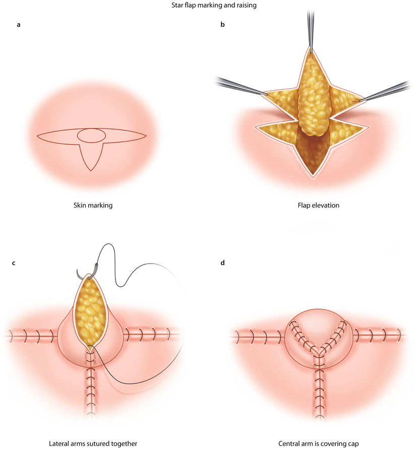

Fig. 33.2
a Star flap marking up. b The three arms of the star are raised full thickness towards the base; the central part has substantial subcutaneous tissue. c The lateral arms are wrapped around the central core and sutured together. d The central arm is used as covering cap. Primary closure of donor sites
33.4.3 CV Flap
The C-V flap (◘ Fig. 33.3), which evolved from the skate flap, consists of a central C flap and two lateral V flaps. The flap is drawn around the chosen site for the new nipple. The base of the V flaps provides the projection of the neo-nipple, whilst the width of the C flap will be equivalent to the final nipple diameter. As the flap is elevated, care must be taken not to divide the base of the C flap as this will impair the blood supply to the whole flap. The V flaps and the distal end of the C flap should include the subdermal vascular plexus and some fatty tissue but are left rather thin, whilst more subcutaneous tissue is left centrally to create the bulk of the nipple.
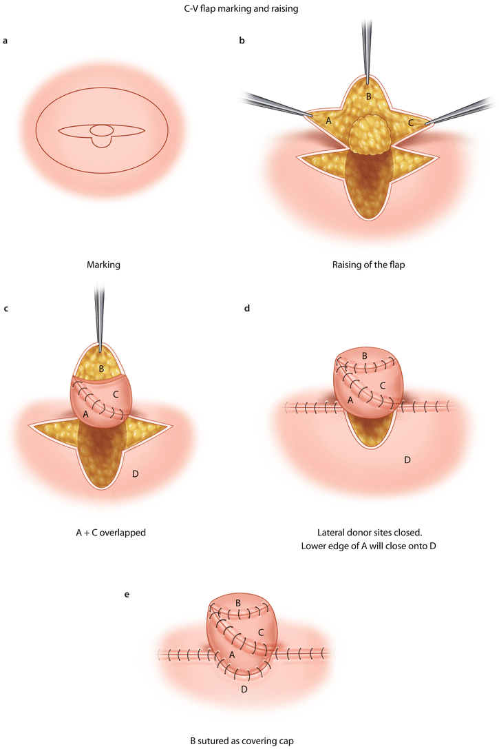

Fig. 33.3
a C-V flap marking up. b Elevation of the flap: lateral arms and distal central arm left thin; more subcutaneous tissue is raised centrally to create the bulk of the nipple. c Lateral arms A and C overlapped and sutured together. d Primary closure of the donor site will make D to close in to provide the base of the new nipple. Lower border of A is sutured to the donor site D. e B is sutured in as covering cap
The donor sites of the V flaps are then closed primarily. This will allow the donor site of the C flap to close in and provide a semicircular edge where the new nipple will be sutured into.
The V flaps are then overlapped onto each other whilst the C flap provides the covering cap.
Good results are obtained with 4/0 monofilament absorbable sutures to create these flaps and 3/0 absorbable sutures for the closure of the V flap donor sites (◘ Fig. 33.7a).
Several minor modifications to this technique are described in the literature, a common one being a fish tail flap where the two lateral flaps are more angulated rather than being diametrically opposite to each other.
33.4.4 S-Flap
In cases where the ideal new nipple position lies within the site of a scar, S-flaps (◘ Fig. 33.4) have proved very useful. Two equal-sized flaps with opposing bases are drawn at each side of the scar. The length and base widths of the flaps will determine both the diameter and projection of the nipple. The flaps are then elevated with some subcutaneous tissue and the tips of each flap opposed and sutured together. The donor sites are then closed primarily. In Cronin’s original description [18], a full thickness skin graft was used to cover both the neo-nipple and areola; however the advent of tattooing has made this requirement obsolete.
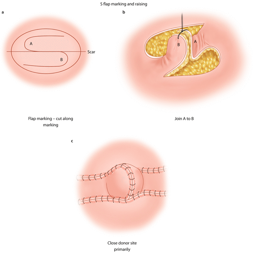

Fig. 33.4
a S flap marking and elevation. b Flaps A and B sutured together to form the new nipple. c Primary closure of donor site
33.4.5 Local Flaps with Augmentation
Surgeons have always battled to recreate a nipple that will continue to keep its shape and projection with time. At the time of surgery, the nipple can be augmented by inserting within the flap autologous, allogeneic or synthetic filler materials – alone or in combination. There is no strong evidence to support one type of filler over another [19]. Recognized autologous grafts include auricular or costal cartilages, bone, fat and dermal grafts. The use of synthetic materials (e.g. silicone, polyurethane, polytetrafluoroethylene) poses the risk of foreign body reactions, migration and possibly extrusion [20] but allow for sustained nipple projection.
Acellular dermal matrices have been reported [21] to provide good long-term nipple projection with low rates of complications but do impose a significant financial burden. The patient’s own dermis may also be used as an internal de-epithelialised graft.
33.4.6 Nipple Sharing (Composite Nipple Graft)
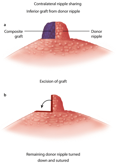
The contralateral nipple is the obvious choice for a donor site as it provides a perfect match in terms of colour and texture but has the major disadvantage of interfering with a perfectly normal contralateral nipple. Composite nipple grafts are indicated in women who have large, projecting nipples and would welcome a nipple reduction. It can also be used in patients whose breast reconstruction is obtained by tissue expansion, meaning that there is very little skin laxity and subcutaneous tissue left to recreate a nipple of adequate size and projection.
Patients who do not fit either of these criteria tend to be reluctant to sacrifice a well-formed and functioning nipple, especially when considering the potential side effects such as scarring, chronic pain, loss of sensation and partial loss of the lactiferous ducts which might result in breast-feeding impairment.
If the projection of the donor nipple is larger than its diameter, the use of the lower half (◘ Fig. 33.5) of the nipple as the graft is optimal. The inferior edge of the donor site is then folded and sutured onto the areola. Alternatively, the composite graft can be taken from the distal part (◘ Fig. 33.6) of the donor nipple which is then closed with either a purse-string suture or by simply opposing the edges.
