Multiple endocrine neoplasia type 2A (MEN2A) is an autosomal dominant disorder caused by the mutations in the rearranged during transfection (RET) protein. Patients with MEN2A present with medullary thyroid carcinoma, pheochromocytomas, and primary hyperparathyroidism as a result of parathyroid hyperplasia.
Case Report
The patient was a 39-year-old man who presented with a 2-year history of spells, manifested by a headache, palpitations, and paroxysmal hypertension (as high as 240/120 mmHg). Occasionally these episodes were accompanied by tremor, nausea, and diaphoresis. The spells lasted 10–15 minutes, occurring one to four times a day. Over time, the spells became more intense and more frequent—ultimately leading to his presentation to his primary care physician. Pheochromocytoma was suspected, and workup confirmed catecholamine excess ( Table 40.1 ). He was initiated on lisinopril and amlodipine for hypertension and referred to our institution for further evaluation. He had no family history of pheochromocytoma, thyroid neoplasm, or hypercalcemia. On physical examination, his blood pressure was 148/95 mmHg, heart rate 73 beats per minute, and body mass index 23.3 kg/m 2 . His physical examination was normal.
| Biochemical Test | Result | Reference Range |
| Plasma metanephrine, nmol/L | 9 | <0.5 |
| Plasma normetanephrine, nmol/L | 15 | <0.9 |
| Calcitonin, pg/mL | 52 | <14.3 |
| Parathyroid hormone, pg/mL | 179 | 15–65 |
| Calcium, mg/dL | 11.4 | 8.6–10 |
| 24-Hour urine: | ||
| Calcium, mg | 410 | <250 |
| Metanephrine, mcg | 7729 | <400 |
| Normetanephrine, mcg | 6011 | <900 |
INVESTIGATIONS
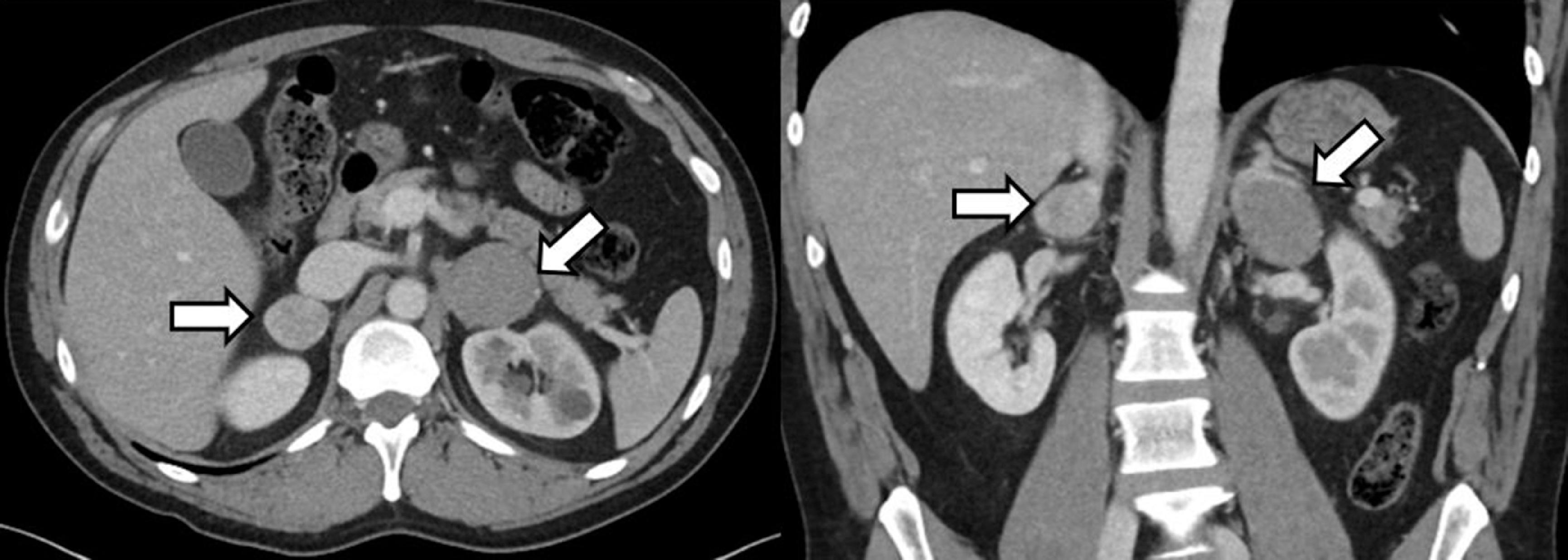
Abdominal computed tomography (CT) revealed bilateral adrenal tumors: a 3.9 × 4.2 × 5.2–cm left adrenal mass and a 2.4 × 2.9 × 2.4–cm right adrenal mass ( Fig. 40.1 ). Additional CT findings included bilateral renal cysts and a 5-mm obstructive stone in the left ureter. Review of outside records demonstrated a history of hypercalcemia. Additional laboratory studies were obtained (see Table 40.1 ) . The levels of metanephrines in the blood and urine were diagnostic of adrenergic pheochromocytomas. In addition, calcitonin concentrations were elevated, and workup confirmed primary hyperparathyroidism. Thyroid ultrasound demonstrated a 0.9 × 0.6 × 0.7–cm solid hypoechoic irregular nodule with punctate echogenic foci, with high suspicion for malignancy. Genetic testing confirmed a pathogenic variant in the RET gene (c.19OOT>C; p.Cys634Arg).

