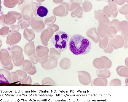
V.D.001 Band and Segmented Neutrophil
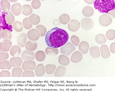
V.D.001
Band and segmented neutrophil. Marrow film. The band neutrophil has the same cytoplasmic appearance as its predecessor, the neutrophilic metamyelocyte, that is a tan coloration throughout the cytoplasm, reflecting the neutral staining properties of the neutrophilic granules, not individually discernible because they are less than 0.5 μm in diameter. The sole distinguishing feature of this state of maturation is the sausage shaped nucleus with a thickness that is nearly identical from one end of the nucleus to the other with no hint of segmentation. A segmented neutrophil sits adjacent to the band neutrophil. Note the clear segmentation with one lobe being connected to the remainder of the nucleus by a very thin strand. Note irregularities in the main body of the nucleus with minor and major constrictions and another developing segment apparent at the right end of the nucleus. The nuclear chromatin is more condensed and darker than that of the band neutrophil. A small lymphocyte is at the bottom of the field. In supravital preparations nuclear segmentation is a dynamic event with changing lobe numbers and areas of segmentation.
V.D.002 Basophilic Myelocyte

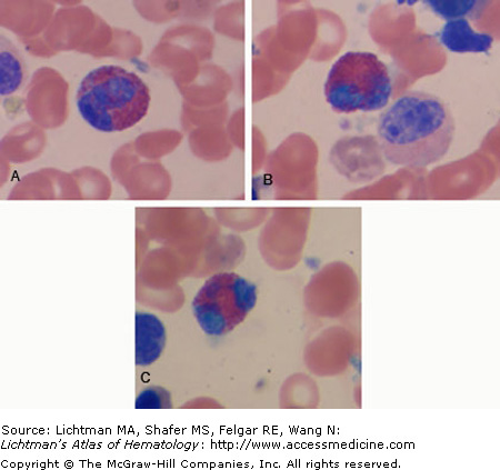
V.D.003 Eosinophilic Metamyelocytes
V.D.003
Eosinophilic metamyelocytes. (A and B). Marrow film. Two eosinophilic metamyelocytes. Note reniform nucleus. Neutrophilic myelocyte in (B). Note marked distinction of cytoplasmic coloration of eosinophil (orange-red) compared to neutrophil (tan). (C) Segmented eosinophil. The transition from metamyelocyte to segmented eosinophil is rapid, making identification of band forms infrequent in normal marrow. Normal eosinophils rarely have more than two nuclear segments.
V.D.004 Eosinophilic Myelocyte
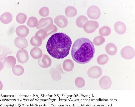
V.D.005 Eosinophilic Myelocytes, Early
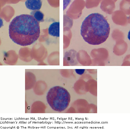
V.D.005
Eosinophilic myelocytes, early. Marrow film. (A–C). Three early myelocytes with cytoplasm partially filled with eosinophilic granules. Note also the relatively immature nuclei with little condensation of chromatin, especially in (A) and (B). This is the earliest stage of marrow eosinophilopoiesis identifiable by light microscopy. A band neutrophil is also present in (C).
V.D.006 Eosinophilic Myelocytes, Late
V.D.007 Granulocyte Precursor Maturation
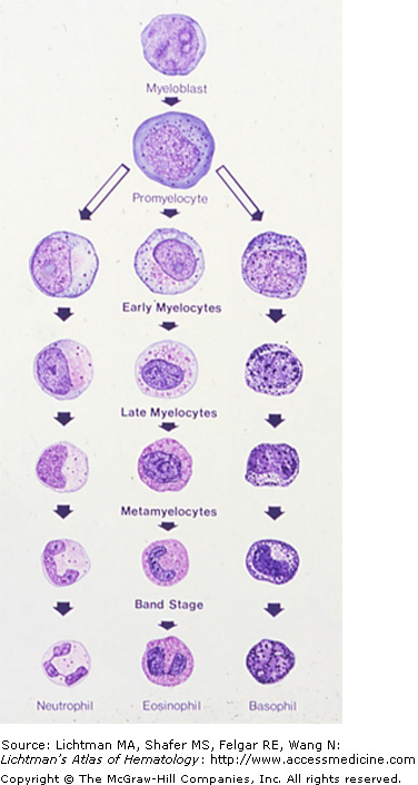
V.D.007
Granulocyte Precursor Maturation. Granulocyte precursor maturation. Diagrammatic representation of the stages of granulocyte precursor maturation. Three lineages, neutrophils, eosinophils, and basophils are shown. Myeloblast. The earliest precursor identifiable by light microscopy is the myeloblast, an agranular cell with a very high nuclear to cytoplasmic ratio, an immature nuclear chromatin pattern, diffuse euchromatin, with little to no blocks of heterochromatin, and, in this case, two nucleoli. There is no way of distinguishing whether a myeloblast is destined to enter one of the three pathways of granulocyte development. On a simple probabilistic basis, most are pre-neutrophils. Promyelocyte. The next developmental stage after the myeloblast is the promyelocyte. The cell enlarges, contains a higher proportion of cytoplasm and the onset of the synthesis of primary granules, rich in peroxidase, results in large azurophilic granules scattered throughout the cytoplasm and often overlying the nucleus on a stained marrow film. Nucleoli may still be evident as in this case. The abundant cytoplasm is usually basophilic. Early Myelocyte. This stage of development is marked by the appearance of specific neutrophilic, eosinophilic, or basophilic granules. The neutrophilic granule is too small to be resolved by the light microscope and their appearance is evident by the tan zone that appears at the site of the Golgi apparatus in the hilum of the nucleus, sometimes likened to a sunburst. The presence of a few eosinophilic or basophilic granules marks the early myelocyte in those lineages. A nucleolus may be evident as in this case. Late Myelocyte. At this stage, the cytoplasm is engorged with the specific granules characteristic of each lineage. The nuclear chromatin is more mature and has less euchromatin and more heterochromatin and nucleoli are not evident. The cell is decreased in size to that of the mature cell. Mitotic capacity has been lost. Metamyelocyte. The nucleus is more elongated than circular and takes on a reniform shape with a clear indentation on one side. Band Form. The nucleus takes on a shape that is long in one axis and narrow in the other: sausage-shaped. Nuclear thickness is about the same throughout the nucleus. Segmented Form. The nucleus segments into lobes. Characteristically two lobes in eosinophils and basophils and two to five in neutrophils. The kinetics of each lineage is different and the time at each stage may be longer or shorter, thus band eosinophils or basophils are less frequent than in the neutrophilic lineage. Basophil granules often obscure the nuclear silhouette.
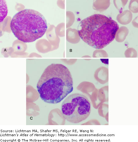
V.D.008 Granulocyte Precursor Maturation, Marrow Films
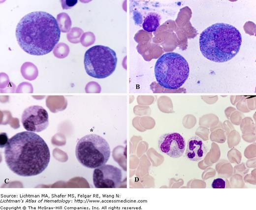
V.D.008
Granular Precursor Maturation, Marrow Films. (A) Myeloblast is the smaller cell to the lower right. It is the first recognizable precursor in the granulocytic series. Relatively high nuclear:cytoplasmic ratio. Note nucleoli and agranular cytoplasm. Promyelocyte in upper left. This cell is the largest granulocyte precursor in the marrow. It often has overt nucleoli, usually more cytoplasm, and azurophilic (primary) granules scattered throughout the cytoplasm and overlying the nucleus. (B) Two very early neutrophilic myelocytes. They are very similar to the promyelocyte in appearance with nucleoli and scattered azurophilic granules throughout the cytoplasm. The distinguishing feature is the burst of tan coloring at the site of the Golgi zone, indicating the initial synthesis of neutrophilic granules. (C) Large cell to the left is an early neutrophilic myelocyte with more neutrophilic granules evident spreading from the Golgi zone at the hilus of the nucleus. It still has some features of the promyelocyte. The cell beneath the asterisk is a late neutrophilic myelocyte. The cell has decreased in size, the nuclear chromatin has condensed. Nucleoli are not evident and the cytoplasm is nearly filled with neutrophilic granules. Below the neutrophilic myelocyte is a neutrophilic metamyelocyte, characterized by its reniform nucleus and cytoplasm filled with neutrophilic granules. The cell above the large early myelocyte on the left is a band neutrophil. The nucleus has reached the shape of a sausage and is about equal indiameter through its length. (D) A band neutrophil (left) and a segmented neutrophil (right). Neytrophilic granules, because of their small size, are not resolvable by the light microscope and are inferred by the characteristic tan staining quality of the cytoplasm.
V.D.009 Megaloblastic Anemia. Giant Band Neutrophils
V.D.009
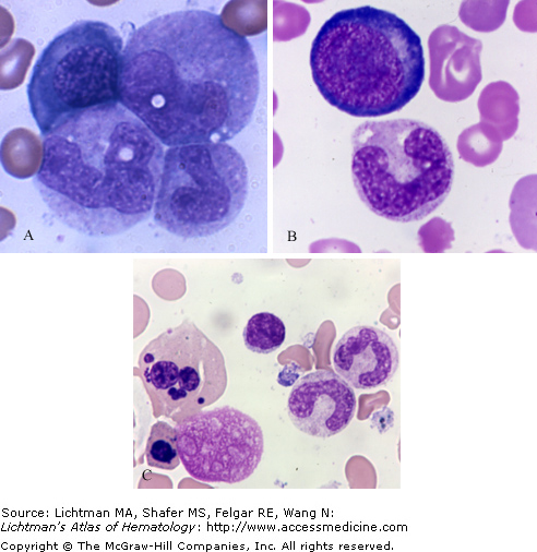
Megaloblastic Anemia. Giant Band Neutrophils. Marrow Films. (A) Upper left, a polychromatophilic megaloblast. Upper right, a giant metamyelocyte. Lower left and right, giant band neutrophils. (B). Late promegaloblast and giant band neutrophil. (C) Far right, two giant band neutrophils. Far left, an orthochromatic erythroblast of gigantic size with nuclear fragmentation.
V.D.010 Myeloblast
V.D.010
Myeloblast. Marrow film. The earliest granulocyte precursor recognizable by light microscopy, derived from the granulocyte colony-forming cell or unit (CFU-G). This cell is about 10 to 15 μm in diameter on a blood film and has a very high nuclear to cytoplasmic ratio. The nucleus always contains at least one clearly demarcated nucleolus and may have two or three. The nucleus has a fine chromatin pattern. The cytoplasm is a pale gray or light blue color with uneven distribution, usually lighter near the nucleus and darker near the cytoplasmic membrane. Cytoplasmic granules cannot be discerned by light microscopy, but using transmission electron microscopy the peroxidase stain reacts at the site of the rough-surfaced endoplasmic reticulum and the Golgi and, sometimes, with immature primary granules. The a) high nuclear to cytoplasmic ratio, b) presence of a nucleolus, and c) the absence of cytoplasmic granules by light microscopy are three essential features of this cell.
V.D.011 Myeloblast
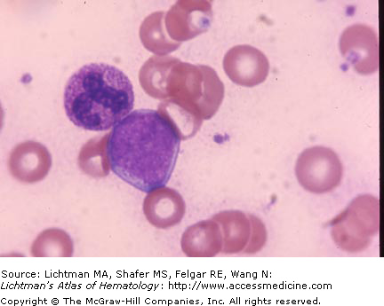
V.D.011
Myeloblast. Myeloblast. Marrow film. The earliest granulocyte precursor recognizable by light microscopy, derived from the granulocyte colony-forming cell or unit (CFU-G). This cell is about 10 to 15 μm in diameter on a blood film and has a very high nuclear to cytoplasmic ratio. The nucleus always contains at least one clearly demarcated nucleolus and may have several. The nucleus has a fine chromatin pattern. The cytoplasm is a pale gray or light blue color with uneven distribution, usually lighter near the nucleus and darker near the cytoplasmic membrane. Cytoplasmic granules cannot be discerned by light microscopy but using transmission electron microscopy the peroxidase stain reacts at the site of the rough-surfaced endoplasmic reticulum and the Golgi and, sometimes, with immature primary granules. The a) high nuclear to cytoplasmic ratio, b) presence of nucleoli, and c) the absence of cytoplasmic granules by light microscopy are three essential features of this cell. A segmented neutrophil abuts the myeloblast.
Related posts:

Stay updated, free articles. Join our Telegram channel

Full access? Get Clinical Tree


