I.A.001 Acanthocytes
I.A.001
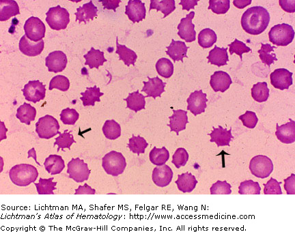
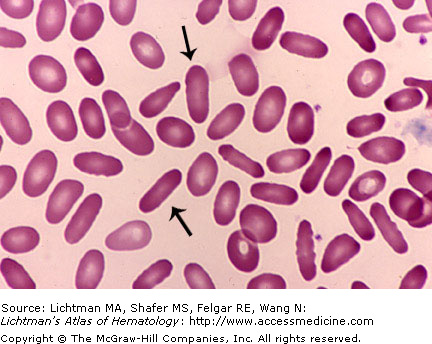
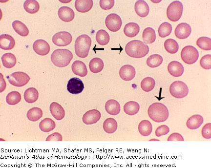
Acanthocytes. Blood film. The image contains many spiculated cells that represent acanthocytes. Two are highlighted by the arrows. Characteristically, acanthocytes have irregularly arranged spicules that project from the red cell surface and are often bent back on themselves. Although superficially they resemble echinocytes, the latter have more numerous projections with rounded, rather than sharply pointed tips, evenly distributed over the red cell surface.
I.A.002 Agglutination
I.A.002
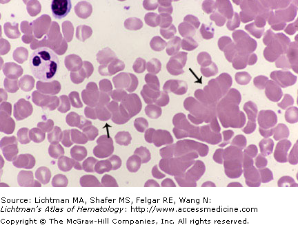
Agglutination. Blood film. Several clumps of agglutinated red cells, two are marked by arrows. These clumps may be seen if cold agglutinins have a thermal amplitude that permits them to be red cell agglutinins at the temperature of the preparation of the blood film or, occasionally with warm agglutinins, if they do not require addition of antiglobulins to agglutinate (complete agglutinins).
I.A.003 Avian Red Cells
I.A.004 Echinocytes
I.A.005 Elliptocytes
I.A.006 Hypochromia
I.A.006
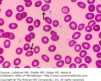
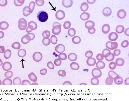
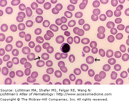
Hypochromic red cells. Blood film with severely hypochromic cells, two of which are marked by arrows. The cells are virtually all pathologically hypochromic, most containing only a narrow rim of cytoplasmic hemoglobin. The lymphocyte nucleus is often used as a rough expectation of the diameter of normal sized red cells. These appear smaller on average. Microcytosis is commonly paired with hypochromia as a dual manifestation of hypoferremic erythropoiesis seen in iron deficiency and the anemia of inflammation.
I.A.007 Keratocyte
I.A.007
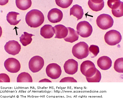
Keratocyte. Blood film. Synonyms include horn, helmet or bite cells. This red cell has lost a piece of peripheral cytoplasm, which leaves two horns on either side of the lost piece. Two examples are marked by arrows. It differs from a schizocyte by both its normal volume and retention of its biconcave shape. The bite cell is often subcategorized as a different type of keratocyte because it results from the removal of an intracellular hemoglobin precipitate by partial splenic phagocytosis.
I.A.008 Knizocyte
I.A.008
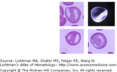
Knizocyte. Blood film and scanning micrograph. A cell with three concavities. The prefix knizo- is derived from ruffle or ridge, the latter separating the concavities. In these images the third concavity is not in the perspective of the image but these images are the characteristic appearance of such cells on a blood film (three stained images) and in a scanning micrograph.
I.A.009 Macrocytes
I.A.009
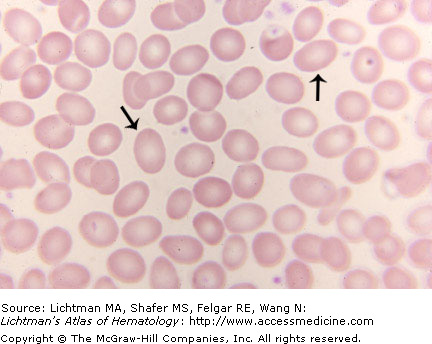
Macrocytes. Blood film. These are red cells that appear to be larger than an average sized normal red cell. If one uses the nucleus of a small lymphocyte as an approximate reference object, macrocytes would be larger than that of the nuclear silhouette. There are several macrocytes in this blood film, three of which are noted by arrows. The horizontal arrows point to oval macrocytes, which are characteristic of megaloblastic anemias.
I.A.010 Microcytes
I.A.010
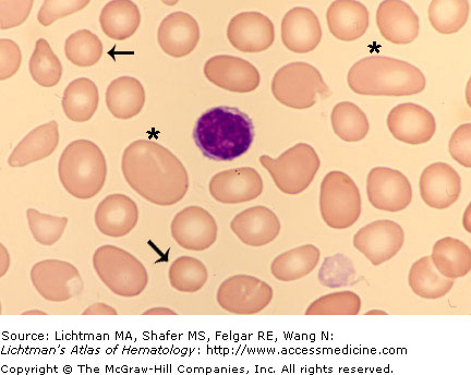
Microcytes. Blood film. These are red cells that appear to be smaller than the average sized normal red cell. If one uses the nucleus of a small lymphocyte as an approximate reference subject, microcytes would be smaller than that circumference. This blood film has cells range from macrocytes, two of which are marked by asterisks, to microcytes, two of which are marked by arrows. It can be difficult to estimate the mean red cell size from a blood film because a) the variation among cells from large to small may be very wide and b) one is trying to infer the red cell volume (three dimensions) from the area occupied by the cell (two dimensions). The thickness of the cell cannot be discerned but is often inferred. As a general rule, if striking macrocytes are present, the population mean red cell volume is usually increased even though microcytes are also present.
I.A.011 Neonatal Blood
I.A.011
Neonatal blood. High hematocrit and thus dense red cell background. The findings in the blood film of “normal” neonates are variable. Neutrophilia, rapidly changing into a mild lymphocytosis, an occasional nucleated red cell or immature granulocyte, mild red cell shape changes, including spherocytes and fragments, can occur but all these abnormalities disappear in the few weeks after birth at which time the blood film is normal. The marked variation in the blood film is the result of unpredictable events at birth that place stress on the hematopoietic system and are reversible.
I.A.012 Normochromia
I.A.012
Normochromic red cell. Blood film. This designation refers to a red cell that appears to have a normal hemoglobin content. This inference can be made by inspection of the size of the pale (hemoglobin-poor) central area of the cells on a blood film. The two arrows point to cells that have a normal sized pale area. Moderate and marked deviations from normal can usually be discerned by the experienced observer.
I.A.013 Normocytosis
I.A.013
Normocytic red cell. Blood film. This designation refers to a red cell that appears to have a normal size. If one uses the nucleus of a small lymphocyte as a reference subject, the normocyte is about the area of that nucleus. The two arrows point to cells that have normal size. It can be difficult to estimate the mean red cell size from a blood film because a) the variation among cells from large to small may be wide and b) one is trying to infer the red cell volume (three dimensions) from the area occupied by the cell (two dimensions). The thickness of the cell cannot be discerned but is inferred. Moderate and marked deviations from normal can usually be discerned by the experienced observer.
I.A.014 Nucleated Red Cells
I.A.014
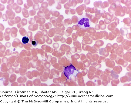
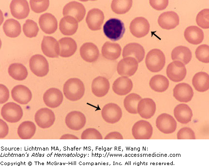
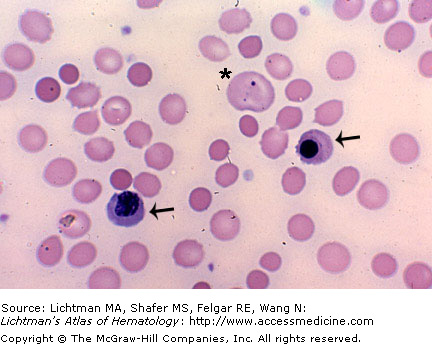
Nucleated red cells. Blood film. Under various pathological conditions, nucleated red cells may be observed in the blood. Examples of such conditions include hemolytic anemia, hypoxic states, and metastatic cancer involving marrow. This image depicts a spherocytic hemolytic anemia (warm antibody type). Two nucleated red cells are indicated by the arrows. Usually the nucleated cells are late orthochromatic erythroblasts with very dense, small nuclei. The asterisk indicates a macrocyte containing a very small nuclear remnant, called a Howell-Jolly body. Many of the cells are microspherocytes, small dense circular cells without pale centers.
I.A.015 Ovalocyte
I.A.015
Ovalocyte. Blood film. A red cell in which its length exceeds its width giving it the shape of an American football. The ovalocyte differs from the elliptocyte in that the width to length ratio is greater. Since the ovalocyte and elliptocyte can be considered parts of a continuum, some have suggested using a single term, elliptocyte, and grading it from I to III, with the lower numbers being cells more oval in shape and the higher numbers representing cells more elliptical in shape.
I.A.016 Poikilocytes, Types of
I.A.016
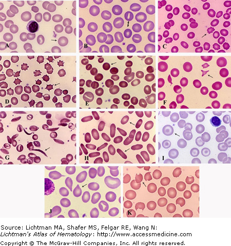
Types of Poikilocytes. Bloods films. (A) Normal blood. Arrow points to a normochromic-normocytic discocyte. (Normal shape). (B) Stomatocytosis. The double arrow points to the two morphologic type of stomatocytes. The upper cell with a slit-shaped pale area and the lower cell with a small central circular pale area. Two different types of “stoma”. (C) Echinocytes. The field has several such cells. The arrow points to one example of a red cell with evenly distributed, blunt, short, projections, circumferentially positioned. (D) Acanthocytes. Several in the field. The arrow points to one example with a few spike-shaped projections, unevenly distributed and of different length projecting from the surface. (E) Spherocytes. Small circular cells with dense staining (hyperchromic), and in the extreme without any central pallor. (F) Schistocytes (fragmented red cells). These usually small (microcytic) cells may assume varied shapes. The arrow points to a triangular shape but two other schistocytes are in the field with different shapes. Despite being damaged and very small they frequently maintain a biconcave appearance as witnessed by their central pallor. (G) Drepanocytes (sickle cells). Numerous sickle cells are shown. Two are in the classic shape of a blade on a farm instrument, the sickle (arrow). Many red cells that have undergone the transformation to a “sickle” cell take the slightly less extreme form of elliptical cells with a very narrow diameter with condensed hemoglobin in the center (paracrystallization). About eight such cells are in the field. (H). Elliptocytes and Ovalocytes. The lower arrow points to an elliptocyte (cigar-shaped). The length/diameter ratio is very large compared to a discocyte. The upper arrow points to an ovalocyte (football-shaped) in which the length/diameter ratio is lower than normal. Because both forms may be seen together in a case of inherited disease (same gene mutation resulting in both shapes), as shown here, it has been proposed that all such shapes be called elliptocytes with a Roman numeral to designate the severity of the shape change toward the elliptical, i.e. elliptocytes I, II, III. (I). Codocytes (target cells). The arrow points to one characteristic example, among several in the field. The distribution of hemoglobin gives the cell the appearance of an archery target. (J) Dacryocytes (tear drop cells). Three dacryocytes are in this field. One example is indicated by the arrow. (K) Keratocyte (horn cell). Several examples are in the field. The arrow points to a typical such cell with two sharp projections.
I.A.017 Poikilocytic Alphabet. Scanning Electron Micrographs
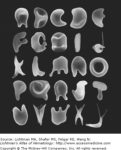
I.A.017
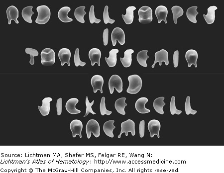
Poikilocytic alphabet. Scanning electron micrographs. The English alphabet is simulated using poikilocytes from the blood of a single individual with beta-thalassemia minor and a patient with sickle cell anemia. Here the alphabet is displayed from A to Y. No Z was found. Can you determine which letters are derived from sickle cells? They are more electron-dense from the paracrystallization of the hemoglobin in sickled cells.
I.A.018 Poikilocytic, Alphabetic. Red Cell. Scanning Electron Micrographs
Related posts:

Stay updated, free articles. Join our Telegram channel

Full access? Get Clinical Tree


