Multiple Myeloma


Figure 30-1 An outline of the main factors leading from multiple myeloma (MM) to monoclonal gammopathy of undetermined significance (MGUS); the distinction between HD and NHD MM; and finally additional frequent aberrations reported in MGUS and MM patients.


Chromosomal Translocations in Multiple Myeloma


Chromosomal translocations in B-cell tumors including MM do not give raise to fusion chimeric proteins, but usually lead to the dysregulated expression of oncogenes through the juxtaposition of their promoters with Ig regulatory elements, via the B-cell–specific mechanisms of switch recombination and somatic hypermutation. 12,18 The primary translocations most frequently identified in MM patients include t(11;14)(q13;q32), which dysregulates the expression of the cyclin D1 gene, 26 with an incidence of approximately 15% in MGUS 27,28 and in MM. 29,30 Another translocation, t(6;14)(p21;q32), induces the dysregulation of cyclin D3 in 2% to 3% of MM patients. 31 t(4;14) is detected in approximately 15% of patients 32 and presents peculiar features. As a result of the translocation, the enhancers Eμ (at the 5′) and Eα (3′) are separated and dysregulate the expression of the juxtaposed genes on both derivative chromosomes, at der(4) and der(14). Hence, on der(14), the 3′ Eα enhancer increases the expression of the receptor tyrosine kinase fibroblast growth factor receptor 3 (FGFR3) gene. On der(4), the 5′ Eμ enhancer drives the expression of the histone methyltransferase Wolf-Hirschhorn syndrome candidate 1 gene (WHSC1, also called Multiple Myeloma Set Domain [MMSET]). MMSET is almost always upregulated in t(4;14) patients, 33 whereas FGFR3 is not overexpressed in 25% of these patients, suggesting that MMSET represents the main oncogenic culprit of t(4;14) and not FGFR3. Of note, approximately 10% of t(4;14) patients develop activating mutations on FGFR3. 34,35 Finally, the t(14;16)(q32.3;q23) is present in approximately 5% to 10% of MM patients and 12,36 induces overexpression of the oncogene MAF, 37 whereas the (14;20)(q32;q11) has been reported in 2% to 5% of MM cases and affects the family member MAFB. 12,38


Mutated Genes in Multiple Myeloma


A recent next-generation sequencing (NGS) effort has led to the identification of novel somatic mutations in MM. 39 This survey has identified on average 35 amino acid–altering point mutations and 21 chromosomal rearrangements per sample, a level of genomic rearrangements again more in line with the degree of mutation rate seen in epithelial cancers such as melanoma and lung carcinomas than in hematological cancers. 40 In addition, as in other blood and epithelial cancers, genes in MM are often mutated at low frequency, suggesting a remarkable and somehow disconcerting intertumor heterogeneity. 40 In fact, in MM, only 10 genes were recurrently mutated. 39 The list included genes where mutations have already been described in MM, such as NRAS (23%), KRAS (26%), and TP53 (8%). Intriguingly mutations were reported also in cyclin D1 (CCND1; 5%), indicating that not only is it frequently translocated and its expression often dysregulated in MM, but it is also mutated (see also later discussion). The remaining six genes include FAM46C (13%), which is also focally deleted; DIS3 (10%); PNRC1 (5%); ALOX12B (8%); HLA-A (5%); and MAGED1 (5%). Mutations affecting these genes have not been previously identified in cancer and reveal novel pathways potentially involved in MM pathogenesis. In particular, two of these genes, DIS3 and FAM46C, are thought to be involved in RNA metabolism and protein homeostasis.

As in other cancers, 41 however, mutations affecting specific genes in MM are in most cases rare; such mutated genes tend to coalesce into specific pathways. In the case of MM, four pathways were significantly enriched in somatic mutations. Confirming previous results, 42,43 genes belonging to the NFκB pathway were frequently mutated. Also, frequent mutations affecting histone-modifying genes such as MLL, MLL2, MLL3, UTX, MMSET, and WHSC1L1 were reported. Mutations affecting the same nucleotide were also found in the IRF4 transcription factor and in its target, PRMD1, confirming previous functional data reporting a prominent role of IRF4 for MM survival. 44 Surprisingly, a significant enrichment in five genes involved in blood coagulation was identified in MM patients. Although a role for this pathway in MM is unknown, thrombin and fibrin have been shown to serve as mitogens in other cell types and have been implicated in metastasis.


Prognostic Implications of Genetic Lesions


Extensive studies in the past 15 years have identified a link between specific genetic lesions and prognosis. HD patients present a better prognosis when compared with NHD MM. 21, 4547 However, if HD patients acquire chromosome 13 loss and/or gains in the long arm of chromosome 1, patient overall survival worsens considerably. 10, 48, 49 Other acquired genetic lesions in HD have been linked to poor prognosis in this group—for example, IgH translocations, especially those involving unknown partners. 50 Finally, HD patients could evolve toward a pattern characterized by expression of genes associated with high proliferation and poor prognosis. 11

As for NHD patients, the prognostic outlook varies widely depending on the chromosomal translocation present. Overall, patients with cyclin D translocations tend to present a better prognosis than NHD patients with t(4;14), t(14;16), and t(14;20).

Gains/amplifications of 1q are associated with t(4;14), t(14;16), and possibly chromosome 13 deletion, 10,51 and in general with more proliferative disease states. 52 Several studies have proposed an association between gains/amplification of this region and poor prognosis, based on cytogenetic analysis, 53 fluorescence in situ hybridization (FISH), 38 and expression profiling, 52 as well as aCGH. 10 Importantly, Zhan and colleagues have shown that among a list of 70 genes linked to early disease-related death, there was an enrichment for overexpressed genes mapping to chromosome 1q. 54

Hemizygous deletions of chromosome 13 are present in MGUS and MM with a similar overall incidence of around 50%. 28,5558 Given the strong correlation between the presence of 13 loss and other genetic lesions, 30,59 including t(4;14), t(14;16), and NHD, its role as an independent prognostic factor has been questioned. 28,30,46,56,57,6064

Finally, aCGH analysis has identified focal regions associated with poor survival containing amplifications on chromosome 8 (involving MYC) and deletion on ch17 (including TP53)— genetic events that have been previously linked to poor prognosis in MM.

It should be emphasized, however, that the significance of any prognostic marker relies heavily on the treatment regimen. In fact, most of the studies just mentioned assessing the prognostic relevance of genetic lesions included patients treated with high-dose chemotherapy followed by bone marrow transplant. A reassessment of the significance of these prognostic and predictive factors is ongoing, in light of the introduction of novel drugs for patient treatment. For example, several of the most well-established genetic prognostic markers failed to show any correlation with prognosis in patients treated with the proteasome inhibitor bortezomib. 65


The Microenvironment in Multiple Myeloma
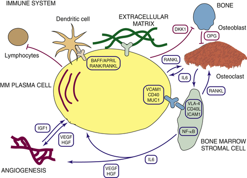

Blood cancers develop in secondary lymphoid organs and in the bone marrow (BM). For several acute hematological cancers, such as Burkitt’s lymphoma, the genetic lesions underlying these tumor cells are sufficient to promote tumorigenesis; the role of the microenvironment is for the most part marginal. In contrast, mature B-cell malignancies including MM rely heavily on their milieu for their growth and survival 6668 (Figure 30-2 ). Indeed, MM cells twist to their advantage the physiological mechanisms underlying healthy plasma cell homing to the bone marrow and the pathways supporting long-lived plasma cells. MM plasma cells establish tight interactions with essentially all the BM components. The severance of these ties has become an essential therapeutic tool. Indeed, the main mechanism of action of the novel drugs recently introduced into the clinic (thalidomide, lenalidomide, and bortezomib) is their impact on the interactions between MM cells and their cellular counterpart.


image

Figure 30-2 A simplified view of the interactions between the multiple myeloma plasma cell and the surrounding environment.

The BM microenvironment includes an extracellular matrix (collagen, laminin, fibronectin, and osteopontin) and a rich cellular component. The cellular BM compartment consists of hematopoietic and mesenchymal progenitor and precursor cells, including hematopoietic stem cells (HSCs); BM-derived circulating endothelial precursors (CEPs) and endothelial cells (BMECs); immune cells (dendritic cells, various populations belonging to the B and T lymphocytes, NKT and NK cells, monocytes, and macrophages); erythrocytes; megakaryocytes and platelets; and nonhematopoietic cells, including an ill-defined group of cells labeled fibroblasts/bone marrow stromal cells (BMSCs). Also included are cells involved in bone homeostasis, such as chondroclasts, osteoclasts (OCs), and osteoblasts (OBs).

MM cells interact with BMSC and the extracellular matrix either directly, via adhesion molecules that include LFA1, VLA4, NCAM, ICAM1, and CD44, or indirectly, through chemokines, cytokines, and growth factors released by tumor cells and BMSC, such as interleukin 6 (IL6), insulin-like growth factor 1 (IGF1), tumor necrosis factor-α (TNFα), transforming growth factor-β (TGFβ1), and VEGF. As a result of these multiple-layered interactions, several cancer-relevant pathways become activated in both the tumor and stromal cells, such as NFκB, JAK/STAT, PI3K, and MAPK, establishing powerful positive feedback loops that further increase MM growth and survival. IL6 represents a central cytokine for the growth and survival of MM cells, although IL6-independence could ensue in the late stage of the disease. IL6 engages its receptor (IL6R), leading to the activation of the Janus kinase/signal transducer and activator of transcription (JAK/STAT) pathway, the proliferation-associated MAPK cascade, and the PI3K/AKT pathway. Moreover, interaction of MM cells with BMSC induces the secretion of IL6 from BMSC through the activation of NFκB. Another paracrine factor that has recently emerged as crucial for MM development is IGF1. Stimulation of MM cell lines with IGF enhances cell proliferation and prevents apoptosis, again through the activation of the MAPK, PI3/AKT, and NFκB pathways. Of note, inhibitors against the receptors of these cytokines and growth factors have been developed, and clinical trials are ongoing to test their effectiveness as novel drugs.

The homing of MM cells to the BM triggers a strong angiogenic response. 69,70 Indeed, it has been shown that angiogenesis correlates with high MM proliferation index, with the more advanced stages of the disease, and ultimately with prognosis. The adhesion of MM cells to the BMECs increases the secretion of several pro-angiogenic cytokines, most importantly of VEGF, basic fibroblast growth factor (bFGF), and matrix metalloproteinases. Conversely, BMECs secrete growth factors, including VEGF, IL6, and IGF1.

Osteolytic bone lesions are a hallmark of MM and are associated with pathologic fractures, bone pain, and diffuse osteoporosis. MM cells increase the number and the activity of OCs, while reducing the number of OBs, tilting the balance toward increased bone reabsorption. An array of factors that activates the OC is produced by both tumor as well as stromal cells. These factors include macrophage inflammatory protein-1a (MIP-1a) and receptor of NFκB ligand (RANKL). OC activity in turn modulates MM cell growth and survival via the secretion of IL6. Conversely, MM cells impair the maturation of OB cells from mesenchymal stem cells, through the secretion of the WNT-signaling antagonist DKK1, an inhibitor of OB differentiation. In addition, the binding of VLA4 on MM cells to VCAM1 on osteoblast progenitors reduces the levels of the transcription factor RUNX2, essential for OB maturation. Osteoblasts not only preserve the bone structure, but also inhibit MM growth both in vitro and in vivo. Therefore, restoring the number and activity of OBs may increase bone formation as well as provide an antitumoral effect.


image

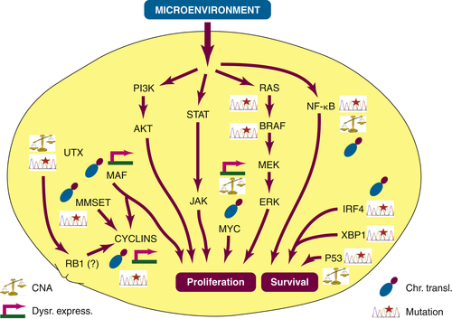
Figure 30-3 Main signaling pathways and genes activated or genetically altered in multiple myeloma. Four different types of aberrations are included: somatic mutations, chromosomal translocations (Chr. transl), copy number aberrations (CNA), and dysregulated expression (Dysr. express.).

Immuno-based tumor surveillance is severely impaired in MM. 71 Both the T and B responses are affected. Specifically, the expansion of regulatory T cells (Treg), reduced T-cell cytotoxic activity and responsiveness to IL2, and defects in B-cell immunity have been reported. These effects are the results of cytokines produced by BMSC, including VEGF, HGF, fibroblast growth factor (FGF), and stromal-cell–derived factor (SDF)-1α. Recently Chauhan and associates have demonstrated a significant enrichment of plasmacytoid dendritic cells (pDCs) in the BM of MM patients. These cells mediate immune deficiency characteristics of MM and are able to promote MM cell growth, survival, and migration and to enhance drug resistance. 72


Deregulated Pathways in Myeloma and the Opening of Novel Therapeutic Opportunities


The knowledge accumulated in recent years allows a clearer definition of the pathways activated in MM tumor cells, either due to genetic lesions inside the tumor cells or emerging from activating signals from the surrounding microenvironment (Figure 30-3 ). In several cases, this knowledge has been turned to the patient’s advantage, because it has allowed the design of novel targeted therapies specifically addressing single lesions.


MYC


Genetic rearrangements involving the MYC (c-MYC) locus are frequent in the more advanced stages of MM. The anatomy of these lesions has been defined. 7375 In 25% of cases, the IgH or IgL locus is juxtaposed with the MYC sequence. 76 The pattern, however, is more complex than the classical reciprocal translocations present in the so-called primary translocations. 18,76 Amplifications, inversions and insertions without apparent translocations, 10,23,74,76 or translocations not involving the Ig loci 74,75 have also been described. In a large patient population, rearrangements affecting c-MYC have been reported with a frequency of 3% in MGUS and 10% to 16% in MM. 73,76 MM cell lines have more frequent rearrangements of the c-MYC locus, ranging from 55% 76 to more than 90%, depending on the study. 73,74 Hence, genetic rearrangements affecting the MYC locus likely represent late events in the course of the disease.

Recent lines of evidence suggest that MYC overexpression might represent an early event, at the critical junction between MGUS and MM. This dysregulated expression seems independent from genetic rearrangements. MYC is overexpressed in a large proportion of MM patients, when compared with plasma cells derived from MGUS patients and healthy individuals, in the absence of evident genetic rearrangements affecting the MYC locus. 10,77,78 Indeed, Chng and colleagues have developed an MYC activation signature and have demonstrated that MYC is activated in up to 67% of MM patients, but not in MGUS. Intriguingly, the MYC activation signature was present in almost all tumors with RAS mutations and was associated with hyperdiploid MM and shorter survival. Importantly, bortezomib treatment was able to overcome the survival disadvantage in patients with MYC activation. 77 In a mouse model of MM, the Vk∗MYC model, somatic hypermutation activates an MYC transgene inducing a phenotype closely resembling indolent MM. 78 These data support the notion that dysregulated expression of MYC might be sufficient for MGUS to convert into MM.

MYC is a transcription factor, therefore considered refractory to direct pharmacological inhibition. 79 Recently, Delmore and co-workers have ingeniously devised a method to inhibit MYC target genes. 80 A compound, JQ1, was designed, interfering with the acetyl-lysine recognition domains (bromodomains) of putative coactivator proteins implicated in transcriptional initiation and elongation. Surprisingly, JQ1 downregulated the transcription of the MYC gene itself, followed by genome-wide downregulation of Myc-dependent target genes, ultimately leading to potent antiproliferative effect associated with cell-cycle arrest and cellular senescence.


MAF


Two chromosomal translocations involve the MAF family gene, namely, MAF in the t(14;16)(q32.3;q23), 37 and MAFB in the (14;20)(q32;q11). 12,38 Despite their low incidence, the study of MM patient cells endowed with these translocations has provided important perspectives on the biology of the MM cell and the modality of its interaction with the microenvironment. Both translocations share an overlapping gene expression signature, 11 suggesting a similar repository of downstream targets. The oncogenic role of c-MAF in MM is established. Forced overexpression of MAF enhances myeloma proliferation, probably through the increased expression of cyclin D2, a MAF target gene consistently overexpressed in this group of patients. 9 In contrast, MAF knockdown reduced tumor formation in immune-deficient mice. Another direct MAF target, integrin β7, increases MM cell adhesion to the BM stroma and induces high levels of VEGF. Interestingly, therefore, MAF not only has a direct oncogenic activity within the MM tumor cell, but also affects the microenvironment to the tumor’s advantage. An additional report has proposed ARK5, an AMP-activated protein kinase (AMPK)-related protein kinase mediating AKT signals, as a target of MAF and MAFB signaling. 81

Surprisingly, overexpression of c-MAF has been reported even in the absence of the translocation in up to 30% of patients. 82,83 A recent report has suggested that this upregulation results from the activation of the MEK/ERK/AP-1 axis, downstream of MMSET, in the t(4;14) patient group. 84 Intriguingly, treatment of MM cell lines with a MEK inhibitor selectively induced apoptosis in MAF-expressing MM, providing a molecular rationale for the clinical evaluation of MEK inhibitors in this subgroup of patients.

Previous reports have suggested a reduced incidence of bone disease in this group of patients. 9,11 Two genes might be responsible for this phenotype. The gene DKK1, whose overexpression has been implicated in MM-related bone disease, 85 is significantly downregulated specifically in patients presenting with MAF and MAFB translocations. 11 Moreover, Robbiani and colleagues have reported that the gene osteopontin (OPN) inversely correlates with MM bone disease and is specifically overexpressed in patients with translocations affecting the MAF genes. 86


Cyclins


Cyclin D dysregulation is a universal phenomenon in MGUS and MM, somehow surprisingly given the exceedingly low proliferation index of both conditions. Among the most recurrent chromosomal rearrangements, two involve cyclins directly: t(11;14)(q13;q32), which occurs in 15% to 20% of MM patients and induces the overexpression of cyclin D1, 26,8789 and t(6;14)(p21;q32), present in 2% to 3% of MM cases, which increases the expression of cyclin D3. 31 In addition, patients presenting with t(4;14), t(14;16), and t(14;20) demonstrate cyclin D2 overexpression, which in the t(14;16) and t(14;20) patients has been directly linked to MAF/MAFB dysregulation. 9 By unknown mechanisms, HD patients usually show overexpression of cyclin D1, alone or in combination with overexpression of cyclin D2, whereas a subset of HD patients instead produce cyclin D2 alone. It should be noted that the few patients who do not show an evident cyclin dysregulation present RB1 inactivation, supporting the notion that the axis RB1/cyclin is universally derailed in MGUS and MM patients. 8


Chromatin Remodeling Genes


Chromatin remodeling genes are among the most frequently mutated genes in the cancer genome. Remarkably frequent genetic lesions have been identified in genes affecting DNA methylation (e.g., DNMT3A in AML 90 ), nucleosome remodelers (ARID1A in ovarian cancer 91 ), and histone-modifying genes in a variety of tumors. 92 In particular, histone posttranslational modifications affecting the N termini of histones 3 and 4, such as acetylation and methylation, are heritable changes that profoundly affect chromatin structure and gene expression. Genetic lesions involving enzymes that add or remove methylation marks from histone tails have been described, including, as mentioned, MMSET and WHSC1L1, UTX, MLL, MLL2, and MLL3.


MMSET


The recurrent translocations t(4;14)(p16;q32) involve FGFR3 93 and MMSET genes. 94 As mentioned earlier, MMSET seems to represent the real target of this translocation—a notion further confirmed by loss-of-function studies, where downregulation of MMSET with specific shRNA decreased growth and viability of t(4;14) MM cells. 95,96 MMSET presents several alternative spicing variants. Intriguingly, within the gene, the breakpoint position can vary, giving rise to different overexpressed transcripts in t(4;14) samples. MMSET interacts and is likely involved in pathways with a clear relevance in carcinogenesis. MMSET interacts with repressors including SIN3A and histone deacetylases HDAC1 and HDAC2. 95,97,98 However, the oncogenic mechanism underlying MMSET tumorigenic activity remains incompletely understood. Recently it has been shown that t(4;14) MM cases demonstrate an open, permissive chromatin state, associated with high H3K36 di- and trimethylation with concomitant low levels of the trimethylated H3K27. 96 This altered chromatin status was associated with the dysregulated expression of genes involved in cell cycle, apoptosis, DNA repair, and adhesion. Along these lines, t(4;14)-driven MM cases present a remarkable increase in DNA methylation across the genome, a pattern unique in MM. 99

Finally, a connection between MMSET and the DNA damage response has been recently uncovered. 100 H4K20 methylation increases locally on the induction of double-strand breaks (DSB). Pei and associates have demonstrated that MMSET is responsible for this methylation, in turn facilitating 53BP1 recruitment. MMSET therefore represents a central hub in the DNA damage checkpoint activation and response.

MMSET belongs to a gene family that includes two other genes, WHSC1L1 and NSD1, with ties to cancer. Indeed, somatic mutations in WHSC1L1 have been identified in MM patients as well. 39 This gene is also involved in a chromosomal translocation in acute myeloid leukemia, t(8;11)(p11.2;p15) 101 ; is amplified in breast cancer 102 and in lung cancer; and is endowed with oncogenic activity. 103 NSD1 participates in a fusion protein resulting from a chromosomal translocation in acute myeloid leukemia. 104


UTX


UTX represents the first histone demethylase found mutated or deleted in cancer, including multiple myeloma, 105 renal carcinoma, transitional cell carcinoma of the bladder, chronic myelomonocytic leukemia, acute lymphoblastic leukemia, and prostate cancer. Intriguingly, MM is the tumor where this gene is most frequently inactivated, because truncating mutations or deletions in MM are present in up to 30% of patients 105 (also G. Tonon, unpublished data).

UTX is a JmjC-class enzyme that demethylates di- and trimethylated H3K27me, counteracting the activity of Polycomb complexes (PcGs) 106 —in particular of the histone methyltransferase enhancer of zeste homologue 2 (EZH2), which mediates H3K27 methylation, a transcription-repressive mark. Although limited information is available, one of the main tumorigenic results stemming from UTX inactivation likely results from the unbridled activity of EZH2, leading to enhanced H3K27 trimethylation. EZH2 is highly expressed in several cancer types, including breast, prostate, and lymphomas, and its expression levels correlate with advanced stages of tumor progression and poor prognosis. 107,108 In MM, EZH2 is often overexpressed. It was reported as one of the 30 genes able to distinguish normal plasma cells from MGUS and aggressive myeloma. 109 More recently, activating oncogenic mutations affecting EZH2 were identified in lymphoma. 110,111 In MM, EZH2 is induced by IL6 and enhances proliferation in MM cell lines, whereas its inhibition induces apoptosis. 112

In addition, UTX has been implicated in cellular differentiation and growth control through transcriptional regulation of the RB1 pathway. 113 Chromatin immunoprecipitation (ChIP)-on-chip experiments have revealed that UTX is present on the promoters of RB1 pathway genes, exerting a transcriptional control on cellular proliferation and mediating cell cycle arrest of primary human fibroblasts, 113 although it remains to be demonstrated whether a similar, RB1-mediated proliferation effect is present also in MM cases with UTX inactivation.

Finally, UTX regulates HOX gene expression, whose orderly activation is essential for normal hematopoiesis. 114 HOXA genes are abnormally expressed in MM. 115 In particular, HOXA9 is normally silenced by trimethylation of H3K27 during hematopoietic differentiation and is consistently upregulated in MM patients. On HOXA9 knockdown, MM cells exhibit a competitive disadvantage. 39 These data suggest that UTX inactivation may dysregulate the expression of HOXA genes, in particular of HOXA9, contributing to the unrestrained proliferation of MM cells.


RB1, P18, and Other Members of the RB1 Pathway


The comprehensive cyclin D dysregulation detected in MM suggest a derailed activity of the RB1 axis. The tumor suppressor RB1 is located on chromosome 13, and a significant fraction of MM patients present with one copy of this chromosome. Nevertheless, a direct role of RB1 inactivation in MM is uncertain, as inactivating mutations of this gene have not been consistently identified among patients with chromosome 13 hemizygous deletions, or focal homozygous deletions affecting the RB1 region. 12

Other members of the RB1 pathway are altered in MM, but the pathogenetic and clinical relevance of these aberrations has not been fully elucidated. For example, the tumor suppressor CDKN2A (p16) is usually not deleted in MM, 10,2224,116 but is methylated in 20% to 30% of MGUS and MM cases. 117121

On the other hand, the role of CDKN4C (p18) is more established. Overexpression of p18 in MM cell lines lacking p18 reduced proliferation, whereas it had no effect in a cell line where p18 is normally expressed. 122 p18 is homozygously deleted in up to 38% of MM cell lines and in 2% of MM tumors, but this percentage goes up to 10% in tumors with highest proliferation, as evaluated with an expression profiling signature surrogate. 122,123 Intriguingly, p18 is often overexpressed in the most proliferative MM, 122 and focal deletions at 1p32.3, where p18 resides, are associated with poor prognosis. 124


NFκB


Lesions involving genes belonging to both the classical and alternative NFκB pathways are frequently detected in MM, namely, in 17% of patients and in approximately 40% of MM cell lines. 39,42,43 The two pathways are tightly interconnected: many signals activate both branches, and many effector proteins or target genes are shared by both cascades. As for the genetic lesions, the most common event consists of inactivating mutations of TRAF3, occurring in 13% of MM patients. 43 Other negative regulators such as TRAF2, BIRC2 and BIRC3, and CYLD carry inactivating mutations or focal homozygous deletions. Chromosome translocations and amplifications involving NFκB-inducing kinase (NIK), 42 CD40, LTBR, TACI, NFKB1, and NFKB243 were reported, and all resulted in constitutive activation of either canonical or noncanonical pathways. A more recent study demonstrated that compensatory and/or cooperative effects occur in MM cell lines harboring such alterations. 125 Interestingly, constitutive activation of either canonical or alternative pathways resulted in the regulation of similar sets of genes and biological pathways, 125 conferring increased autonomy from the microenvironment.

The high prevalence of mutations in NFκB family members in MM cell lines compared to MM patients suggests that alterations in the NFκB pathway are relatively late events. Moreover, NFκB dysregulation mainly occurs in NHD compared to HD patients. Of note, part of the activity of the proteasomal inhibitor bortezomib, widely used in MM treatment, is related to the inactivation of the NFκB pathway. 65


TP53 Deletion and Mutations


Mutations in the TP53 tumor suppressor gene have been reported in MM, with a frequency ranging from 2% to 20%. 39,126129 Although the TP53 mutation frequency has been reported to be low in MGUS, 129 it increases during the progression of the disease, approaching 80% in MM cell lines, 130 suggesting that TP53 inactivation is a late event in MM. A strong association is present between TP53 mutations and poor prognosis. 131

Other studies have used deletions in 17p13 (mostly hemizygous) as a surrogate for the inactivation of the TP53 pathway and reported a frequency that in most cases is around 10%, 56,132134 with a strong association with poor prognosis. 10,56,133135 Half of MM patients with TP53 mutations had concomitant hemizygous losses at 17p13, whereas only 16% of patients with 17p13 hemizygous deletions had mutations in the remaining copy of TP53. 131 Therefore, it is still unclear whether the TP53 pathway is silenced in MM cases with hemizygous 17p13 deletions when no mutations are detected in the remaining copy of TP53. Moreover, whether deletions on 17p13 are indeed a surrogate for TP53 inactivation or are associated with other, yet undetermined, tumor suppressor pathways is still unclear.


The Proteasome Achilles’ Heel


MM cells present a high protein turnover. Protein metabolism in MM cells is finely tuned to prevent overloading that could lead to apoptosis. Compounds targeting different steps of this metabolic process have become the cornerstone of the treatment of this disease, prominent among them the proteasome inhibitor bortezomib, a major breakthrough in the treatment of MM. Among the targets of the proteasome inhibitors are the IL6 136 and NFκB pathways. 137 Although mutations or, more generally, genetic lesions directly affecting the proteasome have not been reported, a recent study has suggested that 1q amplification in MM could be associated with a general dysregulation of genes belonging to the proteasome pathway, in particular of PSMD4, leading to increased resistance to bortezomib. 90 It has been proposed that the recently identified mutations in DIS3, FAM46C, XBP1, and LRRK2 may directly affect the control of protein homeostasis, 39 although this hypothesis needs to be confirmed experimentally.


The Molecular Basis of the Evolution from MGUS to MM


The mechanisms underlying the progression from MGUS to MM are still incompletely understood. 8 From a genetic standpoint, the two conditions are remarkably similar. Both present either HD or NHD karyotypes, 16 although with modest differences in frequencies with MM; IgH/IgL chromosomal translocations; and deletions of chromosome 13. 27,28,138 Also at the transcriptome level, the two conditions are intriguingly similar, including the pattern of cyclin D expression. Indeed, gene expression profiling has repeatedly failed to discriminate between MGUS and MM. 109,139 Of interest, Zhan and colleagues have been able to show how a subset of MM, featuring a better prognosis, had an expression signature similar to MGUS. On the other hand, a small subset of MGUS clustered together with MM, 140 pointing to a subset of MGUS patients whose disease is potentially more prone to evolve into MM.

Notwithstanding, in the absence of defined phenotypic or genetic differences between MGUS and MM, it has been difficult to stratify MGUS patients, to predict their progression toward MM. Multivariate models have been proposed to address this issue, of crucial relevance from a clinical standpoint. 141,142 In particular, one study has shown that the progression risk ranges from 0.40% to 12%, based on BM plasma–cell flow cytometric immunophenotypic profiles. 141 More extensive studies will be needed to definitively validate these approaches and provide a robust tool that will be invaluable to patients and doctors to predict the progression of MGUS and dictate the treatment to be chosen.

Among the few genetic changes reported in MM and not present (or present with a lower incidence) in MGUS are mutations of two members of RAS family (NRAS and KRAS) at codons 12, 13, and 61 in 40% to 55% of MM, but in only a minority of MGUS patients, 143148 suggesting a major role for the activation of the MAPK pathway in the progression from MGUS to MM. KRAS has never been reported to be mutated in MGUS, whereas NRAS is mutated in 7% of cases, pointing to a different role of KRAS versus NRAS in MGUS progression. 8

t(4;14) seems to be more often present in MM than in MGUS. 9, 28, 30, 140, 149, 150 Moreover, the activating mutations affecting FGFR3 are mutually exclusive of the RAS mutations. These data point to MAPK pathway activation as a critical step at the transition from MGUS to MM, mediated by RAS or FGFR3 mutations, or, when neither of these mutations is present, by still unknown mechanisms.

Finally, as mentioned earlier, the dysregulated expression of MYC likely represents an additional universal mechanism driving the MGUS-to-MM progression.

Final Remarks


The general outline of the early events occurring in MM has been greatly elucidated in the past decade. Several questions, however, of critical relevance for the understanding of the pathogenesis of this disease and for the design of novel, more effective treatments, remain to be fully addressed. For example, little is known about the role of the primary events, including cyclin D overexpression and MMSET activity, in the early pathogenesis of the disease. Indeed, the emerging role of epigenetics in MM, and more generally in cancer, warrants extensive studies, given the potential therapeutic implications. Knowledge of the additional lesions promoting the progression of MGUS to MM and, within MM, toward a more aggressive and proliferative disease are still largely incomplete and deserve additional inquiries, given the potential for patient stratification and novel clinical approaches.

The tight interactions between MM cells and BM in the past few years have been extensively studied. Indeed, it could be argued that a considerable part of the success obtained by new compounds introduced in the clinic, such as thalidomide, lenalidomide, and bortezomib, is due to their activity in interrupting the flow of positive signals that the MM cell receives from its surrounding environment. More focused, targeted approaches to further develop compounds tackling these interactions represent a largely untapped treasure chest for novel therapies.

The more extensive application of next-generation sequencing to single patients, at different stages of their disease and of their treatment history, will likely lead to a significant revolution in the MM field—not only in the elucidation of the pathogenetic events responsible for this disease, but also for defining in much finer detail issues such as clonality and drug resistance in individual patients, ultimately driving therapeutic choices and approaches, as recently proposed in pilot studies. 151,152 In fact, even more importantly, the systematic and comprehensive integration of the data emerging from analyses of the genome, transcriptome, methylome, and miRNome should provide a paradigm shift in diagnosis, prognosis, and treatment, initially with available therapies and in the long term with more personalized therapies.


Acknowledgments


Dr. G. Tonon acknowledges the support from the Associazione Italiana per la Ricerca sul Cancro (AIRC), the Association for International Cancer Research (AICR), and the Cariplo Foundation and is a recipient of a Marie Curie International Reintegration Grant. Dr. K. C. Anderson is an American Cancer Society Clinical Research Professor.


References


1. Richardson P.G. , Barlogie B. , Berenson J. et al. Clinical factors predictive of outcome with bortezomib in patients with relapsed, refractory multiple myeloma . Blood . 2005 ; 106 : 2977–2981 .


2. Richardson P.G. , Mitsiades C.S. , Hideshima T. et al. Novel biological therapies for the treatment of multiple myeloma . Best Pract Res Clin Haematol . 2005 ; 18 : 619–634 .


3. Barlogie B. , Shaughnessy J. , Tricot G. et al. Treatment of multiple myeloma . Blood . 2004 ; 103 : 20–32 .


4. Palumbo A. , Anderson K. Multiple myeloma . N Engl J Med . 2011 ; 364 : 1046–1060 .


5. Landgren O. , Kyle R.A. , Pfeiffer R.M. et al. Monoclonal gammopathy of undetermined significance (MGUS) consistently precedes multiple myeloma: a prospective study . Blood . 2009 ; 113 : 5412–5417 .


6. Weiss B.M. , Abadie J. , Verma P. et al. A monoclonal gammopathy precedes multiple myeloma in most patients . Blood . 2009 ; 113 : 5418–5422 .


7. Kyle R.A. , Durie B.G. , Rajkumar S.V. et al. Monoclonal gammopathy of undetermined significance (MGUS) and smoldering (asymptomatic) multiple myeloma: IMWG consensus perspectives risk factors for progression and guidelines for monitoring and management . Leukemia . 2010 ; 24 : 1121–1127 .


8. Zingone A. , Kuehl W.M. Pathogenesis of monoclonal gammopathy of undetermined significance and progression to multiple myeloma . Semin Hematol . 2011 ; 48 : 4–12 .


9. Bergsagel P.L. , Kuehl W.M. , Zhan F. et al. Cyclin D dysregulation: an early and unifying pathogenic event in multiple myeloma . Blood . 2005 ; 106 : 296–303 .


10. Carrasco D.R. , Tonon G. , Huang Y. et al. High-resolution genomic profiles define distinct clinico-pathogenetic subgroups of multiple myeloma patients . Cancer Cell . 2006 ; 9 : 313–325 .


11. Zhan F. , Huang Y. , Colla S. et al. The molecular classification of multiple myeloma . Blood . 2006 ; 108 : 2020–2028 .


12. Kuehl W.M. , Bergsagel P.L. Multiple myeloma: evolving genetic events and host interactions . Nat Rev Cancer . 2002 ; 2 : 175–187 .


13. Smadja N.V. , Fruchart C. , Isnard F. et al. Chromosomal analysis in multiple myeloma: cytogenetic evidence of two different diseases . Leukemia . 1998 ; 12 : 960–969 .


14. Morgan G.J. , Walker B.A. , Davies F.E. The genetic architecture of multiple myeloma . Nat Rev Cancer . 2012 ; 12 : 335–348 .


15. Pui C.H. , Relling M.V. , Downing J.R. Acute lymphoblastic leukemia . N Engl J Med . 2004 ; 350 : 1535–1548 .


16. Chng W.J. , Van Wier S.A. , Ahmann G.J. et al. A validated FISH trisomy index demonstrates the hyperdiploid and nonhyperdiploid dichotomy in MGUS . Blood . 2005 ; 106 : 2156–2161 .


17. Chng W.J. , Winkler J.M. , Greipp P.R. et al. Ploidy status rarely changes in myeloma patients at disease progression . Leuk Res . 2006 ; 30 : 266–271 .


18. Bergsagel P.L. , Kuehl W.M. Chromosome translocations in multiple myeloma . Oncogene . 2001 ; 20 : 5611–5622 .


19. Bergsagel P.L. , Chesi M. , Nardini E. et al. Promiscuous translocations into immunoglobulin heavy chain switch regions in multiple myeloma . Proc Natl Acad Sci U S A . 1996 ; 93 : 13931–13936 .


20. Debes-Marun C.S. , Dewald G.W. , Bryant S. et al. Chromosome abnormalities clustering and its implications for pathogenesis and prognosis in myeloma . Leukemia . 2003 ; 17 : 427–436 .


21. Fonseca R. , Debes-Marun C.S. , Picken E.B. et al. The recurrent IgH translocations are highly associated with nonhyperdiploid variant multiple myeloma . Blood . 2003 ; 102 : 2562–2567 .


22. Largo C. , Saez B. , Alvarez S. et al. Multiple myeloma primary cells show a highly rearranged unbalanced genome with amplifications and homozygous deletions irrespective of the presence of immunoglobulin-related chromosome translocations . Haematologica . 2007 ; 92 : 795–802 .


23. Largo C. , Alvarez S. , Saez B. et al. Identification of overexpressed genes in frequently gained/amplified chromosome regions in multiple myeloma . Haematologica . 2006 ; 91 : 184–191 .


24. Walker B.A. , Leone P.E. , Jenner M.W. et al. Integration of global SNP-based mapping and expression arrays reveals key regions, mechanisms, and genes important in the pathogenesis of multiple myeloma . Blood . 2006 ; 108 : 1733–1743 .


25. Dickens N.J. , Walker B.A. , Leone P.E. et al. Homozygous deletion mapping in myeloma samples identifies genes and an expression signature relevant to pathogenesis and outcome . Clin Cancer Res . 2010 ; 16 : 1856–1864 .


26. Seto M. , Yamamoto K. , Iida S. et al. Gene rearrangement and overexpression of PRAD1 in lymphoid malignancy with t(11;14)(q13;q32) translocation . Oncogene . 1992 ; 7 : 1401–1406 .


27. Fonseca R. , Bailey R.J. , Ahmann G.J. et al. Genomic abnormalities in monoclonal gammopathy of undetermined significance . Blood . 2002 ; 100 : 1417–1424 .


28. Avet-Loiseau H. , Facon T. , Daviet A. et al. 14q32 translocations and monosomy 13 observed in monoclonal gammopathy of undetermined significance delineate a multistep process for the oncogenesis of multiple myeloma. Intergroupe Francophone du Myelome . Cancer Res . 1999 ; 59 : 4546–4550 .


29. Fonseca R. , Blood E.A. , Oken M.M. et al. Myeloma and the t(11;14)(q13;q32): evidence for a biologically defined unique subset of patients . Blood . 2002 ; 99 : 3735–3741 .


30. Avet-Loiseau H. , Facon T. , Grosbois B. et al. Oncogenesis of multiple myeloma: 14q32 and 13q chromosomal abnormalities are not randomly distributed, but correlate with natural history, immunological features, and clinical presentation . Blood . 2002 ; 99 : 2185–2191 .


31. Shaughnessy Jr. J. , Gabrea A. , Qi Y. et al. Cyclin D3 at 6p21 is dysregulated by recurrent chromosomal translocations to immunoglobulin loci in multiple myeloma . Blood . 2001 ; 98 : 217–223 .


32. Keats J.J. , Reiman T. , Belch A.R. et al. Ten years and counting: so what do we know about t(4;14)(p16;q32) multiple myeloma? Leuk Lymphoma . 2006 ; 47 : 2289–2300 .


33. Keats J.J. , Maxwell C.A. , Taylor B.J. et al. Overexpression of transcripts originating from the MMSET locus characterizes all t(4;14)(p16;q32)-positive multiple myeloma patients . Blood . 2005 ; 105 : 4060–4069 .


34. Chesi M. , Brents L.A. , Ely S.A. et al. Activated fibroblast growth factor receptor 3 is an oncogene that contributes to tumor progression in multiple myeloma . Blood . 2001 ; 97 : 729–736 .


35. Intini D. , Baldini L. , Fabris S. et al. Analysis of FGFR3 gene mutations in multiple myeloma patients with t(4;14) . Br J Haematol . 2001 ; 114 : 362–364 .


36. Rasmussen T. , Knudsen L.M. , Dahl I.M. et al. C-MAF oncogene dysregulation in multiple myeloma: frequency and biological relevance . Leuk Lymphoma . 2003 ; 44 : 1761–1766 .


37. Chesi M. , Bergsagel P.L. , Shonukan O.O. et al. Frequent dysregulation of the c-maf proto-oncogene at 16q23 by translocation to an Ig locus in multiple myeloma . Blood . 1998 ; 91 : 4457–4463 .


38. Hanamura I. , Iida S. , Akano Y. et al. Ectopic expression of MAFB gene in human myeloma cells carrying (14;20)(q32;q11) chromosomal translocations . Jpn J Cancer Res . 2001 ; 92 : 638–644 .


39. Chapman M.A. , Lawrence M.S. , Keats J.J. et al. Initial genome sequencing and analysis of multiple myeloma . Nature . 2011 ; 471 : 467–472 .


40. Stratton M.R. Exploring the genomes of cancer cells: progress and promise . Science . 2011 ; 331 : 1553–1558 .


41. Jones S. , Zhang X. , Parsons D.W. et al. Core signaling pathways in human pancreatic cancers revealed by global genomic analyses . Science . 2008 ; 321 : 1801–1806 .


42. Annunziata C.M. , Davis R.E. , Demchenko Y. et al. Frequent engagement of the classical and alternative NFkappaB pathways by diverse genetic abnormalities in multiple myeloma . Cancer Cell . 2007 ; 12 : 115–130 .


43. Keats J.J. , Fonseca R. , Chesi M. et al. Promiscuous mutations activate the noncanonical NFkappaB pathway in multiple myeloma . Cancer Cell . 2007 ; 12 : 131–144 .


44. Shaffer A.L. , Emre N.C. , Lamy L. et al. IRF4 addiction in multiple myeloma . Nature . 2008 ; 454 : 226–231 .


45. Calasanz M.J. , Cigudosa J.C. , Odero M.D. et al. Hypodiploidy and 22q11 rearrangements at diagnosis are associated with poor prognosis in patients with multiple myeloma . Br J Haematol . 1997 ; 98 : 418–425 .


46. Fassas A.B. , Spencer T. , Sawyer J. et al. Both hypodiploidy and deletion of chromosome 13 independently confer poor prognosis in multiple myeloma . Br J Haematol . 2002 ; 118 : 1041–1047 .


47. Smadja N.V. , Bastard C. , Brigaudeau C. et al. Hypodiploidy is a major prognostic factor in multiple myeloma . Blood . 2001 ; 98 : 2229–2238 .


48. Agnelli L. , Fabris S. , Bicciato S. et al. Upregulation of translational machinery and distinct genetic subgroups characterise hyperdiploidy in multiple myeloma . Br J Haematol . 2007 ; 136 : 565–573 .


49. Chng W.J. , Kumar S. , Vanwier S. et al. Molecular dissection of hyperdiploid multiple myeloma by gene expression profiling . Cancer Res . 2007 ; 67 : 2982–2989 .


50. Chng W.J. , Santana-Davila R. , Van Wier S.A. et al. Prognostic factors for hyperdiploid-myeloma: effects of chromosome 13 deletions and IgH translocations . Leukemia . 2006 ; 20 : 807–813 .


51. Fonseca R. , Van Wier S.A. , Chng W.J. et al. Prognostic value of chromosome 1q21 gain by fluorescent in situ hybridization and increase CKS1B expression in myeloma . Leukemia . 2006 ; 20 : 2034–2040 .


52. Chang H. , Yeung J. , Xu W. et al. Significant increase of CKS1B amplification from monoclonal gammopathy of undetermined significance to multiple myeloma and plasma cell leukaemia as demonstrated by interphase fluorescence in situ hybridisation . Br J Haematol . 2006 ; 134 : 613–615 .


53. Wu K.L. , Beverloo B. , Lokhorst H.M. et al. Abnormalities of chromosome 1p/q are highly associated with chromosome 13/13q deletions and are an adverse prognostic factor for the outcome of high-dose chemotherapy in patients with multiple myeloma . Br J Haematol . 2007 ; 136 : 615–623 .


54. Shaughnessy Jr. J.D. , Zhan F. , Burington B.E. et al. A validated gene expression model of high-risk multiple myeloma is defined by deregulated expression of genes mapping to chromosome 1 . Blood . 2007 ; 109 : 2276–2284 .


55. Shaughnessy J. , Tian E. , Sawyer J. et al. High incidence of chromosome 13 deletion in multiple myeloma detected by multiprobe interphase FISH . Blood . 2000 ; 96 : 1505–1511 .


56. Fonseca R. , Blood E. , Rue M. et al. Clinical and biologic implications of recurrent genomic aberrations in myeloma . Blood . 2003 ; 101 : 4569–4575 .


57. Zojer N. , Konigsberg R. , Ackermann J. et al. Deletion of 13q14 remains an independent adverse prognostic variable in multiple myeloma despite its frequent detection by interphase fluorescence in situ hybridization . Blood . 2000 ; 95 : 1925–1930 .


58. Konigsberg R. , Ackermann J. , Kaufmann H. et al. Deletions of chromosome 13q in monoclonal gammopathy of undetermined significance . Leukemia . 2000 ; 14 : 1975–1979 .


59. Fonseca R. , Oken M.M. , Greipp P.R. The t(4;14)(p16.3;q32) is strongly associated with chromosome 13 abnormalities in both multiple myeloma and monoclonal gammopathy of undetermined significance . Blood . 2001 ; 98 : 1271–1272 .


60. Desikan R. , Barlogie B. , Sawyer J. et al. Results of high-dose therapy for 1000 patients with multiple myeloma: durable complete remissions and superior survival in the absence of chromosome 13 abnormalities . Blood . 2000 ; 95 : 4008–4010 .


61. Tricot G. , Barlogie B. , Jagannath S. et al. Poor prognosis in multiple myeloma is associated only with partial or complete deletions of chromosome 13 or abnormalities involving 11q and not with other karyotype abnormalities . Blood . 1995 ; 86 : 4250–4256 .


62. Facon T. , Avet-Loiseau H. , Guillerm G. et al. Chromosome 13 abnormalities identified by FISH analysis and serum beta2-microglobulin produce a powerful myeloma staging system for patients receiving high-dose therapy . Blood . 2001 ; 97 : 1566–1571 .


63. Fonseca R. , Harrington D. , Oken M.M. et al. Biological and prognostic significance of interphase fluorescence in situ hybridization detection of chromosome 13 abnormalities (delta13) in multiple myeloma: an Eastern Cooperative Oncology Group study . Cancer Res . 2002 ; 62 : 715–720 .


64. Shaughnessy J. , Jacobson J. , Sawyer J. et al. Continuous absence of metaphase-defined cytogenetic abnormalities, especially of chromosome 13 and hypodiploidy, ensures long-term survival in multiple myeloma treated with Total Therapy I: interpretation in the context of global gene expression . Blood . 2003 ; 101 : 3849–3856 .


65. Mulligan G. , Mitsiades C. , Bryant B. et al. Gene expression profiling and correlation with outcome in clinical trials of the proteasome inhibitor bortezomib . Blood . 2007 ; 109 : 3177–3188 .


66. Burger J.A. , Ghia P. , Rosenwald A. et al. The microenvironment in mature B-cell malignancies: a target for new treatment strategies . Blood . 2009 ; 114 : 3367–3375 .


67. Podar K. , Chauhan D. , Anderson K.C. Bone marrow microenvironment and the identification of new targets for myeloma therapy . Leukemia . 2009 ; 23 : 10–24 .


68. Hideshima T. , Mitsiades C. , Tonon G. et al. Understanding multiple myeloma pathogenesis in the bone marrow to identify new therapeutic targets . Nat Rev Cancer . 2007 ; 7 : 585–598 .


69. Vacca A. , Ribatti D. , Presta M. et al. Bone marrow neovascularization, plasma cell angiogenic potential, and matrix metalloproteinase-2 secretion parallel progression of human multiple myeloma . Blood . 1999 ; 93 : 3064–3073 .


70. Vacca A. , Ribatti D. Bone marrow angiogenesis in multiple myeloma . Leukemia . 2006 ; 20 : 193–199 .


71. Rutella S. , Locatelli F. Targeting multiple-myeloma-induced immune dysfunction to improve immunotherapy outcomes . Clin Dev Immunol . 2012 ; 2012 : 196063 .


72. Chauhan D. , Singh A.V. , Brahmandam M. et al. Functional interaction of plasmacytoid dendritic cells with multiple myeloma cells: a therapeutic target . Cancer Cell . 2009 ; 16 : 309–323 .


73. Fabris S. , Storlazzi C.T. , Baldini L. et al. Heterogeneous pattern of chromosomal breakpoints involving the MYC locus in multiple myeloma . Genes Chromosomes Cancer . 2003 ; 37 : 261–269 .


74. Shou Y. , Martelli M.L. , Gabrea A. et al. Diverse karyotypic abnormalities of the c-myc locus associated with c-myc dysregulation and tumor progression in multiple myeloma . Proc Natl Acad Sci U S A . 2000 ; 97 : 228–233 .


75. Hollis G.F. , Gazdar A.F. , Bertness V. , Kirsch I.R. Complex translocation disrupts c-myc regulation in a human plasma cell myeloma . Mol Cell Biol . 1988 ; 8 : 124–129 .


76. Avet-Loiseau H. , Gerson F. , Magrangeas F. et al. Rearrangements of the c-myc oncogene are present in 15% of primary human multiple myeloma tumors . Blood . 2001 ; 98 : 3082–3086 .


77. Chng W.J. , Huang G.F. , Chung T.H. et al. Clinical and biological implications of MYC activation: a common difference between MGUS and newly diagnosed multiple myeloma . Leukemia . 2011 ; 25 : 1026–1035 .


78. Chesi M. , Robbiani D.F. , Sebag M. et al. AID-dependent activation of a MYC transgene induces multiple myeloma in a conditional mouse model of post-germinal center malignancies . Cancer Cell . 2008 ; 13 : 167–180 .


79. Darnell Jr. J.E. Transcription factors as targets for cancer therapy . Nat Rev Cancer . 2002 ; 2 : 740–749 .


80. Delmore J.E. , Issa G.C. , Lemieux M.E. et al. BET bromodomain inhibition as a therapeutic strategy to target c-Myc . Cell . 2011 ; 146 : 904–917 .


81. Suzuki A. , Iida S. , Kato-Uranishi M. et al. ARK5 is transcriptionally regulated by the Large-MAF family and mediates IGF-1-induced cell invasion in multiple myeloma: ARK5 as a new molecular determinant of malignant multiple myeloma . Oncogene . 2005 ; 24 : 6936–6944 .


82. Hurt E.M. , Wiestner A. , Rosenwald A. et al. Overexpression of c-maf is a frequent oncogenic event in multiple myeloma that promotes proliferation and pathological interactions with bone marrow stroma . Cancer Cell . 2004 ; 5 : 191–199 .


83. Lombardi L. , Poretti G. , Mattioli M. et al. Molecular characterization of human multiple myeloma cell lines by integrative genomics: insights into the biology of the disease . Genes Chromosomes Cancer . 2007 ; 46 : 226–238 .


84. Annunziata C.M. , Hernandez L. , Davis R.E. et al. A mechanistic rationale for MEK inhibitor therapy in myeloma based on blockade of MAF oncogene expression . Blood . 2011 ; 117 : 2396–2404 .


85. Tian E. , Zhan F. , Walker R. et al. The role of the Wnt-signaling antagonist DKK1 in the development of osteolytic lesions in multiple myeloma . N Engl J Med . 2003 ; 349 : 2483–2494 .


86. Robbiani D.F. , Colon K. , Ely S. et al. Osteopontin dysregulation and lytic bone lesions in multiple myeloma . Hematol Oncol . 2007 ; 25 : 16–20 .


87. Chesi M. , Bergsagel P.L. , Brents L.A. et al. Dysregulation of cyclin D1 by translocation into an IgH gamma switch region in two multiple myeloma cell lines . Blood . 1996 ; 88 : 674–681 .


88. Akiyama N. , Tsuruta H. , Sasaki H. et al. Messenger RNA levels of five genes located at chromosome 11q13 in B-cell tumors with chromosome translocation t(11;14)(q13;q32) . Cancer Res . 1994 ; 54 : 377–379 .


89. Kobayashi H. , Saito H. , Kitano K. et al. Overexpression of the PRAD1 oncogene in a patient with multiple myeloma and t(11;14)(q13;q32) . Acta Haematol . 1995 ; 94 : 199–203 .


90. Shaughnessy Jr. J.D. , Qu P. , Usmani S. et al. Pharmacogenomics of bortezomib test-dosing identifies hyperexpression of proteasome genes, especially PSMD4, as novel high-risk feature in myeloma treated with Total Therapy 3 . Blood . 2011 ; 118 : 3512–3524 .


91. Jones S. , Wang T.L. , Shih Ie M. et al. Frequent mutations of chromatin remodeling gene ARID1A in ovarian clear cell carcinoma . Science . 2010 ; 330 : 228–231 .


92. Chi P. , Allis C.D. , Wang G.G. Covalent histone modifications—miswritten, misinterpreted and mis-erased in human cancers . Nat Rev Cancer . 2010 ; 10 : 457–469 .


93. Chesi M. , Nardini E. , Brents L.A. et al. Frequent translocation t(4;14)(p16.3;q32.3) in multiple myeloma is associated with increased expression and activating mutations of fibroblast growth factor receptor 3 . Nat Genet . 1997 ; 16 : 260–264 .


94. Chesi M. , Nardini E. , Lim R.S. et al. The t(4;14) translocation in myeloma dysregulates both FGFR3 and a novel gene, MMSET, resulting in IgH/MMSET hybrid transcripts . Blood . 1998 ; 92 : 3025–3034 .


95. Marango J. , Shimoyama M. , Nishio H. et al. The MMSET protein is a histone methyltransferase with characteristics of a transcriptional corepressor . Blood . 2008 ; 111 : 3145–3154 .


96. Martinez-Garcia E. , Popovic R. , Min D.J. et al. The MMSET histone methyl transferase switches global histone methylation and alters gene expression in t(4;14) multiple myeloma cells . Blood . 2011 ; 117 : 211–220 .


97. Todoerti K. , Ronchetti D. , Agnelli L. et al. Transcription repression activity is associated with the type I isoform of the MMSET gene involved in t(4;14) in multiple myeloma . Br J Haematol . 2005 ; 131 : 214–218 .


98. Kim J.Y. , Kee H.J. , Choe N.W. et al. Multiple-myeloma-related WHSC1/MMSET isoform RE-IIBP is a histone methyltransferase with transcriptional repression activity . Mol Cell Biol . 2008 ; 28 : 2023–2034 .


99. Walker B.A. , Wardell C.P. , Chiecchio L. et al. Aberrant global methylation patterns affect the molecular pathogenesis and prognosis of multiple myeloma . Blood . 2011 ; 117 : 553–562 .


100. Pei H. , Zhang L. , Luo K. et al. MMSET regulates histone H4K20 methylation and 53BP1 accumulation at DNA damage sites . Nature . 2011 ; 470 : 124–128 .


101. Rosati R. , La Starza R. , Veronese A. et al. NUP98 is fused to the NSD3 gene in acute myeloid leukemia associated with t(8;11)(p11.2;p15) . Blood . 2002 ; 99 : 3857–3860 .


102. Angrand P.O. , Apiou F. , Stewart A.F. et al. NSD3, a new SET domain-containing gene, maps to 8p12 and is amplified in human breast cancer cell lines . Genomics . 2001 ; 74 : 79–88 .


103. Tonon G. , Wong K.K. , Maulik G. et al. High-resolution genomic profiles of human lung cancer . Proc Natl Acad Sci U S A . 2005 ; 102 : 9625–9630 .


104. Jaju R.J. , Fidler C. , Haas O.A. et al. A novel gene, NSD1, is fused to NUP98 in the t(5;11)(q35;p15.5) in de novo childhood acute myeloid leukemia . Blood . 2001 ; 98 : 1264–1267 .


105. van Haaften G. , Dalgliesh G.L. , Davies H. et al. Somatic mutations of the histone H3K27 demethylase gene UTX in human cancer . Nat Genet . 2009 ; 41 : 521–523 .


106. Smith E.R. , Lee M.G. , Winter B. et al. Drosophila UTX is a histone H3 Lys27 demethylase that colocalizes with the elongating form of RNA polymerase II . Mol Cell Biol . 2008 ; 28 : 1041–1046 .


107. Chang C.J. , Yang J.Y. , Xia W. et al. EZH2 promotes expansion of breast tumor initiating cells through activation of RAF1-beta-catenin signaling . Cancer Cell . 2011 ; 19 : 86–100 .


108. Varambally S. , Cao Q. , Mani R.S. et al. Genomic loss of microRNA-101 leads to overexpression of histone methyltransferase EZH2 in cancer . Science . 2008 ; 322 : 1695–1699 .


109. Zhan F. , Hardin J. , Kordsmeier B. et al. Global gene expression profiling of multiple myeloma, monoclonal gammopathy of undetermined significance, and normal bone marrow plasma cells . Blood . 2002 ; 99 : 1745–1757 .


110. Sneeringer C.J. , Scott M.P. , Kuntz K.W. et al. Coordinated activities of wild-type plus mutant EZH2 drive tumor-associated hypertrimethylation of lysine 27 on histone H3 (H3K27) in human B-cell lymphomas . Proc Natl Acad Sci U S A . 2010 ; 107 : 20980–20985 .


111. Yap D.B. , Chu J. , Berg T. et al. Somatic mutations at EZH2 Y641 act dominantly through a mechanism of selectively altered PRC2 catalytic activity, to increase H3K27 trimethylation . Blood . 2011 ; 117 : 2451–2459 .


112. Croonquist P.A. , Van Ness B. The polycomb group protein enhancer of zeste homolog 2 (EZH 2) is an oncogene that influences myeloma cell growth and the mutant ras phenotype . Oncogene . 2005 ; 24 : 6269–6280 .


113. Wang J.K. , Tsai M.C. , Poulin G. et al. The histone demethylase UTX enables RB-dependent cell fate control . Genes Dev . 2010 ; 24 : 327–332 .


114. Agger K. , Cloos P.A. , Christensen J. et al. UTX and JMJD3 are histone H3K27 demethylases involved in HOX gene regulation and development . Nature . 2007 ; 449 : 731–734 .


115. Hudlebusch H.R. , Lodahl M. , Johnsen H.E. et al. Expression of HOXA genes in patients with multiple myeloma . Leuk Lymphoma . 2004 ; 45 : 1215–1217 .


116. Elnenaei M.O. , Gruszka-Westwood A.M. , A’Hernt R. et al. Gene abnormalities in multiple myeloma: the relevance of TP53, MDM2, and CDKN2A . Haematologica . 2003 ; 88 : 529–537 .


117. Ng M.H. , Chung Y.F. , Lo K.W. et al. Frequent hypermethylation of p16 and p15 genes in multiple myeloma . Blood . 1997 ; 89 : 2500–2506 .


118. Gonzalez-Paz N. , Chng W.J. , McClure R.F. et al. Tumor suppressor p16 methylation in multiple myeloma: biological and clinical implications . Blood . 2007 ; 109 : 1228–1232 .


119. Dib A. , Barlogie B. , Shaughnessy Jr. J.D. et al. Methylation and expression of the p16INK4A tumor suppressor gene in multiple myeloma . Blood . 2007 ; 109 : 1337–1338 .


120. Ribas C. , Colleoni G.W. , Felix R.S. et al. p16 gene methylation lacks correlation with angiogenesis and prognosis in multiple myeloma . Cancer Lett . 2005 ; 222 : 247–254 .


121. Kramer A. , Schultheis B. , Bergmann J. et al. Alterations of the cyclin D1/pRb/p16(INK4A) pathway in multiple myeloma . Leukemia . 2002 ; 16 : 1844–1851 .


122. Dib A. , Peterson T.R. , Raducha-Grace L. et al. Paradoxical expression of INK4c in proliferative multiple myeloma tumors: bi-allelic deletion vs increased expression . Cell Div . 2006 ; 1 : 23 .


123. Tasaka T. , Berenson J. , Vescio R. et al. Analysis of the p16INK4A, p15INK4B and p18INK4C genes in multiple myeloma . Br J Haematol . 1997 ; 96 : 98–102 .


124. Boyd K.D. , Ross F.M. , Walker B.A. et al. Mapping of chromosome 1p deletions in myeloma identifies FAM46C at 1p12 and CDKN2C at 1p32.3 as being genes in regions associated with adverse survival . Clin Cancer Res . 2011 ; 17 : 7776–7784 .


125. Demchenko Y.N. , Glebov O.K. , Zingone A. et al. Classical and/or alternative NFkappaB pathway activation in multiple myeloma . Blood . 2010 ; 115 : 3541–3552 .


126. Neri A. , Baldini L. , Trecca D. et al. p53 gene mutations in multiple myeloma are associated with advanced forms of malignancy . Blood . 1993 ; 81 : 128–135 .


127. Preudhomme C. , Facon T. , Zandecki M. et al. Rare occurrence of P53 gene mutations in multiple myeloma . Br J Haematol . 1992 ; 81 : 440–443 .


128. Portier M. , Moles J.P. , Mazars G.R. et al. p53 and RAS gene mutations in multiple myeloma . Oncogene . 1992 ; 7 : 2539–2543 .


129. Corradini P. , Inghirami G. , Astolfi M. et al. Inactivation of tumor suppressor genes, p53 and Rb1, in plasma cell dyscrasias . Leukemia . 1994 ; 8 : 758–767 .


130. Mazars G.R. , Portier M. , Zhang X.G. et al. Mutations of the p53 gene in human myeloma cell lines . Oncogene . 1992 ; 7 : 1015–1018 .


131. Chng W.J. , Price-Troska T. , Gonzalez-Paz N. et al. Clinical significance of TP53 mutation in myeloma . Leukemia . 2007 ; 21 : 582–584 .


132. Avet-Loiseau H. , Li J.Y. , Godon C. et al. P53 deletion is not a frequent event in multiple myeloma . Br J Haematol . 1999 ; 106 : 717–719 .


133. Avet-Loiseau H. , Attal M. , Moreau P. et al. Genetic abnormalities and survival in multiple myeloma: the experience of the Intergroupe Francophone du Myelome . Blood . 2007 ; 109 : 3489–3495 .


134. Chang H. , Qi C. , Yi Q.L. et al. p53 gene deletion detected by fluorescence in situ hybridization is an adverse prognostic factor for patients with multiple myeloma following autologous stem cell transplantation . Blood . 2005 ; 105 : 358–360 .


135. Gertz M.A. , Lacy M.Q. , Dispenzieri A. et al. Clinical implications of t(11;14)(q13;q32), t(4;14)(p16.3;q32), and -17p13 in myeloma patients treated with high-dose therapy . Blood . 2005 ; 106 : 2837–2840 .


136. Davies F.E. , Dring A.M. , Li C. et al. Insights into the multistep transformation of MGUS to myeloma using microarray expression analysis . Blood . 2003 ; 102 : 4504–4511 .


137. Hideshima T. , Richardson P. , Chauhan D. et al. The proteasome inhibitor PS-341 inhibits growth, induces apoptosis, and overcomes drug resistance in human multiple myeloma cells . Cancer Res . 2001 ; 61 : 3071–3076 .


138. Kaufmann H. , Ackermann J. , Baldia C. et al. Both IGH translocations and chromosome 13q deletions are early events in monoclonal gammopathy of undetermined significance and do not evolve during transition to multiple myeloma . Leukemia . 2004 ; 18 : 1879–1882 .


139. Hardin J. , Waddell M. , Page C.D. et al. Evaluation of multiple models to distinguish closely related forms of disease using DNA microarray data: an application to multiple myeloma . Stat Appl Genet Mol Biol . 2004 ; 3 (10) .


140. Zhan F. , Barlogie B. , Arzoumanian V. et al. Gene-expression signature of benign monoclonal gammopathy evident in multiple myeloma is linked to good prognosis . Blood . 2007 ; 109 : 1692–1700 .


141. Perez-Persona E. , Vidriales M.B. , Mateo G. et al. New criteria to identify risk of progression in monoclonal gammopathy of uncertain significance and smoldering multiple myeloma based on multiparameter flow cytometry analysis of bone marrow plasma cells . Blood . 2007 ; 110 : 2586–2592 .


142. Rajkumar S.V. , Kyle R.A. , Therneau T.M. et al. Serum free light chain ratio is an independent risk factor for progression in monoclonal gammopathy of undetermined significance . Blood . 2005 ; 106 : 812–817 .


143. Paquette R.L. , Berenson J. , Lichtenstein A. et al. Oncogenes in multiple myeloma: point mutation of N-ras . Oncogene . 1990 ; 5 : 1659–1663 .


144. Neri A. , Murphy J.P. , Cro L. et al. Ras oncogene mutation in multiple myeloma . J Exp Med . 1989 ; 170 : 1715–1725 .


145. Bezieau S. , Devilder M.C. , Avet-Loiseau H. et al. High incidence of N and K-Ras activating mutations in multiple myeloma and primary plasma cell leukemia at diagnosis . Hum Mutat . 2001 ; 18 : 212–224 .


146. Liu P. , Leong T. , Quam L. et al. Activating mutations of N- and K-ras in multiple myeloma show different clinical associations: analysis of the Eastern Cooperative Oncology Group Phase III Trial . Blood . 1996 ; 88 : 2699–2706 .


147. Corradini P. , Ladetto M. , Voena C. et al. Mutational activation of N- and K-ras oncogenes in plasma cell dyscrasias . Blood . 1993 ; 81 : 2708–2713 .


148. Rasmussen T. , Kuehl M. , Lodahl M. et al. Possible roles for activating RAS mutations in the MGUS to MM transition and in the intramedullary to extramedullary transition in some plasma cell tumors . Blood . 2005 ; 105 : 317–323 .


149. Keats J.J. , Reiman T. , Maxwell C.A. et al. In multiple myeloma, t(4;14)(p16;q32) is an adverse prognostic factor irrespective of FGFR3 expression . Blood . 2003 ; 101 : 1520–1529 .


150. Malgeri U. , Baldini L. , Perfetti V. et al. Detection of t(4;14)(p16.3;q32) chromosomal translocation in multiple myeloma by reverse transcription-polymerase chain reaction analysis of IGH-MMSET fusion transcripts . Cancer Res . 2000 ; 60 : 4058–4061 .


151. Egan J.B. , Shi C.X. , Tembe W. et al. Whole-genome sequencing of multiple myeloma from diagnosis to plasma cell leukemia reveals genomic initiating events, evolution, and clonal tides . Blood . 2012 ; 120 : 1060–1066 .


152. Keats J.J. , Chesi M. , Egan J.B. et al. Clonal competition with alternating dominance in multiple myeloma . Blood . 2012 ; 120 : 1067–1076 .

Only gold members can continue reading. Log In or Register to continue

Stay updated, free articles. Join our Telegram channel

Feb 15, 2017 | Posted by in ONCOLOGY | Comments Off on Multiple Myeloma

Full access? Get Clinical Tree
Get Clinical Tree app for offline access