Multidrug-Resistant Organisms: Epidemiology and Control


Multidrug-Resistant Organisms: Epidemiology and Control


Michael Y. Lin

Robert A. Weinstein

Mary K. Hayden


OVERVIEW

The discovery of penicillin in the 1940s heralded an age of antibiotic development and cures for infections encountered in the hospital. However, each antibiotic innovation has been tempered by the subsequent emergence of organisms resistant to the drug. Antibiotic resistance is not a modern phenomenon: highly diverse genes encoding resistance to β-lactam, tetracycline, and glycopeptides antibiotics were present in bacteria recovered from 30,000-year-old permafrosts (1). Yet, the development of “multiply drug-resistant” pathogens—that is, organisms that are resistant to most or all of available antibiotics—is a modern phenomenon and increasingly encountered among patients in hospitals (2). Antimicrobial-resistant pathogen infections create a burden of increased morbidity, mortality, and cost among hospitalized patients (3).

The hospital is an epicenter for colonization and infection by drug-resistant pathogens, due to three forces (4). First, in treating the sickest of patients, hospitals have traditionally been places where the usage and potency of antimicrobial agents is high. Such “antibiotic pressure” provides a driving force for selecting and maintaining organisms that are able to evolve or acquire mechanisms of resistance. Second, patients in hospitals are often severely ill and immune-compromised, increasing the likelihood of acquiring bacterial colonization or infection. Third, hospitals provide a convenient meeting ground for patients to acquire resistant organisms, from reservoirs such as other patients, the environment, shared equipment, or hospital personnel.

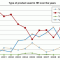
Over the past 50 years, various resistant bacteria have risen to prominence in the hospital setting (Figure 15.1). In the early 1960s, penicillin-resistant Staphylococcus aureus became epidemic and quickly widespread. Subsequent attempts to treat resistant S. aureus infections with methicillin were soon thwarted by the emergence of methicillin resistance. In the 1970s, as vancomycin became available for treatment of infections due to gram-positive cocci, gram-negative bacilli, such as Pseudomonas aeruginosa and Enterobacteriaceae, became dominant healthcare-associated infection (HAI) pathogens. By the 1980s, the introduction of broad-spectrum antimicrobials, such as advanced-generation cephalosporins to better treat gram-negative infections, was countered by the emergence of novel β-lactamases in Enterobacteriaceae. During the same time, the proportion of hospital S. aureus strains resistant to methicillin increased steadily, and vancomycin-resistant enterococci (VRE) surfaced. Other multidrug-resistant (MDR) pathogens of low virulence, such as yeast, methicillin-resistant coagulase-negative staphylococci, and Corynebacterium jeikeium, also became significant HAI pathogens. In the 1990s, resistance to fluoroquinolones and vancomycin emerged among staphylococci, while carbapenem resistance was reported in multiple species of Enterobacteriaceae and became widespread among P. aeruginosa and Acinetobacter baumannii. Since 2000, a group of multidrug-resistant organisms (MDROs) have emerged as dominant in hospitals: Enterococcus faecium, S. aureus, Klebsiella pneumoniae, A. baumannii, P. aeruginosa, and Enterobacter species, sometimes collectively referred to as the “ESKAPE bugs” (5).

The specter of pan-resistant “superbugs” with little to no reasonable antimicrobial treatment has raised concern that, at least for some infections, we have effectively entered a post-antibiotic age (6). Understanding the forces that promote antibiotic resistance is the basis of hospital infection control (IC) efforts. In this chapter, we review the epidemiology of multidrug-resistant pathogens in hospitals and discuss prevention and control strategies.


MECHANISMS AND GENETICS OF RESISTANCE

Bacterial resistance has arisen to each class of new antimicrobials developed to date, often in <3 years from the time of introduction, due to the remarkable genetic diversity and adaptation of bacteria (26,27). Bacteria acquire antibiotic resistance through two main mechanisms: de novo chromosomal mutation or through horizontal gene transfer. Both are important mechanisms and not mutually exclusive. Furthermore, under selective pressure, bacteria frequently aggregate several mechanisms to confer broad resistance to multiple classes of antimicrobials.

Chromosomal antimicrobial resistance usually is the result of genetic mutation and natural selection of strains that survive under the antimicrobial selection pressure. Conditions that favor spontaneous chromosomal mutation include an over-whelming number of organisms (increasing the likelihood of a favorable mutation), incomplete or ineffective antimicrobial therapy (allowing mutants to survive), and a relatively small number of mutations needed for resistance. Some subpopulations of bacteria (e.g., P. aeruginosa) have also been identified as hypermutators, which can increase the likelihood of de novo resistance (28).

Classic examples of chromosomal-mediated antibiotic resistance include genetic mutations of antimicrobial targets leading to resistance for rifamycins (via mutations of the rpoB gene) (29) and for fluoroquinolones (via mutations in DNA topoisomerase) (30). Alteration of promoters of gene expression via chromosomal mutation, thereby changing the production level of antibiotic-inactivating enzymes, antimicrobial targets, or membrane influx or efflux systems, also can lead to resistance. For example, mutations in the promoter controlling the expression of the OprD porin in P. aeruginosa can block the entry of carbapenems, rendering them ineffective (31). Mutations of repressor genes can lead to overexpression of antibiotic-inactivating enzymes, such as the β-lactamase AmpC, leading to third-generation cephalosporin resistance (32).

Horizontal gene transfer is ubiquitous in bacterial communities and represents the major mechanism by which bacteria are able to adapt to hostile natural environments and to antimicrobial innovation. Bacteria may exchange DNA information through transformation (incorporation of exogenous DNA from the surrounding environment), transduction (transfer of genetic material via bacteriophage vectors), or conjugation (direct cell-to-cell transfer of genetic material) (26). Horizontal transfer of resistance genes is facilitated by their location on mobile genetic elements, such as integrons, transposons, and plasmids. The permutations of mobile elements are vast: integrons are found alone, or inside transposons; both integrons and transposons can be carried by plasmids or bacteriophages (33). Such variations allow for gene shuffling and provide bacteria with vast capacity to adapt to changing environments or antimicrobial pressure, particularly in situations where large numbers of bacteria live in communities (such as the gastrointestinal tract or environmental reservoirs) (34).

The rapid spread of extended spectrum β-lactamases among Enterobacteriaceae world-wide has been a closely studied model of horizontal gene transfer. For example, the CTXM extended-spectrum β-lactamase gene (blaCTX-M) is thought to have mobilized multiple times from chromosomal DNA of Kluyvera into plasmids, which have subsequently spread through conjugation into other Enterobacteriaceae species; such events have happened worldwide (35,36). Unfortunately, mobile elements frequently carry multiple resistance mutations simultaneously, allowing for broad resistance to be conferred quickly from one bacterial species to another. Plasmids that carry the carbapenemase gene blaNDM-1 have also been shown to carry aminoglycoside, macrolide, rifampin, and sulfamethoxazole resistance genes, creating Enterobacteriaceae that are nearly pan-resistant (37).


OVERVIEW OF SPECIFIC RESISTANCES

Several sobering trends in resistance have been observed over the years (Figure 15.1, Table 15.3). A surge in aminoglycoside resistance became a chief concern in the 1970s and 1980s, particularly in healthcare-associated Enterobacteriaceae and P. aeruginosa. Although aminoglycoside importance and use has since declined with the development of alternative and often safer antimicrobials, such as advanced-spectrum β-lactam antibiotics and fluoroquinolones, aminoglycoside resistance rates have been stubbornly persistent (38). Rates of tobramycin resistance among Enterobacteriaceae sampled in the United States increased from 1.7% to 8.8% from 1999 to 2008 (39). Aminoglycoside resistance is probably propelled by co-selection with other antibiotic resistance genes, such as fluoroquinolones (40).

The availability of second-generation cephalosporins (such as cefoxitin and cefuroxime), third-generation cephalosporins (such as ceftriaxone and ceftazidime), and of β-lactam-β-lactamase inhibitor combination agents (such as piperacillin-tazobactam) has highlighted an additional set of resistance risks in gram-negative bacilli. For instance, Enterobacter spp. were initially considered susceptible to cephalosporins but frequently developed resistance during therapy. The culprit was a spontaneously derepressed intrinsic chromosomal AmpC β-lactamase (41). Further discovery of plasmid-mediated β-lactamases (such as ESBLs) conferring broad resistance to various penicillins and cephalosporins has made many gram-negatives such as E. coli and Klebsiella spp. difficult to control without turning to “antibiotics of last resort” such as carbapenems. The earliest recognized ESBLs evolved by point mutations from common, older plasmid-borne enzymes and were primarily found among hospital-acquired gram-negative organisms, particularly Klebsiella spp., from the early 1980s to the late 1990s. However, since 2000, the CTX-M family of β-lactamases has become increasingly dominant, displacing other β-lactamases and invading both community and hospital reservoirs through its association with E. coli in addition to Klebsiella spp. (42).


TABLE 15.3 Key Resistance Problems of Select Healthcare-Associated Infection Pathogens


Organism


Key Resistances


Additional resistances


Staphylococcus aureus


Methicillin (all β-lactams), vancomycin


Macrolides, tetracyclines, clindamycin, trimethoprim-sulfamethoxazole, fluoroquinolones, daptomycin, linezolid


Enterococcus faecium, Enterococcus faecalis


Ampicillin (β-lactamase producing), vancomycin, aminoglycosides


Daptomycin, tigecycline, linezolid


Corynebacterium jeikeium


Penicillins, cephalosporins, fluoroquinolones


Macrolides, tetracyclines


Enterobacteriaceae


Cephalosporins (all β-lactams), carbapenems, fluoroquinolones, trimethoprim-sulfamethoxazole


Aminoglycosides


Pseudomonas aeruginosa


Anti-pseudomonal penicillins, anti-pseudomonal cephalosporins, aminoglycosides, fluoroquinolones, carbapenems


Acinetobacter baumannii


Sulbactam, carbapenems, aminoglycosides


Cephalosporins, penicillins, trimethoprim-sulfamethoxazole, fluoroquinolones


Stenotrophomonas maltophilia


Trimethoprim-sulfamethoxazole, ticarcillin-clavulanate


Carbapenems, cephalosporins, penicillins, aminoglycosides, fluoroquinolones


Burkholderia cepacia


Trimethoprim-sulfamethoxazole, carbapenems, fluoroquinolones


Cephalosporins, penicillins, aminoglycosides, tetracyclines


Because carbapenems are currently the broadest-spectrum antimicrobials commercially available, and because they are critical in treating ESBL-producing bacteria as well as other highly resistant gram-negative organisms such as A. baumannii, the increases seen in carbapenem resistance have been alarming. Carbapenem resistance is mediated by various mechanisms, such as loss of the outer-membrane proteins and upregulation of efflux systems (43). In the past decade, many different plasmidmediated carbapenemases—broad-spectrum β-lactamases that hydrolyze carbapenems as well as other β-lactam antibiotics— have emerged and spread worldwide, raising the risk of untreatable infections (37). The emergence of multidrug-resistant strains of gram-negative bacteria that produce New Delhi metallo-β-lactamase (NDM-1) carbapenemase, as well as the remarkable ability of the NDM-1 gene to disseminate across gram-negative species including community-acquired bacteria such as E. coli, Shigella boydii, and Vibrio cholerae, will present perhaps the biggest challenge to IC to date (44,45).

The prevalence of trimethoprim and sulfonamide resistance in gram-negative bacteria remains a concern primarily in outpatient settings, where oral trimethoprim alone or trimethoprim-sulfamethoxazole (TMP-SMX) combination is often prescribed empirically for the treatment of urinary tract infections (UTIs). Resistance to trimethoprim is mediated via alterations in the target enzyme dihidrofolate reductase (46), while resistance to sulfonamides is mediated by alterations in its target enzyme dihydropteroate synthase (47). These resistance
genes are often linked to other resistance genes on transmissible elements, allowing for efficient spread and indirect selective pressure from associated antibiotics. Over the past decades, there has been an increasing trend in TMP-SMX resistance over time; surveillance data from 2000 to 2010 among US urinary E. coli isolates showed an increase from 17.9% to 24.2% (48). Reductions in trimethoprim use alone do not appear to be sufficient to reduce the rate of trimethoprim-resistant E. coli, probably explained by the low fitness cost of trimethoprim resistance and the co-selection of trimethoprim resistance when alternative antibiotics are used (49,50).

Methicillin resistance has been a major concern in staphylococci since the 1980s. In 63 U.S. hospitals from 1974 to 1981, the percentage of S. aureus infections resistant to methicillin increased modestly from 2.4% to 5%, due primarily to epidemics in four large teaching institutions (51). In 1992, the pooled percentage of resistance had risen dramatically to 32.1%, and in 2004, to 53% (52,53). Methicillin resistance in healthcare-associated S. aureus and coagulase-negative staphylococci is endemic in most U.S. hospitals.

Fluoroquinolones target enzymes responsible for bacterial DNA replication, such as DNA gyrase and topoisomerase, giving them broad efficacy against many gram-negative, and some gram-positive, organisms (54). Since the 1980s, their potency and oral bioavailability have made their use widespread in treatment of infections (particularly pulmonary, urinary, and gastrointestinal), as well as in prophylaxis (e.g., in neutropenic patients). Widespread increases in fluoroquinolone resistance have been reported in many bacteria, in particular Enterobacteriaceae, P. aeruginosa, Streptococcus pneumoniae, and Staphylococcus. aureus (55,56,57). Ciprofloxacin resistance among E. coli urinary isolates from the United States increased from 3% to 17% from 2000 to 2010 (48). Although mutations to fluoroquinolone targets and efflux mechanisms explain some of the increase in resistance rates, the discovery of two disseminated classes of plasmid-mediated fluoroquinolone resistance mechanisms (Qnr proteins that interfere with fluoroquinolone interaction with DNA gyrase and fluoroquinolone-modifying enzymes (aac(6′)-Ib-cr)) also accounts for the explosive worldwide increase in fluoroquinolone resistance over time (58).


TABLE 15.4 Examples of Host Factors Associated with Healthcare-Associated Colonization or Infection by an Antimicrobial-Resistant Organism in Selected Case-Control Studies


Factor


Reference


MORE FREQUENTLY IDENTIFIED


Duration of hospital or ICU stay (adjusted or matched in many studies)


(63,64,65,430)


Prior antimicrobials


(65,73,191,431,432,433,434,435,436,437,438,439,440,441,442,443,444,445)


Intensive care unit


(65,433,443,445)


Invasive devices or procedures


(433,438)


central venous catheter


(191,434)


endotracheal intubation


(434,442)


urinary catheter


(436)


nasogastric tube


(435,446)


Underlying comorbidities


(73,191,434,436)


Prior colonization or infection with resistant organism


(73,447)


Prior hospitalization or residence in long-term care facility


(63,64,444)


RISK FACTORS LESS FREQUENTLY IDENTIFIED


Age


(64,443)


Sex


(434,435,444)


Chemotherapy


(448)


Endoscopy


(448)


Surgery (or number of operations)


(64)


Proximity to other patients


(191)


HOST FACTORS PREDISPOSING TO COLONIZATION OR INFECTION WITH ANTIBIOTIC-RESISTANT ORGANISMS

A number of patient risk factors have been associated with acquisition of resistant bacteria (Table 15.4). Our epidemiologic understanding of these factors remains limited because most studies have been retrospective due to practical restraints, limiting the covariates to more easily obtainable data, such as recent antimicrobial exposures. Many of the identified factors are undoubtedly linked and may serve as indirect markers of more difficult-to-measure covariates such as frequency of patient-to-staff contact. It also is important to recognize that risk factors may differ depending on whether epidemic or endemic periods are being studied, and whether the resistant pathogen is isolated during episodes of colonization or infection.

Attention has been directed at specific methodologic issues that may lead to biased estimates of risk for antibiotic resistance (59). First, results of case-control designs, used extensively in studying the risk of acquiring resistant bacteria, are influenced by how the control group is selected. In many studies, control groups are selected either from patients in the population who are uninfected, or from patients who carry the antibiotic-susceptible form of the bacterium of interest (60). Use of either control group may result in slightly differing estimates of risk. When resistant cases are compared to uninfected controls, risk factors for acquisition of both the susceptible and the resistant phenotype of the organism may be identified (60). If instead, controls are selected that are already colonized or infected with the susceptible form of the organism, the odds of association between certain variables and acquisition may be overestimated (61). An alternative study design,
“case-case-control,” uses both types of controls and two separate case-control analyses within a single study in order to differentiate risk factors associated with acquisition of the susceptible and resistant phenotypes of the organism (60).

A second important methodologic principle is adjustment for time at risk (59). Not surprisingly, duration of hospital stay (and specifically, “time at risk” before the index colonization or infection) is often identified as a significant risk factor for acquisition of bacteria (62,63,64). In case-control studies that identify risk factors for antibiotic resistance, time at risk needs to be accounted for via multivariable analysis or by matching cases and controls (59).

A third methodologic principle is adjustment for comorbid conditions (59). Such adjustment is particularly important when the risk factor of interest is antimicrobial use. As a corollary, measurement of comorbid conditions and severity of illness should be performed at a time point before acquisition of the organism of interest, in order to make causal inference valid (65).

A final methodologic concern is the analysis of aggregated antibiotic use data to estimate patient-level risk of acquiring an antibiotic-resistant organism (66). Analysis of aggregated data may not accurately reflect the risk of exposure to an individual patient, since population studies do not link individual outcomes to individual exposures (66). Population studies are useful in allowing the measurement of the total effect of an exposure.

There is a striking commonality of risk factors for colonization or infection with pathogens such as antimicrobial-resistant S. aureus, Enterococcus spp., or gram-negative bacilli (67). These risk factors include advanced age; underlying diseases and severity of illness; inter-institutional transfer of the patient, especially from a nursing home; prolonged hospitalization; gastrointestinal surgery or transplantation; exposure to invasive devices of all types, especially central venous catheters; and exposure to antibiotics, especially cephalosporins (67). Other risk factors are identified in Table 15.4.

The role of antimicrobials in promoting resistant organisms has attracted much attention in the literature, perhaps because of its potential for modification. Although it is accepted that rising antimicrobial consumption promotes resistance, the relation between antimicrobials and resistance at the patient level and population level remains unclear. Thus, it often is difficult for hospitals to decide between diverse antimicrobial stewardship strategies, such as reducing all classes of antimicrobial use, targeting specific antimicrobial classes, or rotating available antibiotics.

For many important drug-resistant organisms, such as Methicillin-resistant S. aureus (MRSA) and VRE, resistance is mediated by complex genes that would unlikely occur spontaneously in any individual patient. In such cases, development of resistance involves horizontal acquisition of either resistant organisms themselves or genetic vectors such as plasmids carrying resistance genes. Furthermore, many important drug-resistant organisms, such as enterococci, colonize the gut or skin, allowing opportunities for indirect exposure to the selective pressures of antimicrobials intended for other pathogens.

Thus, the use of antimicrobials may lead to a rise in the prevalence of resistant organisms on a population level through a variety of indirect mechanisms (68). In the case of VRE, use of cephalosporins eliminates competitive gut flora, promoting opportunities for horizontal acquisition of the organism (69). The use of anti-anaerobic drugs appears to facilitate fecal excretion of resistant enterococci, leading to further opportunities for transmission (70). On a population level, the emergence of VRE in the 1980s was likely due to the accelerating use of vancomycin during that decade (71). However, many classes of antibiotics have been associated with VRE emergence; prior receipt of vancomycin by an individual patient appears to have minimal effect on the risk of VRE acquisition once VRE have become established in a locality (72,73).


SOURCES OF RESISTANT STRAINS

The source of most resistant strains in hospitals appears to be patients who are colonized or infected (74,75,76,77). Because the normal oropharyngeal and intestinal flora of hospital patients may be displaced by multiply resistant enteric bacteria and P. aeruginosa (urine, perineum, and wounds may be similarly affected), there are often many colonized patients for each patient with recognized infection, the so-called “iceberg effect” (Figure 15.2) (74). This shift in flora often occurs within a very few days of hospital admission and affects the older, generally sicker, or more debilitated patients. The causes of shifting commensal bacteria are unclear, and may involve a combination of hospital-related factors (e.g., specific treatment exposures vs. more hands-on care in general) and patient host factors (e.g., possible changes in membrane receptors or ligands, antibiotic suppression of normal flora, potential contribution of biofilm formation on devices such as nasogastric and endotracheal tubes) (78,79,80,81). Some shift in endemic strains may result from emergence of low-count community-acquired flora in the face of antibiotic exposure rather than from true healthcare-associated acquisition (82,83).

It is important to realize that multidrug-resistant bacteria can be recovered from the normal, intact skin of patients (76,84,85,86), as well as from body fluids, secretions, and wounds. While the perineal or inguinal areas of patients usually are most heavily contaminated, the axillae, trunk, arms, and hands also are frequently colonized (87). Pathogens most often found at these sites are A. baumannii, Staphylococcal spp., and Enterococcal spp., perhaps in part because these pathogens are more resistant to desiccation compared to other bacteria (87,88,89). In one study, VRE were cultured from the antecubital fossae of 29% of ventilated patients studied in a medical ICU (76). These findings have implications for control strategies aimed at these pathogens, which are addressed later in this chapter.

Personnel have been documented sources of resistant gram-positive strains, such as MRSA (90,91,92,93) and even coagulase-negative staphylococci (94). However, personnel carriage of resistant gram-negative bacilli (other than transient hand carriage described in the following section) appears to be very unusual. Exceptions include outbreaks reportedly traced to carriers of Acinetobacter spp., Citrobacter spp., or Proteus species. Acinetobacter sp., one of the few gram-negative bacilli that may be among normal skin flora, was noted in one outbreak to recur periodically despite disinfection of the apparent environmental reservoirs. The outbreak was ultimately traced to the colonized hands of a respiratory therapy technician who had dermatitis and apparently contaminated respiratory therapy equipment while assembling it (95). There have also been clusters of Citrobacter spp. infections of the central nervous system
in neonates (96,97), traced to hand carriage by nurses, and an outbreak of Proteus mirabilis infections in newborns traced to a nurse who was a chronic carrier (98). In another study, endemic Pseudomonas aeruginosa infection was maintained in a neonatal ICU by persistent carriage on the hands of healthcare workers; artificial fingernails or nail wraps were both risk factors for hand colonization in this study (99).

Foodborne contamination with multiply resistant gram-negative bacilli has been cited in several investigations (51,100,101) and has been incriminated particularly in oncology units (102). Despite the potential importance of these observations, however, the overall role of food in introducing resistant strains into the general hospital remains unclear.

Environmental sources and reservoirs of resistant strains have been a recurrent problem, especially when patient care equipment becomes contaminated. Extensive outbreaks of UTIs (and respiratory tract, perineal, or intestinal colonization) may result when urine measuring devices, contaminated by enteric bacilli or P. aeruginosa, are shared by many patients (74,103). MRSA contamination of ultrasonic nebulizer filters was linked to an outbreak of infection and colonization in a head and neck surgical ward (104), and electronic thermometers contaminated with vancomycin-resistant Enterococcus faecium were implicated as vehicles of transmission in an outbreak in a medical-surgical ICU (105).

Finally, there has been perennial concern about contamination of many areas of the inanimate environment with which patients do not have regular contact, such as flowerpots and sink traps (106,107,108). These sites, despite sometimes heavy contamination, have not been consistently linked with the spread of bacteria in hospitals.

Contamination of inanimate environmental surfaces that are touched by healthcare workers may be a more important source of transmission, particularly for multidrug-resistant bacteria (such as VRE and Acinetobacter spp.) that survive well on inanimate surfaces (109,110,111). In one report, healthcare workers were shown to transfer VRE from contaminated sites in a patient’s room—such as a blood pressure cuff, bed rail, or soap dispenser—to clean sites in the room or on a patient’s skin via their hands or gloves during routine patient care activities in 10.6% of opportunities (112). Similarly, healthcare workers were found to contaminate their gloves with MRSA by touching only environmental sites in patient rooms (113). For high-risk immunocompromised patients, especially those who have the opportunity for environmental exposures (e.g., the debilitated oncology patient who sits at the sink to wash), strains from sink surfaces have been linked to patient colonization and infection (102).


TABLE 15.5 Relative Importance of Selected Factors in the Emergence of Some Multidrug-Resistant Organisms within Hospitalsa


Multidrug-Resistant Organism, Defined by Key Resistance Marker


Patient-to-Patient Transmission via Contaminated Healthcare Worker Hands


Environment-to-Patient via Contaminated Healthcare Worker Hands


Colonized Healthcare Worker Directly to Patient


Airborne Transmission


Endogenous Selection by Antibiotic Pressure


Methicillin-resistant Staphylococcus aureus


+++


+/++


+


+/−


+


Vancomycin-resistant enterococcus


+++


++


++


Multidrug-resistant Enterobacteriaceae


+++


+


+++


Imipenem-resistant Pseudomonas aeruginosa


+


+


+


+++


Imipenem-resistant Acinetobacter baumannii


+++


++


++


aThe relative importance is an indication of the need to address each factor in control measures for specific resistant pathogens.


MODES OF TRANSMISSION

Traditional teaching is that resistant bacteria are spread in the hospital from an infected patient to a susceptible patient via transient carriage on hands of health care personnel (Table 15.5). Such spread contributes to the iceberg of colonized patients and greatly increases the source and reservoir of resistant strains in the hospital (Figure 15.2). While much of the evidence incriminating hands of personnel is circumstantial and based on finding resistant bacteria colonizing or contaminating healthcare worker hands, both experimental and mathematical models, as well as observational studies in patient care settings have demonstrated that healthcare workers can transfer pathogens from their hands or gloves to patients’ skin or devices (112,114,115,116,117,118). Moreover, the weight of experience, dating back to the successful introduction of hand hygiene as a control measure by Semmelweis, strongly supports this concept.

The contribution of transfer of pathogens from contaminated environmental surfaces to patients via the hands of healthcare workers is receiving renewed attention (112). Given the lower colony counts typically found at environmental sites compared to patient sites (76), for many pathogens, this route of transmission appears to be less important than transfer from patient to patient (Table 15.5).

Healthcare workers who are persistently colonized with antibiotic-resistant bacteria can sometimes transfer these pathogens directly to patients (Table 15.5) (99,119). This is particularly important for MRSA, and may be under-appreciated in settings where MRSA is endemic and cross-transmission is frequent (119), thereby obscuring the contribution of carriers.

Only gold members can continue reading. Log In or Register to continue

Stay updated, free articles. Join our Telegram channel

Jun 16, 2016 | Posted by in INFECTIOUS DISEASE | Comments Off on Multidrug-Resistant Organisms: Epidemiology and Control

Full access? Get Clinical Tree

Get Clinical Tree app for offline access