II.F.001 Monocyte
II.F.001
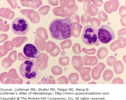
Monocyte, normal. Blood film. Three segmented neutrophils, two small lymphocytes. A monocyte is in the center of the field with a large, reniform nucleus and cytoplasmic color invariably distinctly different from that of neutrophils. The monocyte cytoplasm is blue-gray and the neutrophil is light tan. Note also the lighter staining, lacy nuclear appearance in the monocyte and the denser, condensed chromatin in the neutrophils.
II.F.002 Monocyte
II.F.002
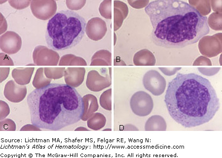
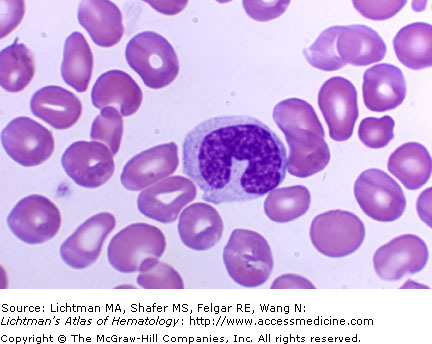
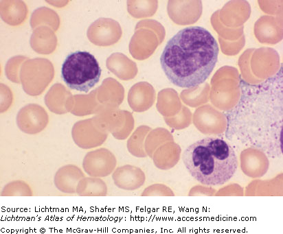
Monocyte, normal. Blood film. A composite of four normal monocytes. (A) Folded nucleus. Modest amount of cytoplasm. (B) Folded nucleus. More ample, gray cytoplasm, cytoplasmic vacuoles (often secondary to exposure to Na2EDTA anticoagulant. (C) Reniform nucleus. Ample cytoplasm, azure granules. (D) Circular nucleus. Azure granules.
II.F.003 Monocyte
II.F.003
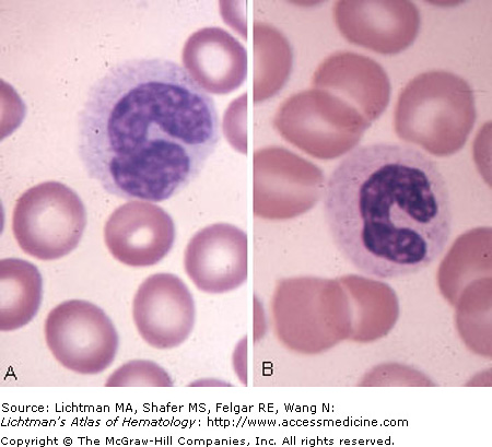
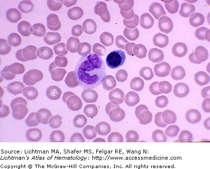
Monocyte, normal. Blood film. (A) Monocyte with horseshoe-shaped nucleus with enlarged, folded end, gray cytoplasm. (B) Band neutrophil with horseshoe-shaped nucleus and pink-orange cytoplasm. The difference in cytoplasmic coloration is an invariable distinction between monocytes and band neutrophils by light microscopy.
