Minimally Invasive and Open Approaches to Mediastinal Nodal Assessment


Minimally Invasive and Open Approaches to Mediastinal Nodal Assessment


CRITICAL ELEMENT


  • Lymph Node Dissection or Systematic Lymph Node Sampling in the Chest

Recommendation: The proper technique enables surgeons to access all lymph node stations using either a minimally invasive or an open approach.

Type of Data: Retrospective.

Strength of Recommendation: Weak.


Rationale

The staging and surgical treatment of lung cancer depends on the evaluation of both N1 and N2 nodal stations. Sampled or dissected nodes should include 9R, 8R, 7, 10R, 4R, and 2R nodes on the right side and 9L, 8L, 7, 6, and 5 nodes (and 4L and 2L nodes, if accessible) on the left side.1 Specific strategies for accessing these nodal stations, as well as some technical maneuvers for avoiding complications during nodal assessment, are described below.1,2,3,4,5,6,7,8,9,10

Adequate lymph node dissection can be achieved using either a minimally invasive or open technique. Surgeons performing video-assisted thoracic surgery (VATS) may find it easier to approach a lymph node examination in an inferior-to-superior fashion. Details on lymph node dissection are outlined in Table 8-1.

Level 8 and 9 lymph nodes, which are adjacent to the inferior pulmonary vein, are exposed by releasing the inferior ligament and extending the dissection anteriorly and posteriorly to the hilum (Fig. 8-1A,B). The number and consistency of the nodal groups, especially those of stations 8 and 9, vary considerably. Nodes may be completely absent, discrete, or lumped in a fibrofatty tissue amenable to block dissection. To expose the 8R nodes, the surgeon opens the pleura between the esophagus and pericardium by extending the dissection from the inferior ligament to the esophageal hiatus (Fig. 8-2). The 8L and 9L stations are approached in a similar manner,
with care taken to avoid any hiatal hernias and preserve the vagus nerve in its medial location to the aorta.


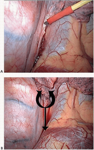
FIGURE 8-1 A,B: Thoracoscopic view of right-sided nodal dissection for stations 8 and 9. (Courtesy of Khalid Amer, FRCS [CTh].)

Station 7 nodes are located between the right and left main bronchi. Nodes on the right are approached between the right main bronchus and the esophagus. The right lung is retracted anteriorly, and the pleural reflection at the back of the hilum is opened from the inferior ligament to the concavity of the azygos vein, medial to the vagus nerve (Fig. 8-3). In this step, all vagal bronchial branches could be divided with
impunity. (If diathermy is used, cutting these branches usually induces a cough reflex.) The bronchus intermedius and right main bronchus are identified and dissected proximally until the left main bronchus is identified. The subcarinal nodes are lifted off the pericardium (usually an avascular plane) and then dissected from their blood supply, with care taken to avoid injury to the membranous bronchus or esophagus (Fig. 8-4).
The nodes should be carefully labeled, as paraesophageal 8R and parabronchial 10R nodes could easily be mistaken for 7R nodes. In this location, injury to the thoracic duct is prevented by tucking the duct under the esophagus; lifting the esophagus off the vertebral bed may injure the thoracic duct. At the completion of this dissection, the right main bronchus, left main bronchus, and subcarinal space should be readily visible (Fig. 8-5).


FIGURE 8-2 Exposure of station 8R by right thoracoscopy. IVC, inferior vena cava. (Courtesy of Khalid Amer, FRCS [CTh].)


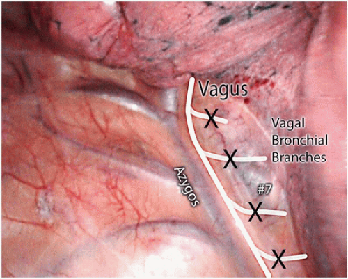
FIGURE 8-3 Right lung retracted forward to expose subcarinal nodes (level 7) via VATS approach. Small vagal branches towards lung can be divided without concern. (Courtesy of Khalid Amer, FRCS [CTh].)


FIGURE 8-4 Mobilization of the subcarinal nodal packet via the right chest. The pericardium is the anterior border of the dissection plane. (Courtesy of Khalid Amer, FRCS [CTh].)


FIGURE 8-5 View of the subcarinal space after complete nodal dissection. (Courtesy of Khalid Amer, FRCS [CTh].)


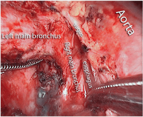
FIGURE 8-6 Subcarinal node dissection from the left chest. (Courtesy of Khalid Amer, FRCS [CTh].)

Accessing subcarinal station 7 nodes from the left is accomplished by following the lower lobe bronchus proximally, as it leads to the subcarinal space and right main bronchus (Fig. 8.6). One useful technique for bringing the carina forward from a deep level is to use a sturdy tape from an anterior port to retract the lower lobe bronchus. Dissection of the posterior aspect of the left hilum facilitates access to the nodes but places the recurrent laryngeal nerve at risk of injury. The location of the separation of the recurrent laryngeal nerve from the vagus nerve varies, and preserving the pleura between the vagus nerve and aorta may lessen the risk of injury to the laryngeal nerve.

Pre- and paratracheal station 2R and 4R nodes are located in a fibrofatty nodal block that can usually be dissected en bloc. These nodes are located in an anatomical triangle bound by the phrenic nerve, vagus nerve, and azygos vein (Fig. 8-7). The apex of the triangle is at the level of the brachiocephalic artery as it crosses the trachea towards the first rib. The lung is retracted downward to expose this triangle, and releasing the pleura between the azygos vein and main bronchus facilitates this exposure. Opening the pleura just lateral to the superior vena cava (SVC) instead of in the middle of the triangle also facilitates this dissection (Figs. 8-8 and 8-9). The right vagus nerve, as well as veins draining directly into the SVC, are present in the triangle, and small veins in this area should be actively sought out and controlled to prevent bleeding complications. In almost all cases, a single vein can be found draining directly into the posterior aspect of the SVC. If inadvertently cut, the vein may retract and bleed profusely, making it difficult to control. The left paratracheal nodes are perhaps the most challenging to expose because the aortic arch, ligamentum arteriosum, and recurrent nerve all hinder dissection. The most lateral of the 4L nodes lie on the tracheobronchial junction and are accessible from the back of the hilum. To expose these nodes, the main pulmonary artery is freed from the bronchus, and
a vessel loop is passed around the artery to facilitate its retraction. Gentle retraction of the carina in a downward direction and the artery in a cephalad direction exposes station 4L.


FIGURE 8-7 View of the right upper mediastinum with anatomical boundaries of paratracheal node dissection. (Courtesy of Khalid Amer, FRCS [CTh].)

Similarly, station 5 and 6 nodes on the left exist in an anatomical triangle whose boundaries are the vagus nerve, phrenic nerve, and aortic arch. Dissection begins by retracting the lung posteriorly and downward. The fibrofatty nodal block is lifted off
the main pulmonary artery into the aortopulmonary space, medial to the vagus nerve (Fig. 8-10). The phrenic nerve is identified on the most medial aspect of the triangle. The nodal block is dissected up to the origin of the left subclavian artery, and all nodes and fatty tissue superficial to the ligamentum are harvested. As long as the dissection is kept to the medial side of the vagus, the recurrent laryngeal nerve is free from potential injury.


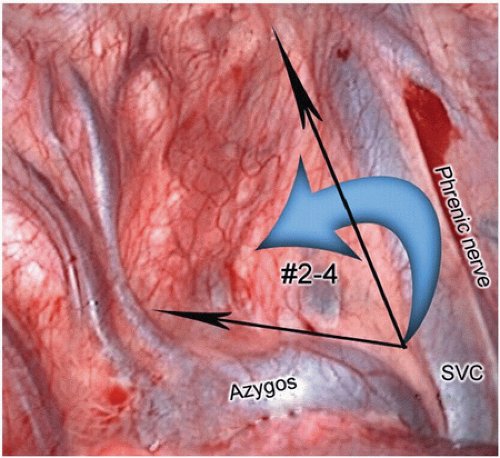
FIGURE 8-8 Black arrows indicate ideal sites for incising the mediastinal pleura, then performing an anterior to posterior mobilization of all soft tissue (blue arrow), including nodes behind the superior vena cava towards the vagus nerve. (Courtesy of Khalid Amer, FRCS [CTh].)


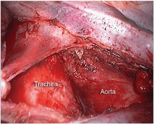
FIGURE 8-9 Right paratracheal node dissection basin after removal of all 2R and 4R nodal tissue. (Courtesy of Khalid Amer, FRCS [CTh].)


FIGURE 8-10 Exposure of level 5 (aortopulmonary window) nodes from behind the left hilum. (Courtesy of Khalid Amer, FRCS [CTh].)


TABLE 8-1 Structures Dissected/Divided


Level


Borders*


VATS Modifications


Comments


2


2R (includes nodes extending to the left lateral border of the trachea):


Upper border: the apex of the right lung and pleural space and in the midline, the upper border of the manubrium


Lower border: the intersection of caudal margin of the innominate vein with the trachea


2L:


Upper border: the apex of the left lung and pleural space and in the midline, the upper border of the manubrium


Lower border: the superior border of the aortic arch


Right dissection:


  • Incise the pleura to create a triangle adjacent to the SVC (posterior to the phrenic nerve), vertebral body (anterior to the vagus nerve), and azygos vein (superior border).


  • Retract the SVC and posterior mediastinum to open space. If performing VATS, a vagal sling and retraction sutures should be used.


  • Excise the 2/4 LN packet starting at the border of the SVC, dissecting posterior toward the trachea and aortic arch.


  • Additional 2R nodes may be found by retracting the brachiocephalic artery anteriorly. Additional 4R nodes may be found beneath the junction of the azygos vein and SVC.


Left dissection:


  • This region is more difficult to approach from the left and generally requires exposure and dissection of the level 7 nodes.


  • Exposure can be improved by passing a silicone tape around the left main pulmonary artery and retracting the artery superiorly and away from the distal trachea, which is depressed inferiorly with a blunt soft retractor such as a peanut sponge.


Right dissection: Control (i.e., clip) veins draining directly into the SVC and large lymphatic channels. Avoid injury to the right recurrent nerve, which is generally 1-2 cm distal to the aortic arch beneath the brachiocephalic artery.


Left dissection: Control (i.e., clip) large lymphatic channels and lymph node arteries. Avoid injury to the left recurrent nerve beneath the aortic arch.


4


4R (includes right paratracheal nodes and pretracheal nodes extending to the left lateral border of trachea):


Upper border: the intersection of the caudal margin of the innominate vein with the trachea


Lower border: the lower border of the azygos vein


4L (includes nodes to the left of the left lateral border of the trachea, medial to the ligamentum arteriosum):


Upper border: the upper margin of the aortic arch


Lower border: the upper rim of the left main pulmonary artery


5


Includes subaortic lymph nodes lateral to the ligamentum arteriosum


Upper border: the lower border of the aortic arch


Lower border: the upper rim of the left main pulmonary artery


  • Incise the pleura to create a triangle adjacent to the phrenic nerve, vagus nerve, and left main pulmonary artery (superior border).


  • Excise the 5/6 LN packet starting at the border of the superior vein, dissecting posterior to the pulmonary artery and anteromedial to the vagus nerve.


  • Dissection of the level 6 packet proceeds to the aortic arch, and the fibrofatty packet is cleaned to the origin of the left subclavian artery. (Placing a tape around the phrenic nerve may protect it during this dissection.)


Nodes overlying the left superior vein belong to station 10L.


6


Includes lymph nodes anterior and lateral to the ascending aorta and aortic arch


Upper border: a line tangential to the upper border of the aortic arch


Lower border: the lower border of the aortic arch


7


Upper border: the carina of the trachea


Lower border: on the left, the upper border of the lower lobe bronchus; on the right, the lower border of the bronchus intermedius


Right dissection:


  • Divide the posterior hilar pleura adjacent to the parenchyma from the inferior pulmonary vein to the azygos vein/superior right mainstem bronchus.


  • Divide the vagus nerve branches to the bronchus, sparing the main trunk.


  • Excise the nodal packet between the right and left mainstem bronchi and esophagus.


Left dissection:


  • Divide the posterior hilar pleura adjacent to the parenchyma from the inferior pulmonary vein to the apex of the hilum anteromedial to the vagus nerve trunk.


  • Divide the vagus nerve branches to the bronchus, sparing the main trunk; to spare the recurrent nerve, avoid dissecting between the vagus nerve and aorta.


  • Retract the lower lobe bronchus upward and anteriorly and simultaneously retract the esophagus posteriorly to expose the subcarinal nodes from the left. (This can be done using retraction tape if VATS is performed.)


  • Excise the nodal packet between the left and right mainstem bronchi and esophagus.


Right dissection: Avoid injury to the membranous bronchus and esophagus. Do not include level 8 and 10R nodes.


Left dissection: Control (i.e., clip) nodal artery branches from the aorta. Do not include level 4L nodes.


8


Includes nodes adjacent to the wall of the esophagus and to the right or left of the midline, excluding subcarinal nodes


Upper border: on the left, the upper border of the lower lobe bronchus; on the right, the lower border of the bronchus intermedius


Lower border: the diaphragm


  • Divide the inferior pulmonary ligament.


  • Excise level 9 nodes within the ligament.


  • Reflect or dissect the pleura from the esophagus.


  • Excise level 8 nodes adjacent to the esophagus from the diaphragm to bronchus intermedius (right) or lower lobe bronchus (left).


Right dissection: Avoid injury to the vagus nerve, thoracic duct, azygos vein, and phrenic nerve.


Left dissection: Avoid injury to the vagus nerve. Watch for hiatal hernia.


Level 9 node quantity varies.


9


Includes nodes lying within the pulmonary ligament


Upper border: the inferior pulmonary vein


Lower border: the diaphragm


* As defined by the International Association for the Study of Lung Cancer.


LN, lymph node; SVC, superior vena cava; VATS, video-assisted thoracic surgery.


From Rusch VW, Asamura H, Watanabe H, et al. The IASLC lung cancer staging project: a proposal for a new international lymph node map in the forthcoming seventh edition of the TNM classification for lung cancer. J Thorac Oncol 2009;4(5):568-577.

Only gold members can continue reading. Log In or Register to continue

Stay updated, free articles. Join our Telegram channel

Jul 22, 2016 | Posted by in ONCOLOGY | Comments Off on Minimally Invasive and Open Approaches to Mediastinal Nodal Assessment

Full access? Get Clinical Tree

Get Clinical Tree app for offline access