Metastatic Brain Tumors
INTRODUCTION
Brain metastasis is a common complication of cancer. Recent population-based data suggest that up to 20% of adults with cancer will develop symptomatic brain metastases during life (1). Autopsy studies indicate that another 25%–30% of patients with disseminated cancer have asymptomatic brain metastases at the time of death. The incidence of brain metastasis varies by primary cancer type, being highest for lung (20%) followed by melanoma (7%), renal (6.5%), breast (5%), and colorectal (1.8%). Prostate, gynecologic, head and neck, and non-melanomatous skin cancers involve the brain parenchyma infrequently. The prevalence of brain metastases has increased in the past three decades. Contributing factors may include more sensitive imaging techniques (gadolinium enhanced MRI), lengthened survival due to more effective systemic therapies, and poor central nervous system (CNS) penetration of many chemotherapeutic and antibody-based agents.
CLINICAL MANIFESTATIONS
Common signs and symptoms of metastatic brain tumors can be classified as either focal or generalized (Table 63-1) (2). Focal symptoms, such as hemiparesis, aphasia, and visual field defects, vary according to location of the tumor. Generalized symptoms, such as headache, confusion, lethargy, nausea, and vomiting, result from increased ICP or hydrocephalus. Metastatic deposits near the ventricular system can cause obstructive hydrocephalus by interruption of normal cerebrospinal fluid (CSF) outflow pathways through the third and fourth ventricles. Obstructive hydrocephalus is of particular concern with posterior fossa tumors. Headaches caused by increased ICP and/or hydrocephalus may have the following characteristics:
• Worse in the morning and with recumbency
• Associated with nausea and vomiting
• Exacerbated by coughing or straining
• Accompanied by confusion or lethargy
The presence of papilledema is very suggestive of increased ICP, although its absence does not exclude it.
EVALUATION
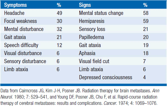
Contrast-enhanced MRI has replaced CT as the study of choice for patients with suspected brain metastasis (Figure 63-1). MRI is more sensitive than CT for small lesions, particularly in the brain stem and posterior fossa (Figure 63-2). In addition, MRI is better able to distinguish metastatic lesions from alternative diagnoses such as abscess or stroke. All cancer patients with new neurological symptoms should be screened for brain metastases. In patients who cannot undergo MRI because of implanted hardware, morbid obesity, or extreme claustrophobia, a contrast-enhanced CT should be obtained instead. If leptomeningeal enhancement is present, lumbar puncture should be strongly considered to rule out leptomeningeal metastases. Lumbar puncture should not be performed in patients with increased intracranial pressure from obstructive hydrocephalus, lateral shift of midline structures, or any evidence of mass effect in the posterior fossa.
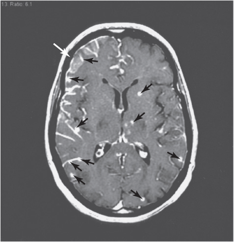
FIGURE 63-1 Gadolinium-enhanced T1-weighted MRI showing multiple small parenchymal metastases (arrowheads) and leptomeningeal enhancement (arrow) in a patient with non-small cell lung cancer. Spinal fluid was positive for malignant cells.
FIGURE 63-2 Contrast-enhanced CT in the same patient showing a single parenchymal lesion (arrowhead) and faint leptomeningeal enhancement (arrow). This image demonstrates the decreased sensitivity of CT compared with MRI for detecting small metastases.
DIFFERENTIAL DIAGNOSIS
The differential diagnosis of a mass lesion in a patient with cancer is broad and includes:
• Primary brain tumor
• Abscess (bacterial, mycobacterial, fungal, parasitic)
• Acute demyelinating plaque
• Subacute cerebral infarction
• Primary intracranial hemorrhage
In a study of 54 patients with cancer who underwent biopsy of a single mass lesion, a diagnosis other than metastatic cancer was obtained in 11% (equally divided between primary brain tumor and infection) (3). This study highlights the importance of obtaining pathologic confirmation before proceeding with definitive therapy if the diagnosis is at all uncertain.
MANAGEMENT
 SYMPTOMATIC THERAPY
Glucocorticoids
Glucocorticoids should be administered to patients with focal deficits, headache, or other symptoms resulting from peritumoral edema or increased ICP. Glucocorticoids act to restore the integrity of the blood-brain barrier and thereby reduce cerebral edema. Dexamethasone is the most widely used glucocorticoid for this purpose because of its long half-life and minimal mineralocorticoid effects. It is typically given as an intravenous 10 mg loading dose followed by 16 mg/day divided in 2–4 doses (either intravenous or oral). Symptoms should improve within 2–3 days, and higher doses (up to 100 mg/day) may be required in some patients. Adverse effects of glucocorticoids are dose dependent, and therefore the dose should always be tapered to the lowest possible dose required to ameliorate symptoms. Patients taking daily glucocorticoids for longer than 2–4 weeks should receive prophylaxis for Pneumocystis jiroveci, typically with trimethoprim/sulfamethoxazole.
Anticonvulsants
Twenty percent to 40% of all brain tumor patients experience a seizure by the time their tumor is diagnosed. In these patients, the need for anticonvulsant therapy is clear. An additional 20%–40% of patients will develop seizures as a complication of their disease. Based on this risk, many physicians choose to start prophylactic anticonvulsants at the time of diagnosis. However, this practice must be weighed against the potential adverse effects of anticonvulsants, which include:
• Allergic reaction, including Stevens-Johnson syndrome
• Hepatic dysfunction
• Cognitive impairment and imbalance
• Myelosuppression
• Alteration of cytochrome P-450 enzyme system
The latter is an important consideration in patients receiving chemother-apeutic and targeted agents that are metabolized by the liver, such as taxanes, vinca alkaloids, and many tyrosine kinase inhibitors. The American Academy of Neurology has published a practice parameter finding no evidence to support the use of prophylactic anticonvulsant medication in patients with brain tumors who have not experienced a seizure (4).
 DEFINITIVE THERAPY
Related posts:

Stay updated, free articles. Join our Telegram channel

Full access? Get Clinical Tree


