, Hans Guski2 and Glen Kristiansen3
(1)
Carl-Thiem-Klinikum, Institut für Pathologie, Cottbus, Germany
(2)
Vivantes Klinikum Neukölln, Institut für Pathologie, Berlin, Germany
(3)
Universität Bonn, UKB, Institut für Pathologie, Bonn, Germany
3.1 Diagnostic Antibody Panel for Tumors of the Upper Respiratory Tract
Cytokeratin profile, CD56, synaptophysin, chromogranin, EBV, NUT, p16
3.2 Diagnostic Antibody Panel for Epithelial Pulmonary Tumors
Cytokeratin profile, TTF-1, napsin A, p63, p40, CD56, and surfactant proteins [1]
3.3 Diagnostic Antibody Panel for Mesenchymal Pulmonary Tumors
CD1a, langerin (CD207), HMB45, STAT6, CD31, CD34, CD99
Thyroid transcription factor-1 (TTF-1) | ||
Expression pattern: nuclear | ||
Main diagnostic use | Expression in other tumors | Expression in normal cells |
Pulmonary carcinoma (adenocarcinoma, bronchioloalveolar carcinoma, and small cell carcinoma), carcinoma of thyroid gland (papillary, follicular, and medullary) | Non-pulmonary small cell carcinoma of different locations, subset of extrahepatic cholangiocarcinomas, glial-ependymal and choroid plexus tumors, pituicytoma, granular cell tumor of the sellar region, meningeal tumors | Type II pneumocytes and Clara cells of the lung, thyroid follicular and parafollicular C cells, parathyroid, pituitary gland, diencephalon |
Positive control: thyroid tissue | ||
Diagnostic Approach
Thyroid transcription factor (TTF-1 also known as NKX2-1 or thyroid-specific enhancer-binding protein) is a homeobox-containing transcription factor that regulates the development, differentiation, and gene expression of the thyroid gland (follicular and parafollicular C cells). TTF-1 plays also an active role in the regulation of development and transcriptional activity of the lung and central nervous system (diencephalon). In adult thyroid gland, TTF-1 is expressed in both follicular and parafollicular cells and controls the synthesis of different thyroid hormones and thyrotropin receptor. In normal lung, TTF-1 is strongly expressed in type II alveolar cells, Clara bronchiolar cells, and, in a lesser degree, in the epithelial cells of tracheal mucosa. In lung tissue, TTF-1 regulates the expression of different surfactant proteins, Clara cell secretory protein, and ATP-binding cassette transporter A3 and other active factors [2].
In routine immunohistochemistry, TTF-1 is widely used as a specific and sensitive marker for the majority of bronchopulmonary adenocarcinomas and pulmonary small cell carcinoma in addition to follicular, papillary, and medullary thyroid carcinomas (Figs. 3.1 and 3.2). A lesser degree of expression is found in large cell carcinoma of the lung and undifferentiated thyroid carcinoma [3, 4]. Pulmonary squamous cell carcinoma is usually negative for TTF-1, but low expression levels in a small percentage of pulmonary squamous cell carcinoma are reported using the TTF-1 clone SPT24.213.
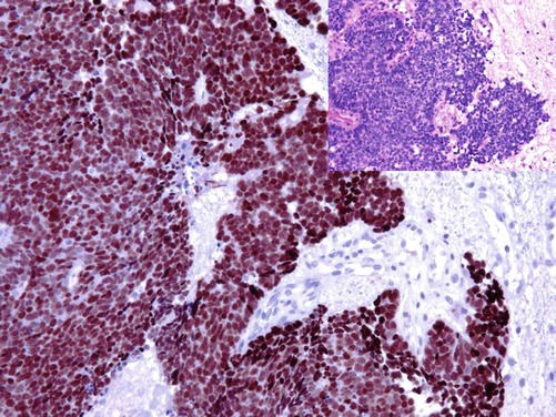
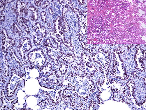

Fig. 3.1
Strong nuclear TTF-1 expression in pulmonary small cell carcinoma

Fig. 3.2
Strong nuclear TTF-1 expression in pulmonary adenocarcinoma
Diagnostic Pitfalls
Despite the known specificity of TTF-1 to lung and thyroid tumors, TTF-1 positivity is also reported in different extrapulmonary tumors such as small cell carcinomas of the urinary bladder and ovaries in addition to Merkel cell carcinoma. The aberrant expression of TTF-1 is also reported in about one half of extrahepatic cholangiocarcinomas including gallbladder adenocarcinoma, while nonneoplastic biliary epithelium lacks the TTF-1 expression [5]. TTF-1 positivity is also found in rare uterine and ovarian tumors such as mixed Müllerian tumor.
The TTF-1 expression in various types of CNS tumors especially those in the third ventricle region is also to take in consideration when searching for the primary of brain metastases [6]. Remarkable is the nuclear TTF-1 expression in tumors of the neurohypophysis including pituicytoma and granular cell tumor of the sellar region [7]. A further interesting observation is the strong cytoplasmic stain found in hepatocytes and hepatocellular carcinoma using the 8G7G3/1 clone, probably due to a cross-reaction with 150–160 KDa mitochondrial protein, which can be used as a diagnostic marker [8].
Napsin A | ||
Expression pattern: cytoplasmic | ||
Main diagnostic use | Expression in other tumors | Expression in normal cells |
Pulmonary adenocarcinoma | Papillary renal cell carcinoma, endometrial and ovarian clear cell carcinoma, subset of cholangiocarcinomas | Type 2 pneumocytes, respiratory epithelium of bronchioles, alveolar macrophages, Clara cells, proximal renal tubules, pancreatic acini and ducts, plasma cells and small subset of lymphocytes |
Positive control: lung tissue | ||
Diagnostic Approach
Napsin A is pepsin like aspartic proteinase, a member of the novel aspartic proteinase of the pepsin family taking part in the proteolytic processing of surfactant precursors. Napsin A is expressed in the majority of pulmonary adenocarcinomas and is used as a specific marker for pulmonary adenocarcinoma, whereas all other primary pulmonary carcinoma types lack the expression of napsin A (Fig. 3.3). Generally, the expression of napsin A correlates with the expression of TTF-1, and only a small percentage of pulmonary adenocarcinomas are napsin positive but TTF-1 negative. All mesothelioma types constantly lack the expression of napsin.
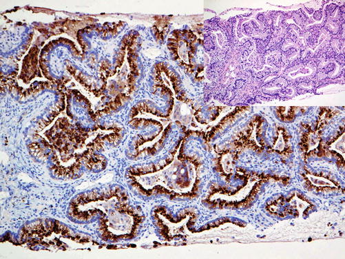

Fig. 3.3
Metastatic pulmonary adenocarcinoma with strong cytoplasmic napsin A expression
Diagnostic Pitfalls
The expression of napsin A may be found in other non-pulmonary tumors. Low expression level of napsin A is observed in papillary renal cell carcinoma and in a small subset of clear renal cell carcinoma. The expression of napsin A is also reported in about 90% of endometrial and ovarian clear cell carcinoma, in about one third of extrahepatic cholangiocarcinomas, and later may be also positive for TTF-1 [5, 9, 10]. Weak napsin expression is also reported in a small subset of colorectal and esophageal and pancreatic adenocarcinomas. As the morphology of the mentioned adenocarcinoma types may be similar to that of pulmonary adenocarcinoma especially in metastatic tumors, a complete diagnostic antibody panel must be used for accurate diagnosis.
Surfactant proteins | ||
Expression pattern: cytoplasmic/membranous | ||
Main diagnostic use | Expression in other tumors | Expression in normal cells |
Pulmonary adenocarcinoma | Type 2 pneumocytes, bronchiolar cells | |
Positive control: lung tissue | ||
Diagnostic Approach
Surfactant proteins including A, B, C, and D in addition to surfactant precursors are lipoproteins synthesized by type II pneumocytes and Clara bronchiolar epithelial cells. Antibodies to surfactant proteins are good markers for pulmonary adenocarcinoma. Pulmonary squamous cell carcinoma, large cell carcinoma, and non-pulmonary adenocarcinomas beside mesothelioma are usually negative for surfactants.
Stay updated, free articles. Join our Telegram channel

Full access? Get Clinical Tree


