, Hans Guski2 and Glen Kristiansen3
(1)
Carl-Thiem-Klinikum, Institut für Pathologie, Cottbus, Germany
(2)
Vivantes Klinikum Neukölln, Institut für Pathologie, Berlin, Germany
(3)
Universität Bonn, UKB, Institut für Pathologie, Bonn, Germany
Diagnostic Antibody Panel for Histiocytic and Dendritic Cell Tumors
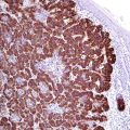
CD1a | ||
|---|---|---|
Expression pattern: membranous | ||
Main diagnostic use | Expression in other tumors | Expression in normal cells |
Langerhans cell histiocytosis | Myeloid leukemia, mycosis fungoides, cutaneous T-cell lymphomas, T-ALL | Cortical thymocytes, Langerhans cells, immature dendritic cells |
Positive control: skin | ||
Diagnostic Approach
CD1a is one of the four isoforms of CD1 (a, b, c, d) expressed on the antigen-presenting cells. CD1a is found on the surface of cortical thymocytes and dendritic cells in addition to Langerhans cells. CD1a is a specific marker for normal and neoplastic Langerhans cells but constantly negative in histiocytic, follicular dendritic, and interdigitating cell tumors. CD1a is also expressed in some types of T-cell lymphoma, chiefly cutaneous T-cell lymphoma.
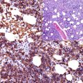
CD21 | ||
|---|---|---|
Expression pattern: membranous | ||
Main diagnostic use | Expression in other tumors | Expression in normal cells |
Follicular dendritic cell sarcoma | Hairy cell leukemia, mantle cell and marginal zone lymphoma | Follicular dendritic cells, mature B-cells, immature thymocytes, skin, pharyngeal and cervical epithelial cells, renal tubule, adrenal cortex, hepatocytes, capillary endothelial cells |
Positive control: lymph node | ||
Diagnostic Approach
CD21 is a C3d receptor on the membrane of the B lymphocytes that also acts as a receptor for EBV. CD21 is also expressed by follicular dendritic cells but constantly negative in monocytes, granulocytes, and T lymphocytes. CD21 is positive in a subset of B-cell lymphoma, namely, chronic lymphocytic lymphoma, and weakly in mantle cell lymphoma and follicular lymphoma. CD21 is rarely expressed in a small subset of T-cell lymphomas [3–6]. CD21, CD35, and podoplanin are very helpful markers for follicular dendritic cell tumors (sarcoma). CD21 is usually negative in histiocytic, Langerhans, and interdigitating cell tumors. The expression of CD21 in pharyngeal and cervical epithelial cells must be considered in the interpretation of the immunostain.
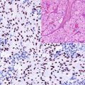
CD68 | ||
|---|---|---|
Expression pattern: cytoplasmic/membranous | ||
Main diagnostic use | Expression in other tumors | Expression in normal cells |
Histiocytic tumors, dendritic cell tumors, AML (FAB-M4/M5), giant cell tumors | Fibrous histiocytoma, nodular fasciitis, villonodular synovitis, granular cell tumor, inflammatory myofibroblastic tumor, mast cell disease, hairy cell leukemia | Macrophage, monocytes, osteoclasts, Kupffer cells, mast cells, synovial cells, microglia, dendritic cells, fibroblasts, Langerhans cells, myeloid cells, CD34+ progenitor cells, neutrophils, B and T cells |
Positive control: appendix | ||
Diagnostic Approach
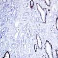
CD68 is a glycoprotein found in the lysosomes and endosomes involved in the regulation of phagocytic activity of macrophages. CD68 is a widely used marker for histiocytes and histiocytic tumors but lacks specificity for these cells [7].
Diagnostic Pitfalls
CD68 has a wide expression range and may be found in different hematologic diseases of B-cell, T-cell, NK, and myeloid lineage.
Stay updated, free articles. Join our Telegram channel

Full access? Get Clinical Tree


