, Hans Guski2 and Glen Kristiansen3
(1)
Carl-Thiem-Klinikum, Institut für Pathologie, Cottbus, Germany
(2)
Vivantes Klinikum Neukölln, Institut für Pathologie, Berlin, Germany
(3)
Universität Bonn, UKB, Institut für Pathologie, Bonn, Germany
8.1 Diagnostic Antibody Panel for Exocrine Pancreatic Tumors
8.2 Diagnostic Antibody Panel for Endocrine Pancreatic Tumors
Cytokeratin profile, chromogranin, synaptophysin, PDX-1, CD56, PAX-6, somatostatin, insulin, gastrin, glucagon, vasoactive intestinal polypeptide (VIP), human pancreatic polypeptide (hPP), and proliferation index (Ki-67) (see also Chap. 14, Endocrine and Neuroendocrine Tumors)
CA19-9 | ||
|---|---|---|
Expression pattern: membranous/cytoplasmic | ||
Main diagnostic use | Expression in other tumors | Expression in normal cells |
Pancreatic and gastrointestinal carcinoma | Ovarian and lung adenocarcinoma, renal cell carcinoma, transitional cell carcinoma, mucoepidermoid carcinoma | Epithelium of the breast ducts, salivary and sweat glands, lung, gastrointestinal tract, hepatobiliary system |
Positive control: Pancreatic tissue | ||
Diagnostic Approach
CA19-9 is a glycoprotein epitope on the sialyl Lewis a structure functioning as a ligand for the adhesion molecule E-selectin. CA19-9 is normally present on the apical surface of the ductal epithelium of the breast, salivary, and sweat glands beside the glands of gastrointestinal mucosa.
CA19-9 strongly stains pancreatic, hepatobiliary, and gastrointestinal adenocarcinomas but lacks the specificity for these carcinoma types. CA19-9 has a very wide expression spectrum as it is found in many other carcinomas of different origin. Consequently, the diagnosis of primary pancreatic carcinoma must be supported by a complete immunohistochemical panel.
PDX-1 | ||
|---|---|---|
Expression pattern: nuclear | ||
Main diagnostic use | Expression in other tumors | Expression in normal cells |
Pancreatobiliary adenocarcinoma, pancreatic and duodenal neuroendocrine tumors | Gastric and colorectal adenocarcinoma, pancreatic acinar cell carcinoma, prostatic carcinoma | Endocrine cells of the pancreas, pancreatic ductal epithelium and centroacinar cells, pyloroduodenal mucosa, Brunner’s glands, enteroendocrine cells |
Positive control: pancreatic tissue | ||
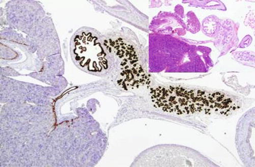
PDX-1 (pancreatic and duodenal homeobox 1) [2] also known as insulin promoter factor 1 is a transcription factor involved in the pancreatic development and maturation of the endocrine β-cells in addition to Brunner’s glands, duodenal papilla, and bile ducts. In adult tissue, PDX-1 is intensely expressed in endocrine cells of the upper gastrointestinal tract and pancreas in addition to pyloroduodenal and pancreatic duct mucosa (Fig. 8.1). PDX-1 strongly labels pancreatic endocrine tumors and pancreatobiliary adenocarcinomas including adenocarcinoma of the gallbladder and cholangiocarcinoma. Weak expression of PDX-1 is also found in a subset of colorectal adenocarcinomas. Focal weak PDX-1 expression may be also found in the prostatic glands, lung and breast epithelium, thyroid, liver, spleen, kidney, and skin.


Fig. 8.1
Section through a 12-week embryo showing PDX-1 highlighting pancreatic ducts, duodenal mucosa, and mucosa of the bile ducts
S100P | ||
|---|---|---|
Expression pattern: cytoplasmic/nuclear | ||
Main diagnostic use | Expression in other tumors | Expression in normal cells |
Pancreatic ductal adenocarcinoma, breast carcinoma | Non-small-cell lung carcinoma, gastrointestinal adenocarcinomas, transitional cell carcinoma, ovarian carcinoma, and melanoma | Myocardium and skeletal muscle, epithelial cells of gastrointestinal and prostatic glands, kidney, bladder, and leukocytes |
Positive control: pancreatic carcinoma | ||
Diagnostic Approach
S100P protein is one of the members of the S100 protein family consists of 95 amino acids primarily isolated from human placenta [4]. Besides the placenta, S100P is also expressed in many other types of normal tissue including the myocardium and skeletal muscle and epithelial cells of gastrointestinal tract and prostatic gland as well as the kidney, bladder, and leukocytes. S100P is also expressed in various tumor types such as non-small-cell lung carcinoma, breast carcinoma, pancreatic carcinoma including pancreatic ductal adenocarcinoma, pancreatic intraductal papillary mucinous neoplasm and preneoplastic cells, gastric and colorectal adenocarcinoma, transitional cell carcinoma, ovarian carcinoma, and melanoma [5–8]. Normal breast tissue and normal and inflamed pancreatic tissue lack the expression of S100P. This wide expression profile makes S100P a useful marker for the diagnosis of pancreatic and breast adenocarcinomas especially on small biopsies and FNP. S100P is negative in pancreatic endocrine tumors and acinar cell carcinoma. Prostatic carcinoma and renal cell carcinoma are usually negative for S100P. The expression of S100P is usually associated with a poor prognosis.


