, Hans Guski2 and Glen Kristiansen3
(1)
Carl-Thiem-Klinikum, Institut für Pathologie, Cottbus, Germany
(2)
Vivantes Klinikum Neukölln, Institut für Pathologie, Berlin, Germany
(3)
Universität Bonn, UKB, Institut für Pathologie, Bonn, Germany
Normal breast tissue consists of mesenchymal and epithelial components, which in their turn includes ductal and acinar (lobular) and myoepithelial components, each cell type having its characteristic immunoprofile. The immunoprofile of breast tumors depends on the origin of neoplastic cells.
10.1 Diagnostic Antibody Panel for Breast Carcinoma
Cytokeratin profile, estrogen and progesterone receptors, GATA-3, mammaglobin, GCFPD-15, E-cadherin, NY-BR-1, S100P, and HER-2.
10.2 Diagnostic Antibody Panel for Fibroepithelial Tumors
Cytokeratin profile, proliferation index (Ki-67).
10.3 Diagnostic Antibody Panel for Mesenchymal Tumors
See panels of other mesenchymal tumors.
Estrogen receptor | ||
|---|---|---|
Expression pattern: nuclear | ||
Main diagnostic use | Expression in other tumors | Expression in normal cells |
Breast and endometrial carcinoma | Ovarian serous, mucinous, and endometrioid carcinoma, transitional cell carcinoma, hepatocellular carcinoma, gastric adenocarcinoma, skin adnexal tumors, uterine leiomyoma and leiomyosarcoma | Breast, endometrium, myometrium, and endometrial stromal cells, fallopian tube mucosa, sweat glands, salivary glands, hepatocytes, pituitary gland |
Positive control: normal breast tissue | ||
Diagnostic Approach
Estrogen receptors (ER) are a member of the steroid family of ligand-dependent transcription factors and include two types encoded by two different genes on different chromosomes, the alpha type (ER-α) and beta type (ER-β); each type includes different splice variants. Both types have different distributions in different organs and different tissue types [1]. The ER-α type is mainly expressed in both epithelial and stromal cells of the breast, uterus, placenta, liver, CNS, endothelium, and bone, whereas the ER-β type is mainly expressed in the prostate, testes, ovary, spleen, thymus, skin, and endocrine glands including thyroid and parathyroid glands, adrenal glands, and the pancreas. Anyway, many tissue types show the expression of both receptor types.
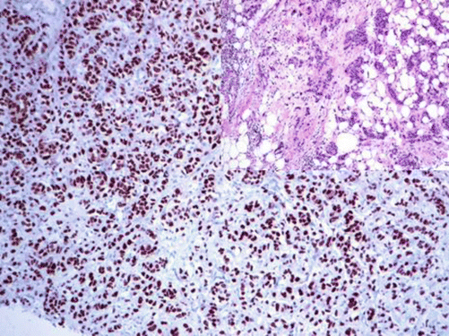
The expression of estrogen receptors (ER) is a good marker for the majority of breast carcinomas in addition to tumors of uterine and ovarian origin. Adequate and rapid tissue fixation with buffered neutral formalin is required for optimal stain results. For all steroid receptors, any stain pattern other than nuclear must be interpreted as negative. The expression of ER-α type is an important predictor for the response to the antihormone therapy (Fig. 10.1) [2]. Few scoring systems were suggested for semiquantitative estimation of estrogen and progesterone receptors. The modified scoring system suggested in 1987 by Remmele, the modified scoring system suggested in 1985 by McCarty, and the Allred scoring system proved to be the most practical and simplest systems. The three systems depend on the evaluation of the nuclear stain intensity and the percentage of positive tumor cells.


Fig. 10.1
Strong nuclear expression of estrogen receptors in breast carcinoma
Remmele Scoring System
This simple scoring system [2–4] has a 12-point scale (0–12). To calculate the score, one of the numbers 0, 1, 2, or 3 is given according to the intensity of the nuclear stain, and one of the numbers 0, 1, 2, 3, or 4 is given according to the percentage of positive tumor cells (see table). The score is calculated by multiplying the number reflecting the dominant stain intensity by the number reflecting the percentage of these positive tumor cells with a maximum score value of 12 (3 × 4). Tumors with a score of less than 3 show usually a poor response to the antiestrogen therapy.
Calculation of Remmele score
Percentage of positive cells | Intensity of the stain | ||
|---|---|---|---|
0 | No positive cells | 0 | No detectable stain |
1 | Positive cells less than 10% | 1 | Weak nuclear stain |
2 | Positive cells 10–50% | 2 | Moderate nuclear stain |
3 | Positive cells 51–80% | 3 | Strong nuclear stain |
4 | Positive cells more than 80% | ||
McCarty Scoring System
This scoring system [5] has a 300-point scale (0–300). The McCarty histoscore is the total value of each percentage of positive cells (0–100) multiplied by the number reflecting the intensity of the immunohistochemical stain (0, no detectable staining; 1, weak nuclear staining; 2, moderate nuclear staining; 3, strong nuclear staining) and calculated as the following:
Percentage of tumor cells with strong positivity X 3 = A
Percentage of tumor cells with moderate positivity X 2 = B
Percentage of tumor cells with weak positivity X 1 = C
The value of the histoscore = A + B + C.
The clinical significance of this histoscore is explained as the following:
50 or less: negative (−)
51–100: weakly positive (+)
101–200: moderately positive (++)
201–300: strongly positive (+++)
Allred Scoring System
The Allred scoring system has an 8-point scale (0–8). This scoring system is calculated by adding the number representing the proportion of positive cells 0, 1, 2, 3, 4, or 5 to the number reflecting the intensity of the nuclear stain 0, 1, 2, or 3 (see table below). Tumors with a score of less than 3 show usually a poor response to the antiestrogen therapy.
Calculation of Allred score
Percentage of positive cells | Intensity of the stain | ||
|---|---|---|---|
0 | No positive cells | 0 | No detectable stain |
1 | Positive cells less than 1% | 1 | Weak nuclear stain |
2 | Positive cells 1–10% | 2 | Moderate nuclear stain |
3 | Positive cells 10–33% | 3 | Strong nuclear stain |
4 | Positive cells 33–66% | ||
5 | Positive cells more than 66% | ||
Diagnostic Pitfall
The expression of ER depends on the histological type and differentiation grade of the breast tumor. Additionally, the expression of ER is not restricted to the abovementioned organs and tissue types but also can be found in other tumors such as hepatocellular carcinoma and transitional cell carcinoma. Additional markers such as GATA-3, mammaglobin, GCDFP15, and progesterone receptors as well as the cytokeratin profile are helpful to confirm the diagnosis of primary breast carcinoma.
Progesterone receptor | ||
|---|---|---|
Expression pattern: nuclear | ||
Main diagnostic use | Expression in other tumors | Expression in normal cells |
Breast carcinoma, endometrial carcinoma | Skin adnexal tumors, meningioma, solid pseudopapillary tumor of pancreas, stroma of mixed epithelial stromal tumor of the kidney, stromal tumors of the prostate | Breast and endometrial cells, endometrium stromal cells |
Positive control: normal breast tissue | ||
Diagnostic Approach
Progesterone is a steroid hormone involved in the differentiation of breast parenchyma and endometrium in addition to milk protein synthesis. Progesterone receptors (PgR) are good marker for breast carcinomas and have more specificity than estrogen receptors as they are expressed only in a limited number of tumors such as endometrial carcinoma. The progesterone receptor status is one of the important prognostic factors in breast, endometrial, and ovarian cancers [2]. A high expression level of both estrogen and progesterone hormone receptors is a positive prognostic factor for breast and endometrial cancers and predicts good response to antiestrogenic therapy.
Diagnostic Pitfalls
Similar to the estrogen receptors, the expression of PgR depends on the grade of tumor differentiation. High-grade carcinomas are often negative for steroid receptors.
GATA-3 | ||
|---|---|---|
Expression pattern: nuclear | ||
Main diagnostic use | Expression in other tumors | Expression in normal cells |
Breast carcinoma, transitional cell carcinoma of the urinary tract, tumors of skin adnexa, yolk sac tumor | Endometrioid carcinoma, trophoblastic tumors/choriocarcinoma, basal cell carcinoma, mesothelioma, pancreatic ductal adenocarcinoma, colorectal adenocarcinoma, different salivary gland tumors, chromophobe renal cell carcinoma, bladder small cell carcinoma, paraganglioma, neuroblastoma, pheochromocytoma, adrenal cortical carcinoma, squamous cell carcinoma of different locations, peripheral T-cell lymphoma | Adult breast, terminal ducts of parotid gland, urinary bladder and renal pelvis mucosa, prostatic basal cells and seminal vesicle epithelium, cortex and medulla of adrenal gland, ductal epithelium of skin adnexa and salivary glands, trophoblasts, T-lymphocytes |
Positive control: normal breast tissue | ||
Diagnostic Approach
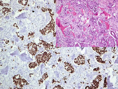
GATA-3, also known as endothelial transcription factor 3, is one of the six members of the GATA family of transcription factors taking part in the regulation of proliferation and differentiation of luminal epithelium of breast glands. GATA-3 is also involved in the differentiation of T-lymphocytes and skin adnexa. In diagnostic immunohistochemistry, GATA-3 is widely used as a marker for primary and metastatic breast carcinoma and transitional cell carcinoma (Fig. 10.2) [6, 7]. In breast carcinomas, the expression of GATA-3 strongly correlates with the expression of the estrogen receptors but lacks the therapeutic and prognostic value. The expression of GATA-3 is found in up to 90% of breast carcinoma while the lowest expression level is found in triple negative breast carcinomas as well as metaplastic and sarcomatoid breast carcinomas (<70%). Only one third of male breast carcinomas are positive for GATA-3 [8]. Generally, high expression levels of GATA-3 in breast cancer predict a good prognostic outcome. GATA-3 as a marker for urothelial tumors is discussed in a later section.


Fig. 10.2
Bone metastases of invasive ductal breast carcinoma. Tumor cells with strong nuclear GATA-3 expression
Diagnostic Pitfalls
Stay updated, free articles. Join our Telegram channel

Full access? Get Clinical Tree


