Management of Myeloproliferative Disorders and Chronic Myeloid Leukemia


Management of Myeloproliferative Disorders and Chronic Myeloid Leukemia


Ayalew Tefferi


The myeloproliferative neoplasms (MPN) are considered separate from the myelodysplastic syndromes (MDS) and are further classified into classic and nonclassic MPN (Table 46-1).1 MPN represent stem cell-derived clonal myeloproliferation, and the disease initiating mutation has been defined in some but not all cases. The current chapter will focus on the classic MPN: essential thrombocythemia (ET), polycythemic vera (PV), primary myelofibrosis (PMF), and chronic myelogenous leukemia (CML).


ESSENTIAL THROMBOCYTHEMIA

ET is currently a diagnosis of exclusion and represents an acquired nonreactive thrombocythemic state that is not accounted for by another myeloid malignancy. The World Health Organization (WHO) diagnostic criteria requires evaluation of a bone marrow (BM) biopsy to establish a morphologic diagnosis of ET.2 Approximately 60% of patients with ET carry the JAK2V617F mutation, which is a reliable clonal marker and thus can distinguish ET from reactive/secondary thrombocytosis.3 However, the particular mutation cannot distinguish ET from another MPN.


Clinical Aspects

The incidence of ET is estimated to be 2.5 per 100,000.5 Because life expectancy is near normal,6 the prevalence of the disease is severalfold higher. Several studies have reported a female preponderance in ET by approximately 2-fold compared with males.7 The median age at diagnosis is approximately 55 years, and as many as 20% of patients may be younger than 40 years. The disease is rare in children. One-fourth to one-third of the patients with ET may be totally asymptomatic at presentation; the remainder may report “vasomotor” symptoms or manifest thrombohemorrhagic complications. In addition, one-third to one-half may present with palpable splenomegaly.

Vasomotor symptoms occur in one-third of patients and include headache, lightheadedness, syncope, atypical chest pain, acral paresthesia, visual disturbances, livedo reticularis, and erythromelalgia. The last-named disorder refers to burning pain of the hands or feet associated with erythema and warmth. These events are usually controlled by treatment with low or standard doses of acetylsalicylic acid (ASA) (40 to 325 mg per day).8 Reported incidence rates of thrombosis and hemorrhage at diagnosis range from 9% to 22% and 3% to 37%, respectively.9 After diagnosis, these events occur in 7% to 17% and 8% to 14% of patients, respectively.

In the Mayo Clinic experience, transformation of the disease into PV, PMF, and acute myeloid leukemia (AML), in the first decade, was observed in 2.7%, 4%, and 1.4%, respectively.7 Reported incidence figures of leukemic conversion range from 0.6% to 5%.10 Leukemic conversion in ET has also occurred in the absence of previous therapy,11 suggesting that the event may be a natural sequela of the disease that depends more on disease biology and duration than on specific therapy.


Prognosis

In a previously reported case-control study, the overall risk of thrombotic episodes in patients with ET was 6.6% per patient-year compared with 1.2% per patient-year in the control group.12 A history of thrombosis (31.4% per patient-year) and age >60 years (15.1% per patient-year) were significantly associated with a high risk of thrombosis. The experience of many other investigators supports the above observations (Table 46-3).10 The lack of a significant correlation between platelet count and the risk of thrombosis has been consistently observed.13 Bleeding manifestations at initial presentation of ET have been significantly associated with extreme thrombocytosis (platelet counts >1 to 2 million per µl) and with the use
of antiplatelet therapy.14 On the other hand, the risk of major bleeding episodes during the clinical course of the disease is very low (<5%).13


Table 46-1 WHO Classification of Chronic Myeloid Malignancies


1.


MPN


Classic MPN


  1. CML, BCR-ABL1 positive


  2. PV


  3. PMF


  4. ET


Nonclassic MPN


  1. Chronic neutrophilic leukemia


  2. Chronic eosinophilic leukemia not otherwise specified


  3. Mastocytosis


  4. MPN, unclassifiable


2.


MDS


Refractory cytopenia with unilineage dysplasia


— Refractory anemia


— Refractory neutropenia


— Refractory thrombocytopenia


Refractory anemia with ring sideroblasts


Refractory cytopenia with multilineage dysplasia


Refractory anemia with excess blasts


MDS with isolated del(5q)


MDS, unclassifiable


Childhood MDS


Refractory cytopenia during childhood


3.


MDS/MPN


Chronic myelomonocytic leukemia


Atypical chronic myeloid leukemia, BCR-ABL1 negative


Juvenile myelomonocytic leukemia


MDS/MPN, unclassifiable


Refractory anemia with ring sideroblasts and thrombocytosis


4.


Myeloid and Lymphoid Neoplasms Associated with Eosinophilia and Genetic Abnormalities


Myeloid and lymphoid neoplasms associated with PDGFRA rearrangement


Myeloid neoplasms associated with PDGFRB rearrangement


Myeloid and lymphoid neoplasms associated with FGFR1 abnormalities


POLYCYTHEMIA VERA

PV is a clonal stem cell disease characterized by an increased red cell mass.23 Erythrocytosis in PV is independent of the erythroid growth factor, erythropoietin (Epo). In 2005, a novel gain-of-function JAK2 mutation (JAK2V617F) was identified in almost all patients with PV, and the precise pathogenetic role of the specific mutation is currently under investigation.24
The incidence of PV is estimated to be 2.3 per 100,000 per year, and the median age at diagnosis is approximately 60 years.25 The disease affects both men and women equally but is rare in children.


Table 46-2 Causes of Thrombocytosis
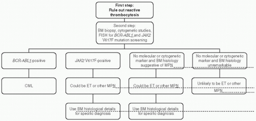


Primary Thrombocytosis


Reactive Thrombocytosis


▪ ET


▪ PV


▪ Myelofibrosis with myeloid metaplasia (overt)


▪ Myelofibrosis with myeloid metaplasia (cellular phase)


▪ Chronic myeloid leukemia


▪ MDS


▪ Acute leukemia


▪ Infection


▪ Tissue damage


▪ Chronic inflammation


▪ Malignancy


▪ Rebound thrombocytosis


▪ Renal disorders


▪ Hemolytic anemia


▪ Post-splenectomy


▪ Blood loss


From Tefferi A, Barbui T. bcr/abl-negative, classic myeloproliferative disorders: diagnosis and treatment. Mayo Clin Proc. 2005;80:1220-1232.


Clinical Aspects

Approximately 20% of patients with PV present with thrombotic events, including strokes, transient ischemic attacks, retinal artery or venous occlusions, coronary artery ischemia, pulmonary embolism, hepatic or portal vein thrombosis, deep vein thrombosis, and digital ischemia.9 The incidence of recurrent thrombosis depends on age, history of thrombosis, and the particular form of therapy. Bleeding complications are less frequent and less serious and have been significantly associated with the use of ASA.


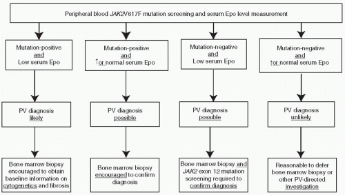
Figure 46.2 A diagnostic algorithm for PV.

Leukemic conversions occur in <5% of patients with PV that is treated with phlebotomy alone. Treatment with certain agents, especially alkylating agents, increases the risk of acute leukemia. In contrast, specific therapy may or may not modify the risk of fibrotic transformation, which occurs in 10% to 30% of patients with PV who are followed for 10 to 25 years. Aquagenic pruritus (postbath) is a characteristic manifestation of PV that is difficult to treat.


Prognosis

Age and history of previous thrombosis are the most powerful predictors of recurrent thrombosis in PV.29 Neither the degree of thrombocytosis nor the presence of platelet function abnormalities has been correlated with thrombotic risk. Patients with PV, similar to those with ET, can be stratified into defined risk groups that are managed differently (Table 46-3).

Jun 19, 2016 | Posted by in ONCOLOGY | Comments Off on Management of Myeloproliferative Disorders and Chronic Myeloid Leukemia

Full access? Get Clinical Tree

Get Clinical Tree app for offline access