Malignant Non-Hodgkin Lymphomas in Children


Malignant Non-Hodgkin Lymphomas in Children


Carl E. Allen

Kala Y. Kamdar

Catherine M. Bollard

Thomas G. Gross


INTRODUCTION

Non-Hodgkin lymphoma (NHL) of childhood is a diverse collection of different lymphomas that includes all of the malignant lymphomas that are not classified as Hodgkin lymphoma (HL). Advances in histopathology, immunology, cytogenetics, and molecular biology have resulted in enormous progress in our understanding of the biology of the NHL, leading to more rational classification of these diseases. The study of malignant lymphomas has, in turn, contributed substantially to our comprehension of normal lymphocyte development.

Clinically, it has been recognized for many years that childhood NHL is a much more systemic disease than HL, with hematologic dissemination that is more akin to what is observed in leukemia. Studies to detect minimal disease have now demonstrated that NHL in children involves cells that usually traffic throughout the body and tend to be systemic diseases from the outset, even in cases in which disease appears clinically limited.1 This systemic nature of childhood NHL has led to a different clinical staging system for childhood NHL and treatment strategies that differ from HL and NHL observed in adult patients. As recently as the 1970s, fewer than 20% of children with NHL were projected to survive their disease, and the majority of affected children died within 2 years of diagnosis. Virtually all of the survivors were patients who presented with localized disease. Progress in therapy for childhood NHL is one of the stunning success stories of the past two decades. In developed countries, over 80% of children with NHL can now be cured with modern therapy, even patients with widely disseminated disease. It is notable, however, that these extraordinary advances in treatment have not come from the development of new more effective agents; as all of the drugs featured in modern treatment protocols were available in the early 1970s. Rather, more rational classification systems, improvements in clinical risk stratifications, advances in supportive care to reduce the life-threatening complications of NHL and of therapy, and more rational application of chemotherapy with the empirical development of intensive regimens for children presenting with advanced-stage disease have all contributed to improvements in the outcome for children with NHL. Patients with refractory and recurrent NHL have historically dismal outcomes despite the most aggressive therapies, including stem cell transplant, although some pilot studies with novel targeted agents and immunotherapy indicate potential for improvement. The future requires that new approaches and novel agents be developed to not only improve outcomes for patients with NHL but also reduce acute and long-term consequences of treatment.


OVERALL INCIDENCE/EPIDEMIOLOGY

The incidence of lymphoma in children varies by age and varies considerably in different world regions.1,2 In the United States and in developed countries, malignant lymphoma (including NHL and HL) is the third most common group of malignancies in children after leukemias and brain tumors and account for 15% of all childhood malignancies in children younger than 20 years. NHL is more common than HL in children younger than 10 years, but the relative incidence of HL increases rapidly in children older than 10 years, making the incidence of HL almost twice that of NHL in children between the ages of 15 and 19. Approximately 750 to 800 new cases of childhood NHL are diagnosed in the United States each year. There is a marked male predominance in all age groups, but particularly in children younger than 15 years, in whom three-fourths of the cases occur in males. The incidence of NHL varies considerably by age; NHL is uncommon in children younger than 5 years, accounting for only 3% of cancers; but the incidence of NHL increases steadily throughout life, accounting for 8% to 9% of cancers in children older than 10 years.2

The etiology of NHL is largely unknown. Epidemiologic studies evaluating prenatal and postnatal exposures have not been fruitful for the most part, and exposures studied to date have not been associated with increased risk of lymphoma. The use of pesticides in the home has been linked to the risk of NHL, although no specific agent has been identified.3 Exposure to drugs and radiation have not been demonstrated to be major risk factors for NHL (except for immunosuppressive drugs—see later and Chapter 24). Therapy-related secondary NHL in children is rare and is primarily lymphoblastic lymphoma (LBL) or diffuse large B-cell lymphoma (DLBCL).4 Phenytoin has been associated with “pseudolymphoma,” which usually regresses when the drug is discontinued. Immunodeficiency, whether inherited or acquired, is clearly related to the development of NHL, increasing the incidence of NHL more than 100-fold compared to age-matched controls.5 The association between Epstein-Barr virus (EBV) and specific NHLs is discussed later.


Initial Evaluation

Unlike adult NHL, which commonly presents as indolent low or intermediate grade, most NHLs in children present as aggressive disseminated disease. Children with NHL benefit from prompt referral to a specialized cancer center for evaluation, staging, and therapy. Potential clinical emergencies in patients with NHL prior to diagnosis include complications from rapidly growing masses: superior/inferior vena cava obstruction, acute airway obstruction, spinal cord compression, pericardial tamponade, intussusception/intestinal obstruction, and central nervous system (CNS) complications. NHL cells frequently have a very high mitotic index, with potential for tumors to grow rapidly and die rapidly, which can result in hyperuricemia and tumor lysis syndrome (TLS). Medical history exploring the infectious history can identify patients at risk for NHL associated with inherited or acquired immune deficiency. Initial studies typically include laboratory studies from peripheral blood to evaluate blood counts, renal function and electrolytes, and lactate dehydrogenase (LDH). If clinically feasible, excisional biopsy is optimal to ensure quality and quantity of tissue for analysis by histology, flow cytometry, cytogenetics, and molecular studies. Once diagnosis is established, radiographic staging of chest, abdomen, and pelvis with a computed tomography (CT) scan is the historical standard, although baseline positron emission tomography (PET)/CT scan may add sensitivity.6 Bilateral bone marrow aspiration and biopsy and cerebrospinal cytology are also needed for staging.


Staging

The goal of staging studies should be to assess rapidly the extent of disease to determine prognosis and to assign appropriate therapy. Development of a staging system with prognostic utility for children with NHL has been challenging because of the unique biology and differing patterns of spread and response to therapy of the four major subtypes of NHL seen in children and adolescents.7 The Ann Arbor staging classification8 does not adequately reflect prognosis in childhood NHL. The progression of disease in childhood NHL does not follow an orderly and predictable pattern of lymphatic spread, as in HL. Extensive extranodal disease is more common in children with NHL than in adults with NHL. Therefore, the clinical staging system proposed at the St. Jude Children’s Research Hospital (Table 23.1) has been widely accepted.7 It relies on noninvasive procedures that can be carried out expeditiously. Similar to the Ann Arbor staging system, the St. Jude staging system takes into account the primary site as well as disease extent is considered in assigning clinical stage. The primary differences between St. Jude staging system and the Ann Arbor staging system are as follows. In the St. Jude stage I disease, localized thoracic and abdominal disease are excluded; other localized extranodal disease, however, is considered stage I. In St. Jude stage II disease, localized (resected) abdominal disease is included, and again any thoracic disease is excluded. St. Jude stage III disease includes any thoracic disease, paraspinal disease, or facial nerve palsy in addition to disease on both sides of the diaphragm. Disease involvement in St. Jude system considered stage IV includes marrow or CNS disease, other than paraspinal or facial nerve palsy. On the basis of the St. Jude staging system, almost 40% of children with NHL present with stage I and II and the remainder with more advanced-stage III and IV disease. The distinction between lymphoma and leukemia is arbitrary and is based simply on the percentage of a bone marrow aspirate that is infiltrated by malignant cells. If the bone marrow has more than 25% blasts or malignant cells, the patient is considered to have acute leukemia rather than NHL. Children with between 5% and 25% marrow involvement are considered to have stage IV NHL, whereas those with fewer than 5% blasts in the marrow are considered to not have tumor involved. As opposed to many leukemia criteria, any identifiable tumor cell in the cerebrospinal fluid (CSF) constitutes CNS disease.


TABLE 23.1 St. Jude Staging System for Non-Hodgkin Lymphoma


Stage I


A single tumor (extranodal) or single anatomic area (nodal) with the exclusion of thoracic or abdomen


Stage II


A single tumor (extranodal) with regional node involvement


Two or more nodal areas on the same side of the diaphragm


Two single (extranodal) tumors with or without regional node involvement on the same side of the diaphragm


A primary gastrointestinal tract tumor that is resectable, usually in the ileocecal area, with or without involvement of associated mesenteric nodes


Stage III


Two single tumors (extranodal) on opposite sides of the diaphragm


Two or more nodal areas above and below the diaphragm


All the primary intrathoracic tumors (mediastinal, pleural, and thymic)


All extensive primary intraabdominal diseases


All paraspinal or epidural tumors, regardless of other tumor site(s)


Stage IV


Any of the aforementioned with initial CNS and/or bone marrow involvement


>25% blasts in the marrow is considered leukemic disease. Any identifiable tumor cell in the CSF constitutes CNS disease.


Modified from Murphy SB. Classification, staging and end results of treatment of childhood non-Hodgkin’s lymphomas: dissimilarities from lymphomas in adults. Semin Oncol 1980;7:332-339.


The French Society of Pediatric Oncology (SFOP) and Berlin-Frankfurt-Munster (BFM) groups have adapted the St. Jude staging system for treatment assignment for children with B-cell lymphomas (Table 23.2). The French risk stratification identifies the very best risk patients (Group A) with completely resected localized disease and worst risk patients (Group C) with CNS disease and/or bone marrow involvement. The rest of the patients are included in a very heterogeneous Group B, consisting of patients with all clinical stages I-IV. The BFM risk stratification subdivides patients into four groups based on clinical stage, as well as LDH at diagnosis, a surrogate marker for total disease burden. R1 and R4 are similar to the French Group A and C, while the majority of patients in the French Group B are divided into R2 and R3, depending on LDH level, in the BFM stratifications. For LBL patients, studies have shown that all patients, regardless of stage, have better outcome when treated in a manner similar to that of acute lymphoblastic leukemia (ALL).9,10 For anaplastic large cell lymphoma (ALCL), the European Intergroup for Childhood NHL (EICNHL) has stratified patients into low-risk versus high-risk based on clinical factors.11 Patients with one of the following risk factors are considered high-risk: visceral (lung, liver, or spleen), skin, or mediastinal involvement.12 However, a Children’s Oncology Group (COG) study could confirm only bone marrow involvement as poor prognostic clinical factor.13

With more sensitive techniques, such as flow cytometry or polymerase chain reaction (PCR), several groups have now demonstrated that systemic disease in marrow and blood is much more
frequent than would be predicted by clinical staging systems. Early reports suggest that minimal disseminated disease or minimal residual disease detection in all the major subtypes of pediatric NHL may have prognostic value.14,15,16,17,18


TABLE 23.2 NHL Risk Stratification Schema


B-CELL NHL (FAB/LMB)


Stratum


Disease Manifestations


A


Completely resected stage I and abdominal stage II


B


Multiple extra-abdominal sites. Nonresected stage I and II, III, IV (marrow <25% blasts, no CNS disease)


C


Mature B-ALL (>25% blasts in marrow) and/or CNS disease


B-CELL NHL (BFM)


Stratum


Disease Manifestations


R1


Completely resected stage I and abdominal stage II


R2


Nonresected stage I/II and stage III with LDH < 500


R3


Stage III with LDH 500-999


Stage IV, B-ALL (>25% blasts), no CNS disease and LDH <1000


R4


Stage III, IV, B-ALL, and LDH ≥1000


Any CNS disease


Therapeutic strategies for NHL are based on stage- and histology-directed multiagent chemotherapy, and 5-year survival for children and adolescents is now over 80%. Prognostic factors include age (older children with generally worse outcomes), sites of disease (high stage with worse outcomes than low stage; mediastinal involvement also with inferior outcomes), tumor burden (elevated LDH with inferior outcomes), and response to initial therapy.


NHL Therapy Overview

Prior to 1975, the prognosis for children with NHL was dismal. Most of the children who survived had surgical removal of early-stage disease with or without radiation therapy (RT). The addition of single cytotoxic agents only slightly improved the results with the exception of African children with Burkitt lymphoma (BL). In the original studies of African BL, single-agent chemotherapy was given because radiation was not readily available. Surprisingly, treatment with one or more doses of cyclophosphamide resulted in some cures.19 Even today about 50% of African BL can be cured with a 28-day course of low-dose cyclophosphamide and prednisone and four intrathecal injections costing less than US$50.20

The morphologic similarity between NHL and ALL and the invariable progression of LBL to a leukemic phase led investigators at St. Jude in the early 1970s to treat NHL with chemotherapy regimens known to be effective in ALL. This novel approach proved to be successful, especially for children with early-stage NHL. However, children with NHL involving the mediastinum had only transient remissions. More intensive ALL regimens such as the Memorial Sloan-Kettering LSA2L2 protocol and the adriamycin (doxorubicin), prednisone, and vincristine (APO) regimen developed at the Dana-Farber Cancer Institute proved to be more successful in children with advanced-stage NHL. During this same time period, Ziegler reported long-term disease control in both American and African patients with BL using a four-drug regimen of cyclophosphamide, methotrexate, vincristine, and prednisone, and Djerassi and Kim demonstrated remissions in children with NHL after moderate-dose methotrexate infusions with citrovorum rescue. The results paved the way for prospective randomized clinical trials comparing leukemia versus lymphoma therapy for NHL in children. These studies provided principles that still hold true today. Leukemia regimens proved to be superior to the lymphoma regimens for LBL and the converse was noted for BL. However, results were roughly equivalent in children with advanced-stage large cell lymphoma.

During the next decade progress continued because of refinements in chemotherapy and advances in supportive care. The 5-year event-free survival (EFS) rates improved to 85% to 95% for children with early-stage NHL and to 70% to 90% for advanced-stage disease. CNS-directed therapy was recognized as an important component of these successful regimens, especially for children with lymphoblastic and Burkitt lymphomas. However, the addition of RT to chemotherapy for both early- and advanced-stage NHL was not shown to improve survival.9,21,22 Since long-term follow-up studies have shown that the most significant factor for late death following treatment of pediatric NHL is RT, it is currently recommended only for select patients, i.e., treating CNS disease in LBL patients and life-threatening emergencies at diagnosis such as airway compression from a mediastinal mass.23 The role of surgery in the treatment of children with NHL is mostly limited to diagnostic biopsies, placement of central venous access devices, and treatment of complications of therapy. However, in children presenting with localized disease, e.g., ileocecal intussusception secondary to BL, complete resection of the involved segment of intestine and associated mesentery is recommended. Otherwise, there is no role for performing major tumor resection or debulking procedures in children with NHL.

Another major advancement is the decrease in early death due to TLS. Particular attention to kidney function and to serum levels of uric acid, potassium, calcium, and phosphorus is critical in these children. These patients, particularly those with BL and LBL, are at high risk for TLS and uric acid nephropathy. Measures should be instituted emergently to reduce the likelihood of uric acid nephropathy, including vigorous intravenous hydration (at least twice maintenance), alkalinization of urine with sodium bicarbonate when allopurinol is administered, and careful monitoring of serum electrolytes. Rasburicase, a recombinant urate oxidase, has been shown in recent clinical trials to rapidly lower serum uric acid levels and prevent the metabolic problems associated with tumor lysis, including hyperphosphatemia and renal failure.24,25 It is not necessary to alkalinize the urine when using rasburicase. Rasburicase should be used in place of allopurinol for children with NHL who have hyperuricemia or who are at high risk for TLS. For the most part, this includes patients with stage III and IV BL and mature B-cell leukemia (primarily Burkitt leukemia), and select patients with bulky LBL and DLBCL. The use of rasburicase has markedly reduced the requirement for dialysis in this population. Additionally, a cytoreduction prophase has been added to many regimens, which also helps achieve tumor control without increasing the risk of clinical deterioration during initiation of therapy, although this does not obviate the need for hydration and uric acid nephropathy prophylaxis.21,26,27,28


NHL Cellular Classifications: Specific Biological and Clinical Considerations

The groups of lymphomas included under the umbrella of “NHL” are linked by the common feature of not being HL, thus representing a wide range of biological features and clinical presentations. This chapter will focus on the most common forms of NHL in children and adolescents—lymphoblastic lymphoma (precursor T-cell or, less frequently, precursor B-cell), mature B-cell lymphoma (Burkitt, Burkitt-like, DLBCL), anaplastic large cell lymphoma (T-cell or null-cell lymphomas)—and will briefly review mature T-cell lymphomas29,30 (Table 23.3).


PRECURSOR LYMPHOMAS

Precursor or lymphoblastic lymphomas are thought to originate from thymic T cells (T-LBL) or precursor B cells in the bone marrow (B-LBL).31 They share many biologic and therapeutic features with ALL, and they are often considered different clinical presentations of the same disease.32,33 The distinction between LBL involving the bone marrow and ALL is rather arbitrary, with leukemia designated if at least 25% lymphoblasts are present in the bone marrow at diagnosis (regardless of other lymphomatous masses in the body). Accordingly, the current World Health Organization (WHO) classification has developed the diagnostic category of lymphoblastic leukemia/lymphoma.34 Morphologically, the cancer cells are indistinguishable and the immunophenotyping profiles overlap considerably.32,35 However, there are some notable differences, suggesting that there may be some underlying biologic differences between LBL and ALL. Precursor T-lymphoblastic subtype constitutes 75% of LBL cases, with the remainder being precursor B-LBL. In contrast, the precursor B subtype is much more common than the precursor T subtype in ALL.36,37 Additionally, molecular profiling demonstrates some differences between T-LBL and T-ALL, and cytogenetic changes seen in T-cell ALL are not commonly detected in T-LBL, although technical considerations may factor in this difference.31,38,39


TABLE 23.3 Molecular and Clinical Features of NHL


Classification


Characteristic Immunophenotype


Characteristic Clinical Features


Chromosome Translocations


Genes Commonly Involved


NHL Subtype Frequency (%)


Precursor B-lymphoblastic


B-cell precursors


Skin, lymph nodes, bone


3


Precursor T-lymphoblastic


Immature T cell


Anterior mediastinal, lymph nodes


TCRαδ-TAL1


TCRαδ-RHOMB2


TCRαδ-RHOMB1


TCRαδ-HOX11


TCRβ-LYL1


TCRαδ-MYC


TCRβ-LCK


15-20


Burkitt and Burkitt-like


Mature B cells (sIg+)


Abdominal, intestinal with intussusception


t(8;14)


t(2:8)


t(8;22)


IgH-cMYC


Igκ-cMYC


Igλ-cMYC


35-40


Diffuse large B cell


B cell (germinal center phenotype)


Lymph nodes, abdominal, bone


15-20


Mediastinal large B cell


B cell (medullary thymus phenotype)


Mediastinal


1-2


Pediatric Follicular


B cell (follicular center)


Lymph nodes


IgH-IRF4


TNFSFR14


Rare


Nodal marginal zone


B cell (marginal zone)


Lymph nodes


Rare


Anaplastic large cell


T cell, NK cell (CD30+) or undifferentiated


Skin lesion, lymph nodes, bone lesions


t(2:5)


t(1;2)


t(2;3)


t(2;17)


t(X;2)


inv 2


NPM-ALK


TPM3-ALK


TFG-ALK


CLTC-ALK


MSN-ALK


ATIC-ALK


15-20


Mature T cell


T cell (TdT+)


Skin, CNS


Rare


Epidemiology

LBL constitutes approximately 20% of childhood NHL40 (Table 23.3). There is a 2:1 male predominance for LBL overall (more pronounced for the T-lymphoblastic subtype), but the incidence of LBL remains constant across the pediatric age group for both boys and girls.36


Biology and Pathology

LBL morphology is similar to that of ALL, with lymphoblasts of small or medium size with scant cytoplasm, round or convoluted nuclei, fine chromatin, and indistinct nucleoli (Fig. 23.1A). TdT is usually expressed, and surface immunoglobulin is usually absent.41 Flow cytometry is essential to distinguish T-LBL and B-LBL.31 T-LBL will show cytoplasmic CD3 positivity and usually CD7 positivity. T-LBL may demonstrate a more mature immunophenotype than that seen in T-ALL. B-LBL demonstrates B-lineage markers such as CD19, CD79a, and CD22. There are no known cytogenetic prognostic factors for LBL as there are for ALL, and recurrent cytogenetic anomalies have not been well described for LBL as they have been for ALL. The most frequent chromosomal aberration reported for T-LBL and T-ALL involves the T-cell receptor (TCR) gene loci at chromosome 14q11 or 7q34. Common partner genes seen in T-ALL and T-LBL include TAL1,MYC,HOXA gene cluster, and MYB.42,43 Other chromosomal abnormalities described for T-cell LBL and T-cell ALL include NOTCH1 mutations, alterations of chromosome 9p containing CDKN2A/CDKN2B loci, and deletions in chromosome 6p.44 BCR-ABL positivity has not been reported but should probably be screened in light of the poor prognosis associated with this in ALL. Gene expression profiling has shown upregulation of genes encoding adhesion molecules and extracellular matrix proteins and does show differences between T-ALL and T-LBL.39


Clinical Presentation and Evaluation

LBL typically presents with rapidly enlarging neck and mediastinal lymphadenopathy. Other possible sites of involvement include bone marrow, CNS (4% to 5%), abdominal organs, other lymph nodes, bone, skin, and, occasionally, testes.45,46 Whereas mediastinal masses are more common in T-LBL than in B-LBL, bone and skin involvement are seen more often in B-LBL than in T-LBL.47 Mediastinal masses may cause significant airway or vascular compression; so caution is warranted when lying the patient supine or giving sedation for imaging or procedures. On occasion, pretreatment with steroids or, less commonly, local radiation may be needed to stabilize the patient and complete the diagnostic workup. Since pretreatment may affect diagnosis and staging, the workup should be completed as soon as safely possible after pretreatment is initiated. Computed tomography (neck, chest, abdomen, and pelvis) is used for staging and to help determine the optimal site for biopsy in some situations. Tissue biopsy should be obtained for histologic diagnosis in the least invasive manner possible, and it is important for immunohistochemistry and flow cytometry to be used in combination to determine the lymphoma subtype. If LBL is diagnosed, bilateral bone marrow evaluation and cerebrospinal fluid evaluation should be done to complete staging workup. Prognosis is affected by bone marrow or CNS involvement. Positron emission tomography may be useful for staging and response evaluation as well, although its utility for this entity is still being evaluated.


Future Directions

Further attempts to refine risk stratification strategies for LBL may be useful. The presence of minimal disseminated disease in the bone marrow at diagnosis and the response to induction therapy are being evaluated as prognostic factors. With regard to therapeutic agents, nelarabine is a promising novel nucleoside analogue with specificity for T lymphoblasts that appears to be effective in relapsed disease. It is currently being evaluated for efficacy for frontline disease in cooperative group trials. Immunotherapy with T-cell-specific antibodies and proteasome inhibitors also merit further evaluation in future trials. Other agents being studied for ALL, such as mTOR inhibitors, may also have efficacy in LBL, and recent trials are including both LBL and leukemia together because of the significant clinical and biologic overlap between these two entities.


MATURE B-CELL LYMPHOMAS

Mature B-cell NHLs in children are most typically BL (including atypical Burkitt lymphoma [aBL]) and DLBCL. BL is relatively common in children (about 40% of all pediatric NHL) but much more rarely seen in adults, particularly in immunocompetent adults.2,29,30 Although rare in children, the new WHO classification now recognizes two low-grade B-cell neoplasms with features that are sufficiently different from that which is typical of adult disease to designate them as separate entities, i.e., pediatric follicular lymphoma (FL) and pediatric marginal zone lymphoma.

One feature of pediatric mature B-cell NHL is the relatively high proportion of germinal center cell-derived neoplasms compared to adults, in whom there is more of a split between germinal center and post-germinal center neoplasms.56,57 This may, in part, be due to the developing immune system and primary responses to antigens in the pediatric age group. Pediatric mature B-cell NHL tends to have a high proliferation rate and overexpression of pro-proliferative proteins (such as cMYC), suggesting that the mechanism of lymphomagenesis is due to abnormal proliferation rather than defective apoptosis as is seen in many adult B-cell lymphomas.

Several of the mature B-cell NHLs in the pediatric age group are associated with specific cytogenetic and molecular characteristics (Table 23.3). Translocations of the c-myc oncogene have been used to define BL, but it should be noted that abnormalities in the c-myc locus are also seen in many cases of DLBCL. Conversely, the t(14;18) translocation (IgH-BCL2) seen in many adult DLBCL is not as frequently seen in pediatric DLBCL and is usually not seen in the rare cases of pediatric FL.58,59

Molecular approaches will usually identify B-cell immunoglobulin gene rearrangements in pediatric B-NHL.60 In some cases, translocations result in a fusion protein, resulting in the expression of a protein that possesses oncogenic capabilities. Many of these translocations and fusion proteins are unique to lymphoma, allowing fluorescence in situ hybridization (FISH) or reverse transcriptase polymerase chain reaction (RT-PCR) to be used for detection. Overexpression of the gene may lead to increased protein expression, detectable by immunohistochemical methods, as well.60,61


BURKITT LYMPHOMA

The history of BL, the “Rosetta stone of cancer,” is central to many seminal discoveries in oncology. It was the first human tumor to be associated with a virus, one of the first to be associated with recurrent chromosomal translocations, and the first reported to be associated with HIV infection.62 It is classified as endemic, sporadic (the type found in nonmalarial areas), and immunodefiency-related.29,30


Epidemiology

For reasons that remain incompletely defined, endemic BL is observed along the equatorial “malaria-belt.”62,63 In high-risk areas, BL accounts for approximately half of pediatric cancer diagnoses, with incidence estimated to be 50-fold higher than sporadic BL.64 The immunodeficiency-associated variant is 1,000 times more frequent in HIV-infected than in uninfected children.65 Across all types, boys are more frequently affected than girls.


Biology and Pathology

BL arises from transformation of germinal center B cells that leads to uncontrolled proliferation. Characteristic translocations including the proto-oncogene c-myc and immunoglobulin gene regulatory elements implicate V(D)J or somatic recombination events in pathogenesis of BL (Table 23.3). Cofactors that likely contribute to the pathologic recombination events include malaria, EBV infection, and HIV infection.


EBV and BL

The relationship between EBV and the pathogenesis of NHL began with the elegant investigations into the epidemiology
of a lymphoma observed in children of equatorial Africa conducted by Denis Burkitt in the late 1950s. Dr. Burkitt demonstrated that the disease was associated with climate, occurring most frequently in areas of equatorial Africa where the rainfall exceeded 20 inches annually and where the mean temperature exceeded 60°F during the coolest months—in short, the regions where malaria is endemic.66 This observation led to a collaboration with a young virologist named Anthony Epstein, who was searching for human oncogenic viruses, and resulted in the discovery of a herpesvirus from cultures of endemic BL tissue, the first human tumor-associated virus.67 Subsequent serologic studies demonstrated that EBV is the causative agent for infectious mononucleosis.68

Since that time, it has been demonstrated that EBV can very efficiently transform human B cells into immortal cell lines in vitro and is associated with numerous human malignancies, especially lymphoproliferative disease of the immunocompromised host. EBV is one of eight known human herpesviruses. Once infected, individuals carry EBV latently in resting B cells for life, although oral epithelium continues to support viral replication and remains the source of virus, which can be transmitted to others through saliva. Only nine viral-encoded proteins (EBNA-1, EBNA-2, EBNA-3A, EBNA-3B, EBNA-3C, EBNA leader protein [LP], LMP [latent membrane protein]-1, LMP-2A, and LMP-2B) plus two nontranslated EBV-encoded RNAs (EBER-1 and EBER-2) are expressed during viral latency. EBNA-2, 3A, 3B, 3C and the LMPs have been shown to promote B-cell proliferation and/or survival, but it is felt that EBNA-2 and LMP-1 expression are essential for B-cell immortalization.69,70

Differential expression of latent EBV genes is observed in B cells of various stages of differentiation and in different malignancies associated with the virus. The site of EBV latency in seropositive healthy individuals is the resting memory B cells, where no viral proteins, only EBER-1 and EBER-2, are expressed (type IV latency). It is felt that this allows infected B cells to escape the host T-cell response. Type III latency is characterized by expression of all nine latent viral proteins and is seen in in vitro immortalized B cells and lymphoproliferative disease of immunodeficiency. Type II latency is characterized by the expression of EBNA-1, LMP-1, LMP-2A & B, EBER-1, and EBER-2 and is observed in nasopharyngeal carcinoma and other EBV-positive lymphomas, such as T- or NK-cell NHL, as well as the Reed-Sternberg cells of HL. In type I latency only EBNA-1, EBER-1, and EBER-2 are expressed, and this is the viral gene expression observed in BL and some EBV-positive gastric carcinoma. The only known function of EBNA-1 is to assure replication and passage of the extrachromosomal viral DNA during cell division. Patients with EBV-positive BL have a higher frequency of somatic mutation in the immunoglobulin variable heavy-chain gene, suggestive of antigen selection. One hypothesis arising from this observation is that EBV may drive BL from memory cells where EBV-negative disease may originate from more immature germinal center B cells.71

It has been over 50 years since BL was first described and EBV was discovered. Deregulation of cMYC is observed in all cases of BL and appears essential for the development of BL. EBNA-1 is the only viral protein expressed in BL, and the presence of EBV is found in only 10% to 20% of BL cases from North America and Europe; so the role of EBV in the pathogenesis of BL remains to be fully defined.69,70


Malaria and Endemic BL

The geographic link between malaria and endemic BL observed by Dr. Burkitt has been confirmed by many subsequent studies. More recently, the risk of BL has been correlated with high antibody titer against EBV and Plasmodium falciparum, and malaria,71,72 and malaria may impact immune function, promoting persistence of EBV.62 Malaria, therefore, is hypothesized to increase the risk of endemic BL by impairing T-cell-mediated control of EBV-infected B cells, leading to increased chance of transformation.


HIV and BL

In contrast to non-BL NHL in which incidence increases with decreasing CD4+ T-cell count, BL incidence is lowest in people with <50 CD4 T cells/µL and highest in those with >250 CD4 T cells/µL.65 Studies of the relative contribution of HIV to the risk of endemic BL are conflicting.62


Pathology

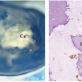
Classic morphologic, immunophenotypic, and genetic features have been described for BL. However, no single parameter can be used as a diagnostic standard, and a combination of diagnostic techniques is required for diagnosis.30 Morphologically, BLs are characterized by intermediate-sized homogeneous cells with round-to-oval nuclei containing multiple, variably prominent basophilic nucleoli with a modest amount of somewhat basophilic cytoplasm, which will appear vacuolated, due to lipid droplets, on cytologic preparations (Fig. 23.1B). These tumors have very high mitotic activity, and tissue sections will often show a “starry sky” appearance that results from reactive macrophages scattered among the malignant lymphoid cells that are engulfing apoptotic debris from the rapidly dividing tumor cells. This starry-sky appearance is not specific for BL, but can be seen in any rapidly dividing NHL.30,61 Histologically, endemic and sporadic BLs are indistinguishable (Fig. 23.2C).

Only gold members can continue reading. Log In or Register to continue

Stay updated, free articles. Join our Telegram channel

Aug 25, 2016 | Posted by in ONCOLOGY | Comments Off on Malignant Non-Hodgkin Lymphomas in Children

Full access? Get Clinical Tree

Get Clinical Tree app for offline access