Malignant Mesothelioma
BACKGROUND
Malignant mesothelioma is a rare malignancy arising from the mesothelial cells of the pleural or peritoneal surfaces. Mesothelioma can arise from the pleura (pleural mesothelioma), peritoneum (peritoneal mesothelioma), pericardium (pericardial mesothelioma), or tunica vaginalis (testicular mesothelioma). In the United States, there are approximately 3000 new cases of mesothelioma reported annually. Eighty percent of these occur as pleural mesothelioma, and this will be the focus of this chapter.
The development of mesothelioma is most clearly associated with prior asbestos exposure (1). Asbestos was (and continues to be in some parts of the world) an important and affordable industrial resource due to its resistance to heat and combustion. Asbestos was used in shipbuilding, in car brakes, in the production of cement, and as insulation. There are two main forms of asbestos known as amphiboles and chrysotile. Amphiboles are long thin asbestos fibers and are felt to be the most carcinogenic of the asbestos fibers. Chrysotile asbestos has also been associated with mesothelioma, although the frequency may be less than with amphibole asbestos (1). The latency period between the time of asbestos exposure to development of mesothelioma can be 20–40 years. These unique features reflect the population of patients who develop mesothelioma including asbestos miners, plumbers, pipefitters, or those who worked in shipbuilding industries. In the United States, mesothelioma is a disease of Caucasian men reflecting the population of asbestos workers in the 1960s and early 1970s. The median age of patients diagnosed with mesothelioma is in the mid-60s, although, in the Surveillance Epidemiology and End Results (SEER) database from the United States, the median age is over 70 years. Approximately 80% of patients who develop mesothelioma are men. Women who develop mesothelioma also may have worked in industries that used asbestos, although there are reports of secondary exposure for example from clothing of spouses who worked directly with asbestos (2). The estimated incidence of mesothelioma worldwide also reflects the use of asbestos in different regions of the world. It is estimated that the cumulative worldwide incidence of mesothelioma will not peak for another 10–20 years. However, in the United States some estimates suggest that this may have already occurred, while in Europe, Australia, and Japan, where common asbestos use occurred until much later than in the United States, the incidence may not peak for another 15–20 years (3). In addition, asbestos is still extensively used in many developing countries suggesting that the worldwide incidence of mesothelioma will continue to rise.
Other etiologic factors besides asbestos can also increase the risk of developing mesothelioma. For example, numerous cases have been reported in patients who have previously received ionizing radiation such as that used to treat Hodgkin lymphoma or other malignancies (4). Genetic factors may also play a role. Germline mutations in the BRCA-associated protein-1 (BAP1) gene were reported in two families with multiple affected generations (5). Mutations in this gene also increase the risk of developing other malignancies such as uveal melanoma and renal cell carcinoma, suggesting this is part of a newly described cancer predisposition syndrome (6–8). The prevalence of BAP1 germline mutations as an underlying cause for mesothelioma remains unknown, however.
CLINICAL PRESENTATION
The most common clinical presentation of malignant pleural mesothelioma includes dyspnea on exertion and shortness of breath. These clinical symptoms often lead physicians to obtain a chest x-ray where a unilateral pleural effusion is noted (Figure 50-1). Mesothelioma is rarely an incidental finding on a routine chest x-ray. Patients can also present with non-pleuritic chest pain. This symptom is important to elicit from patients as those who present with chest pains often have disease extending into the chest wall and thus they are clearly not surgical candidates. Other presenting signs and symptoms include discordant chest wall expansion, weight loss, night sweats, and the presence of a palpable subcutaneous mass (Table 50-1 and Figure 50-2).
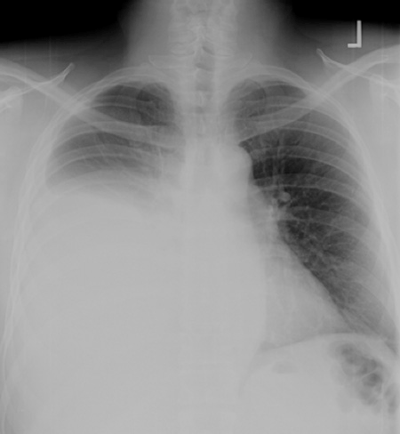
FIGURE 50-1 Anterior-posterior chest radiograph of a patient with newly diagnosed malignant mesothelioma. A large right-sided pleural effusion can be appreciated in this chest x-ray.
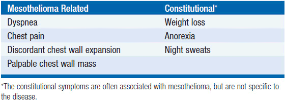
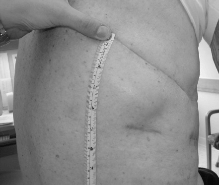
FIGURE 50-2 Subcutaneous chest wall mass in a patient with mesothelioma. This painful subcutaneous mass appeared a few months following surgical exploration.
DIAGNOSTIC EVALUATION
The presentation of a patient with a new pleural effusion often leads to a thoracentesis to evaluate the nature of the effusion. The pleural effusions in mesothelioma are usually cytologically negative, and patients frequently undergo repeated thoracenteses in order to establish the diagnosis of mesothelioma. Due to the frequent nature of cytologically negative effusions, cytology is not a reliable method to diagnose mesothelioma. In addition, cytologic specimens are often insufficient to determine the histologic subtype of mesothelioma, which itself provides prognostic information.
The diagnostic procedure of choice for mesothelioma is a thoracoscopic biopsy. There are several advantages to this approach. First, it provides the surgeon the ability to assess the extent of tumor on the visceral and parietal pleura. Second, a biopsy of the involved area can be obtained under direct visualization. This is critical to make an accurate histologic diagnosis. Finally, thoracoscopy provides the ability to also perform a pleurodesis, which can help control recurrent pleural effusions. It is important to note that mesothelioma can grow along and through previous biopsy sites (e.g., see Figure 50-2). Thus if a patient is being considered for future a pneumonectomy, it will be important to mark and excise en bloc the prior biopsy site.
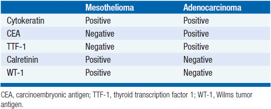
The pathologic evaluation of malignant mesothelioma can be challenging and the disease can sometimes be confused pathologically with adeno-carcinoma of the lung. However, several immunohistochemical markers can help distinguish adenocarcinoma from malignant mesothelioma (Table 50-2) (9). Unlike adenocarcinomas, mesotheliomas do not express thyroid transcription factor 1 (TTF-1) or carcinoembryonic antigen (CEA). In contrast, mesotheliomas do express the Wilms tumor-1 (WT-1) protein and calretinin, which are not expressed by lung adenocarcinomas. In addition to the pathologic diagnosis of mesothelioma, it is important to distinguish the histologic subtype of mesothelioma. The most common histologic subtype is epithelial mesothelioma and accounts for approximately 60% of all mesotheliomas. Other subtypes of mesothelioma include sarcomatoid mesothelioma and mixed (containing components of both epithelial and sarcomatoid) type mesothelioma. The main reason to determine the histo-logic subtype of mesothelioma is that patients with sarcomatoid mesothelioma have a much worse prognosis than those with epithelial mesothelioma and may not be ideal candidates for aggressive surgical resection.
TABLE 50-2 IMMUNOHISTOC HEMICAL MARKERS USED TO DISTINGUISH MALIGNANT MESOT HELIOMA FROM LUNG ADENOCARCINOMA
Three main imaging modalities, computed tomography (CT), positron emission tomography (PET), and magnetic resonance imaging (MRI), are used to evaluate patients with newly diagnosed mesothelioma. Contrast enhanced CT scanning often reveals a thickened lobulated circumferential pleural rind (Figure 50-3) with or without the presence of a concurrent pleural effusion. CT scanning can also help identify the presence of pleural plaques, a sign of previous asbestos exposure, and whether the disease extends into the intralobular fissures. However, CT scanning is not very accurate in determining chest wall invasion or transdiaphragmatic extension of mesothelioma. These features are important to determine, especially if the patient is being considered for surgery. MRI can occasionally be used in this situation to identify chest wall invasion (Figure 50-4) as well as transdiaphragmatic invasion (10), although some studies have not found it superior to CT imaging (11). FDG-PET scanning has also recently been evaluated for its clinical utility as most mesotheliomas are PET avid. Analogous to lung cancer, FDG-PET scanning can identify occult metastatic disease in patients who are otherwise potential surgical candidates (12) and may have prognostic significance (13). However, its utility in defining locoregional disease and the role of FDG-PET scanning in advanced mesothelioma remain to be determined.
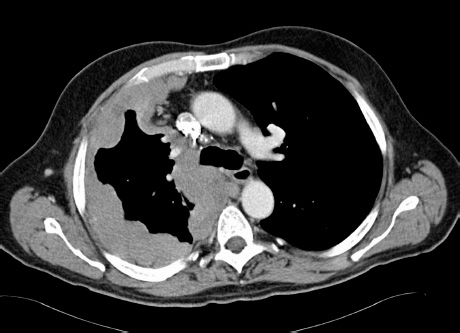
FIGURE 50-3 Computed tomography (CT) appearance of mesothelioma. A thickened circumferential pleural rind is seen on chest CT in this patient with epithelial mesothelioma.
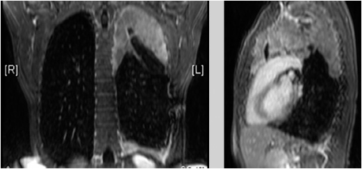
FIGURE 50-4 Coronal (left) and sagittal (right MRI images of the left hemithorax of a patient with newly diagnosed malignant mesothelioma. The MRI images help to define mesothelioma invading into the chest wall on the lateral side in the left hemithorax.
Soluble mesothelin-related protein (SMRP) is a serum marker for mesothelioma. Mesothelin is a cell surface protein present on mesothelial cells. It can remain membrane bound or be shed into the serum and be detected using an ELISA assay (14). In a study using this assay, 37/44 (84%) of patients with mesothelioma had elevated levels of SMRP compared to 3/160 (2%) of patients with other lung or pleural diseases. In addition, 7 of 40 patients exposed to asbestos had elevated levels of SMRP. Interestingly within 1–5 years, 3 of the 7 patients developed mesothelioma, which suggested that SMRP could also serve as a screening test for asbestos-exposed individuals. However, in a larger cohort of exposed patients who developed mesothelioma, SMRP levels from banked serum samples obtained prior to the diagnosis were elevated in only 17 of 106 people (15). SMRP levels do seem to correlate with disease status, declining after surgery or with response to chemotherapy, suggesting a possible utility for monitoring in conjunction with imaging (16).
STAGING
Related posts:

Stay updated, free articles. Join our Telegram channel

Full access? Get Clinical Tree


