Lymphocytes and Plasma Cells


II.G.001 Alder Reilly Anomaly


II.G.001


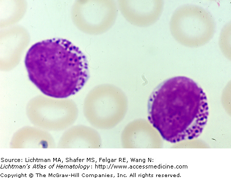
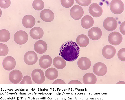
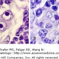
Alder Reilly Anomaly. Blood film. Lymphocyte with large cytoplasmic granules characteristic of the mucopolysacchidoses.


II.G.002 Batten Disease


II.G.002


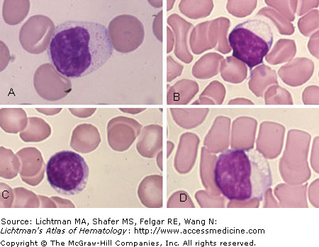
Batten disease. (A-D) Lymphocytes displaying small and large vacuoles in cytoplasm. Batten disease is a fatal, inherited disorder of the nervous system that begins in childhood. In some cases, the early signs are subtle, taking the form of personality and behavior changes, slow learning, clumsiness, or stumbling. Symptoms of Batten disease result from accumulation of lipopigments made up of fats and proteins in the tissues.


II.G.003 Chediak-Higashi Disease


II.G.003


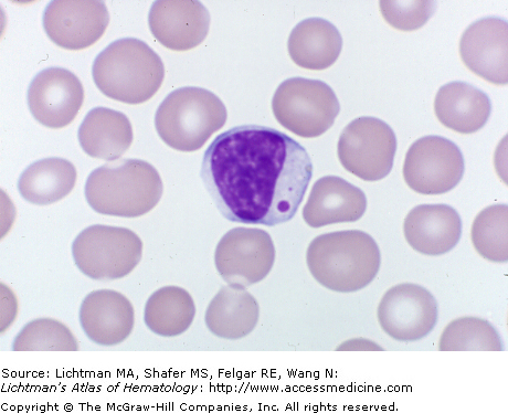
Chediak-Higashi Disease. Blood Film. Large cytoplasmic granule observed in a blood lymphocyte, characteristic of this disorder.


II.G.004 Chediak-Higashi Disease


II.G.004


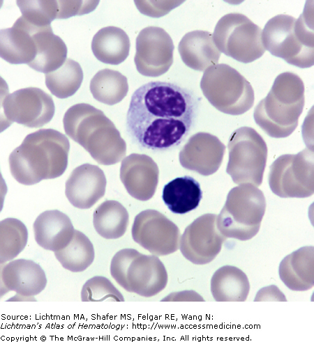
Chediak-Higashi Disease. Blood Film. Two nuclei in lymphocyte. Unusual finding.


II.G.005 Congenital Rubella


II.G.005


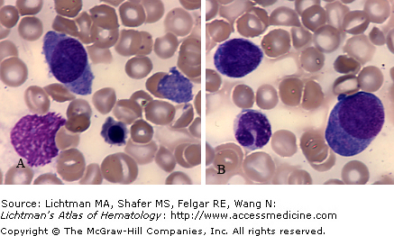
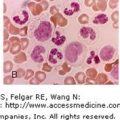
Congenital Rubella. Blood films. (A) Large plasmacytoid cell. Small nucleated red cell. (B) Two plasmacytoid cells with intensely blue cytoplasm.


II.G.006 Drug reaction


II.G.006


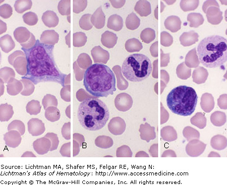
Drug reaction. Blood film. Composite of reactive lymphocytes from patients undergoing drug reactions. (A) Large lymphocyte with nucleoli. (B) Large lymphocyte with nucleoli. (C) Lymphocyte with intensely blue cytoplasm (plasmacytoid lymphocyte).


II.G.007 Hurler Disease


II.G.007


Hurler disease. Blood film. Lymphocyte. Note numerous cytoplasmic granules characteristic of this mucopolysaccharidosis. Presumably these are lysosomal accumulations of glycosaminoglycans, characteristic of this deficiency in enzymes that degrade glycosaminoglycan (α-L-iduronidase in Hurler disease).


II.G.008 Infectious Lymphocytosis


II.G.008


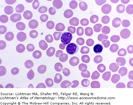
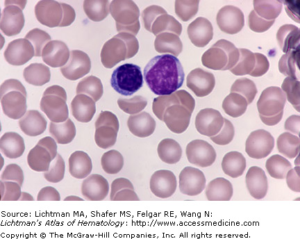
Infectious lymphocytosis. Blood film. Increased lymphocyte count with small lymphocytes as the characteristic cell.


II.G.009 Plasma Cells: Mott Cells


II.G.009


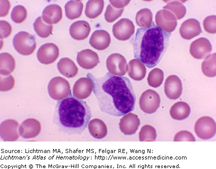
Infectious mononucleosis. Blood film. Three large lymphocytes with abundant cytoplasm. Cytoplasmic periphery conforms to red cell outlines. Nuclear shape not a strict circle but elongated or irregular.


II.G.010 Plasma Cells: Mott Cells


II.G.010


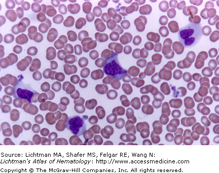
Infectious mononucleosis. Blood film. Four large lymphocytes with abundant cytoplasm. Cytoplasmic periphery conforms to red cell outlines. Nuclear shape not a strict circle but elongated or irregular.


II.G.011 Infectious Mononucleosis


II.G.011


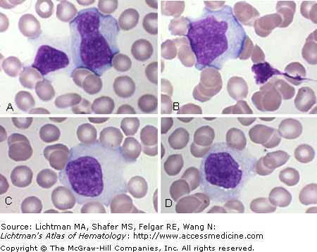
Infectious mononucleosis. Blood film. (A–D) Large lymphocytes with abundant cytoplasm. Cytoplasmic periphery conforms to red cell outlines. Nuclear shape not a strict circle but elongated or irregular.


Jun 12, 2016 | Posted by in HEMATOLOGY | Comments Off on Lymphocytes and Plasma Cells

Full access? Get Clinical Tree

Get Clinical Tree app for offline access