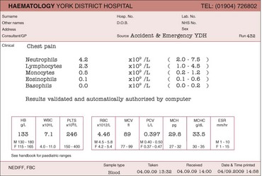
Laboratory haematology I – Blood and bone marrow

9


Laboratory haematology I – Blood and bone marrow


Diagnosis of most blood disorders is possible from a combination of clinical history, clinical examination and relatively routine laboratory tests. Haematology laboratories are heavily dependent on complex electronic machinery. The ubiquitous full blood count (FBC) is the archetypal haematological investigation and is performed by specialised automated cell counters. However, despite the accessibility of modern technology, the more simple traditional techniques of blood and bone marrow film spreading, staining, and light microscopy remain essential parts of the haematologist’s repertoire.


The blood count


Many of the diseases discussed in this book are first suggested by an abnormality in the blood count (often referred to as the full blood count). The test is performed on a small specimen of anticoagulated venous blood; the normal anticoagulant is ethylene diamine tetra-acetic acid (EDTA). A typical report is illustrated in Figure 9.1. As can be seen, it contains a large amount of numerical information pertaining to the three cell lines in the peripheral blood: red cells (and haemoglobin), white cells (with a differential count of each specific cell type) and platelets.


When interpreting the report it is sensible to initially focus on the haemoglobin (Hb) concentration, total white cell count (WBC) and platelet count – most blood abnormalities of clinical significance are associated with a derangement of at least one of these values. Much of the remaining information details the nature of the red cells and their degree of haemoglobinisation, and the precise make-up of the white cell count. The former values are helpful in the diagnosis of anaemia, and the latter in the diagnosis of a variety of diseases of white cells (e.g. leukaemias) and reactions to systemic disease. To understand the role of the automated blood count in clinical practice, and particularly its limitations, it is helpful to understand how the numerical values are generated.


Automated haematology counters


The two essential functions of the automated blood cell counter are the measurement of Hb concentration in the blood and the counting and sizing of blood cells.


Most counters use a modification of the traditional cyanmethaemoglobin method to measure Hb concentration. In essence, blood is diluted in a solution where Hb is converted to cyanmethaemoglobin and then the Hb concentration derived from the light absorbance (optical density) of the resultant solution measured by a spectrophotometer. Automated machines have at least two channels for cell counting. In one, red cells and platelets may be counted and in the other red cells are lysed leaving white cells for analysis. Extra channels are often used for differential white cell and reticulocyte counting.


There are two basic methods for cell counting and sizing: electrical impedance and light scattering. The electrical impedance method relies on blood cells being very poor conductors of electricity. Thus, when the cells are passed in a stream through a narrow aperture across which an electrical current is maintained, the individual cells create an increase in electrical impedance of a size proportional to the cell volume. In the light scattering method the cells deflect a beam of light (often a laser beam) and a detector converts the scatter into pulses proportional to cell size. For sophisticated measurements such as the differential white cell count the two methods can be used together with the addition of other modalities reliant on biochemical reactions and light absorbance.

Stay updated, free articles. Join our Telegram channel

Jun 12, 2016 | Posted by in HEMATOLOGY | Comments Off on Laboratory haematology I – Blood and bone marrow

Full access? Get Clinical Tree

Get Clinical Tree app for offline access