Introduction
Hypertension is common, silent, and often deadly when not successfully treated . Hypertension’s initial presentation can include cardiovascular disease (CVD) or renal disease . Although many sources associate hypertension and headaches, mild (140 to 159/90 to 99 mmHg) or moderate (160 to 179/100 to 109 mmHg) hypertension does not induce headaches . To avoid or reduce the morbidity and mortality of hypertension, blood pressure (BP) measurements must be a routine element of care for individuals of all ages .
To address the laboratory evaluation of endocrine etiologies of elevated BP (i.e., hypertension), this chapter will provide an overview of the current definition of hypertension, risk stratification and treatment, and the general causes and the endocrine causes of hypertension. This chapter is a revision and update of the 2008 American Association for Clinical Chemistry publication by the authors . Hypertension is of tremendous clinical importance because it has adverse effects on many systems of the body. If left untreated, hypertension produces significant morbidity and mortality .
The physiological control of BP is complex. The most basic concept is that BP equals the cardiac output (C.O.) multiplied by the peripheral vascular resistance (PVR) (BP≈C.O.×PVR). If either C.O. or PVR increase, BP will rise. C.O. (mL/min or L/min; C.O.=stroke volume×heart rate) is dependent on preload (related to blood volume and the return of blood to the heart), contractility (also known as the ionotropic state of the heart), afterload, and heart rate. Afterload is determined by the diastolic BP and the resistance of the aortic valve to opening during ventricular systole. PVR (mmHg⋅min/mL) is determined by the blood viscosity ( η ; which is usually not an important variable unless polycythemia is present), the length of the resistance blood vessel (i.e., the small arteries and arterioles) which is fixed, and the radius ( r ) of the resistance blood vessel (i.e., the small arteries and arterioles) that is variable. Because resistance (PVR, or more simply “ R ”) is proportional to the inverse of r to the fourth power ( r 4 ), small changes in r produce large changes in R (resistance). Radius ( r ) is controlled by the contractile state of the vascular smooth muscle surrounded the small arteries and arterioles.
Rαη×Lr4
Some biologic agents act directly on vascular smooth muscle (e.g., both catecholamines and vasopressin cause vasoconstriction). Epinephrine can be released systemically by the adrenal medulla or locally by the sympathetic nervous system. Some agents sensitize tissues to catecholamines (e.g., thyroid hormone increases tissues’ responsiveness to catecholamines). Certain agents act via the central nervous system in changing sympathetic tone (angiotensin II and aldosterone ). Other agents control vascular smooth muscle tone indirectly via nitric oxide (NO) produced by the endothelium of the small arteries and arterioles. In turn NO relaxes the vascular smooth muscle reducing PVR. Histamine (from mast cells), serotonin (from platelets), bradykinin (from the plasma), and platelet-activating factor (from many types of cells) stimulate the endothelium to produce NO causing vasodilation. Prostaglandins (PGI2, prostacyclin) produced by the endothelium can also act as vasodilators.
Blood volume has a major influence on BP. The sympathetic nervous system is regulated by low-pressure baroreceptors that sense blood volume in the atria and pulmonary vessels. Decreased blood volume elicits vasoconstriction via increased sympathetic nervous system discharge. High-pressure arterial baroreceptors in the aortic arch and carotid arteries sense arterial pressure. Increased BP elicits vasodilatation via reduced sympathetic nervous system discharge. The sympathetic nervous system also regulates renal perfusion that affects blood volume by regulating the glomerular filtration rate. The central manager of autonomic function (that anatomically includes the sympathetic nervous system) is the central nervous system that regulates BP by influencing sympathetic activity and fluid balance .
Because free water reabsorption by the kidney influences blood volume, vasopressin [also known as antidiuretic hormone (ADH)] is an important regulator of fluid status. Vasopressin regulates free water reabsorption predominantly in the collecting duct (CD). Via hypothalamic “osmoreceptors,” increased plasma osmolality elicits vasopressin secretion causing increased free water reabsorption. Sympathetic nervous system baroreceptors (as described earlier) also regulate vasopressin release. Decreased volume or BP elicit vasopressin release. Only at supra-physiologic concentrations of vasopressin regulate BP through its direct vasoconstrictor action. Not to be ignored, organs do autoregulate their own perfusion to maintain optimal organ perfusion over a wide range of systemic BPs.
Definition of hypertension
The most recent hypertension guidelines from the American College of Cardiology, Clinical Policy Approval Committee and the American Heart Association, Science Advisory and Coordinating Committee were released in 2017 and published in 2018 . This “2017 ACC/AHA/AAPA/ABC/ACPM/AGS/APhA/ASH/ASPC/NMA/PCNA Guideline for the Prevention, Detection, Evaluation, and Management of High Blood Pressure in Adults” will be referred to as the “2017 ACC/AHA guideline” for simplicity.
This 2017 ACC/AHA guideline updated the seventh report of the Joint National Committee (JNC 7) on the “Prevention, Detection, Evaluation, and Treatment of High Blood Pressure” that was released in August of 2004 . JNC 7 had updated JNC 6 . The 2017 ACC/AHA guideline addressed four questions concerning: (1) self-directed monitoring of BP and/or ambulatory BP monitoring, (2) the optimal BP target for BP during therapy of hypertension, (3) antihypertensive drug classes and outcome, and (4) monotherapy versus initial multidrug therapy and outcome . Of significance for laboratorians, there were changes in the cut points used to describe various BP levels and changes in the recommended laboratory evaluation (to be discussed later).
Adapted from the 2017 ACC/AHA guideline is Table 11.1 . Compared to the 2004 guideline, the category of “prehypertension” was removed and the category of “elevated BP” was substituted. The definition of normal BP was not changed (i.e., <120 mmHg/<80 mmHg). In 2004 prehypertension was defined as 120–139 mmHg/80–90 mmHg. In 2017, “elevated” BP was defined as 120–139 mmHg/<80 mmHg. The cut points for the definitions of stages 1 and 2 hypertension were lowered.
| BP classification | SBP (mmHg) | DBP (mmHg) | |
|---|---|---|---|
| Normal | <120 | and | <80 |
| Elevated BP | 120–129 | and | <80 |
| Stage 1 hypertension | 130–139 | or | 80–89 |
| Stage 2 hypertension | ≥140 | or | ≥90 |
Before the diagnosis of hypertension can be established, BP must be measured two or more times on two or more visits. The 2017 ACC/AHA guideline states that ambulatory BP monitoring (ABPM) and home BP monitoring (HBPM) improve the opportunities for the diagnosis of hypertension and hypertension treatment monitoring. However, better understanding of ABPM and HBPM is required before these monitors are included in routine clinical practice. Older publications advised caution in recommending ABPM as a “gold standard” for the definition of hypertension .
The greatest elevation in BP, either systolic or diastolic, is used to grade the severity of the hypertension. Systolic hypertension is just as much a risk factor for an adverse outcome as diastolic hypertension. For example, in the Framingham study, systolic hypertension was as powerful a predictor of coronary heart disease as diastolic hypertension . Before age 50, diastolic hypertension is more common than either systolic hypertension or combined systolic and diastolic hypertension. After age 50 with increasing age, systolic hypertension becomes predominant. For reference, in the general population from age 30 and onward, systolic BP and pulse pressure (the difference between the systolic and diastolic BPs) progressively rise. Diastolic BP rises until age ~50 and then declines thereafter. Therefore in our senior years, in the absence of hypertension, systolic BP is higher, diastolic is lower and pulse pressure is wider. Note : Treatment recommendations for hypertension are provided in the 2017 ACC/AHA guideline. A detailed discussion of these therapeutic guidelines is outside the scope of this chapter.
Definition of hypertension in children
The current 2017 guidelines regarding hypertension in children update the 2004 guidelines . These guidelines were written in 2016 by the American Academy of Pediatrics. BP in children is assessed using tables based upon the child’s sex, age, and height . The assessment of BP in children is illustrated in Table 11.2 . BP cut points for children age 13 and older are now identical to current adult cut points.
| BP classification | Criteria |
|---|---|
| Normal | <90th %tile |
| Elevated a | ≥90th %tile to <95th %tile [or] 120/80 mm Hg to <95th %tile |
| Stage 1 hypertension a | ≥95th %tile to <95th %tile + 12 mmHg [or] 130/80 mm Hg to 139/89 mm Hg |
| Stage 2 hypertension a | ≥95th %tile plus 12 mm Hg [or] ≥140/≥90 mm Hg |
a Whichever cut point is lower defines the applied cut point.
Causes of hypertension
Most cases of hypertension are without a recognized etiology, which is termed “primary” hypertension . The older term “essential” hypertension is no longer in use. Primary hypertension usually presents clinically between ages 35 and 55 years. The genetic basis of essential hypertension is poorly understood . Whereas hypertension is strongly familial, it is uncommon that individual gene alleles affect BP . A reduction in glomeruli number is one factor proposed as a cause of essential hypertension .
It is estimated that 10% of cases of hypertension in adults have an underlying etiology . The recognition of such cases of “secondary” hypertension is important because treatment of the underlying cause of the hypertension may lead to a cure or marked improvement in the BP with a reduction in risk for CVD. A 2004 study from Japan reported a 9% frequency of secondary hypertension . Hypertension in children is more often secondary to an underlying disorder than in adults .
A leading cause of secondary hypertension is renal disease. Renal disease appears to cause hypertension via: (1) activation of the renin-angiotensin-aldosterone system (RAAS) and (2) sodium retention accompanied by fluid retention. In some cases, the adrenergic nervous system may also be stimulated causing vasoconstriction. It is beyond the scope of this chapter to examine all causes of secondary hypertension. The classification of hypertension by physiology can begin by separating hypertension into: (1) systolic hypertension (e.g., cases where the diastolic BP is normal) and (2) combined systolic and diastolic hypertension ( Table 11.3 ) (early on, diastolic hypertension may be solely present). Systolic hypertension results from aortic stiffness or increased left ventricular contractility (e.g., increased thyroid hormone—hyperthyroidism) and is physiologic (but transient) during exercise. Diastolic hypertension results from increases in PVR. Consequently if diastolic hypertension occurs, systolic hypertension often follows.
|
Laboratory evaluation of hypertension in adults
Routine evaluations recommended by the 2017 ACC/AHA guideline ( Table 11.4 ) provide information about: (1) the etiologies of hypertension (e.g., glucose, urinalysis, creatinine, and potassium, etc.), (2) target organ disease (e.g., creatinine), and/or (3) the risk status of the patient for CVD (e.g., creatinine, glucose, and fasting lipid profile) are listed. Compared to 2004, the 2017 ACC/AHA guideline adds sodium and thyroid-stimulating hormone (TSH) as screening tests and replaces hematocrit (Hct) testing with the complete blood count (CBC). Uric acid is added as an optional test. While not a laboratory test, the 12-lead electrocardiogram (EKG) is again recommended as an assessment for left ventricular hypertrophy, ischemia, previous myocardial infarction, and arrhythmia. The echocardiogram is an optional test that can inform the physician regarding cardiac anatomy (e.g., LV wall thickness), contractility, dyskinesis, and valvular disease.
| Routine tests | Information provided (commentary form the authors) |
|---|---|
| Fasting plasma glucose | A screening test for diabetes mellitus. Not mentioned as a screening test for diabetes is hemoglobin A1c, which is one of three diabetes screening tests recommended by the American Diabetes Association in 2019. a Diabetes may also be observed in Cushing syndrome, acromegaly, and pheochromocytoma. Hypokalemia (potentially from hypermineralocorticoidism) can cause hyperglycemia from reduced insulin secretion b |
| CBC | Polycythemia can cause hypertension. Anemia is a finding in many chronic diseases including chronic renal insufficiency |
| Lipid profile | Assesses the patient’s risk for the development of atherosclerosis. Includes: total cholesterol, triglycerides, HDL cholesterol, and calculated LDL cholesterol. Should be drawn in the fasting state |
| Creatinine | Assessment of renal function. Creatinine can be used to estimate the glomerular filtration rate (eGFR) in persons age 18 years and older |
| Sodium | Useful in monitoring diuretic, ACE inhibitor, or ARB treatment of hypertension |
| Potassium | Elevated K + is seen in advanced renal insufficiency. Low K + is seen in some cases of hypermineralocorticoidism (hyperaldosteronism, hypercortisolism, or elevated DOC), K + -wasting forms of chronic renal failure and as a side effect of potassium-wasting diuretics. A baseline K + is required because diuretics can produce hypokalemia, whereas ACE inhibitors, ARBs, spironolactone, amiloride, and triamterene can cause hyperkalemia |
| Calcium | Elevated calcium can cause hypertension, for example, primary hyperparathyroidism. If hypercalcemia is detected, additional measurements should include ionized calcium, phosphate, alkaline phosphatase, and parathyroid hormone |
| TSH | Hypothyroidism and hyperthyroidism can both cause hypertension. If the TSH is elevated, free (unbound) thyroxine (FT4) should be measured. If the TSH is depressed, FT4 and triiodothyronine (T3) should be measured |
| Urinalysis | Assessment of renal function. Systemic illness can frequently produce an abnormal urinalysis |
| EKG | Screens for LVH, ischemia, previous myocardial infarction, and arrhythmias |
| Optional tests | Information provided |
| Echocardiogram | Cardiac anatomy (e.g., LV wall thickness), contractility, dyskinesis, and valvular disease |
| Urinary albumin to creatinine | Sensitive indicator of diabetic nephropathy and, in nondiabetic individuals, endothelial dysfunction. Increased urinary albumin excretion occurs in stage III diabetic nephropathy in type 1 diabetes prior to the development of ratio (ACR) dipstick-positive proteinuria |
| Uric acid | Frequently elevated in gout and the metabolic syndrome. Elevation in uric acid helps support the diagnosis of insulin resistance |
a American Diabetes Association, Classification and Diagnosis of Diabetes: Standards of Medical Care in Diabetes—2019, Diabetes Care 42 (Supplement 1) (2019) S13–S28.
b J.H. Helderman, D. Elahi, D.K. Andersen, G.S. Raizes, J.D. Tobin, D. Shocken, R. Andres, Prevention of the glucose intolerance of thiazide diuretics by maintenance of body potassium, Diabetes 32 (2) (1983) 106–111.
The basic tests ( Table 11.4 ) are accomplished in all patients when hypertension is first diagnosed. More comprehensive testing should be pursued when: (1) the severity of hypertension worsens, (2) there is an inadequate reduction in BP using standard treatments, (3) there is more target organ damage than anticipated for the severity of the hypertension, or (4) a secondary cause is possible based upon historical or clinical suspicions.
Laboratory evaluation of hypertension in children
The 2017 pediatric guidelines state that: (1) if there is a family history of hypertension, (2) the child has a body mass index≥85 percentile, or (3) the child lacks a history or physical findings indicative of possible secondary hypertension, an extended workup for secondary causes of hypertension is not necessary in children age 6 and older. For those hypertensive children that do warrant laboratory testing, Table 11.5 provides testing recommendations. A 2019 review recommends that the bedside CKiD Schwartz equation be used to calculate a pediatric estimated glomerular filtration rate (eGFR) from the creatinine concentration . The Modification of Diet in Renal Disease (MDRD) calculation of the eGFR is not recommended in children because this calculation has not been validated in children.
| Patient population | Screening tests |
|---|---|
| All children |
|
| Obese children a |
|
| Optional tests b |
|
a BMI (body mass index): ≥95th percentile for age and sex.
b When indicated by history, physical examination, or initial studies.
Endocrine hypertension and mechanisms
In several endocrine conditions, the mechanisms of hypertension are unclear or multifactorial. Nevertheless, there are two major mechanisms that appear to cause hypertension of endocrine origin ( Table 11.6 ). The major proposed mechanisms are sodium retention and vascular smooth muscle contraction (also known as vasoconstriction; with or without cardiac stimulation). These two mechanisms may interact .
| Mechanisms of hypertension | Prototypic examples |
|---|---|
| (1) Sodium retention: | |
| (1a) Activation of ENaC: | |
| (1a1) ENaC activation via the renin-angiotensin-aldosterone system (RAAS), or | Excess mineralocorticoid activity [e.g., excess aldosterone, desoxycorticosterone (DOC), cortisol, or sex steroids] |
| (1a2) ENaC activation via an end-organ disorder. |
|
| (1b) Hyperinsulinism | States of insulin resistance: obesity, type 2 diabetes, metabolic syndrome, acromegaly |
| (2) Vascular smooth muscle contraction (i.e., increased peripheral vascular resistance) and possible cardiac stimulation: | |
| (2a) Direct hormone effects on β1-adrenergic myocardial receptors and β2-adrenergic receptors (e.g., catecholamines, with or without central actions) | Pheochromocytoma |
| (2b) Increased sensitivity to catecholamines | Hyperthyroidism |
| (2b) Divalent hormone (e.g., calcium) effects on vascular smooth muscle | Hypercalcemia |
| (2c) Other direct effects on vascular smooth muscle or the endothelium |
|
Drugs can also perturb these systems . Drugs that activate the renin- RAAS include 9-α-fluorohydrocortisone (fludrocortisone), glucocorticoids, and sex steroids (e.g., androgens). Drugs with catecholamine activity (e.g., methamphetamines, methylphenidate, amphetamine, phenylephrine, and pseudoephedrine) can potentially cause hypertension. Such drugs should be sought by history and urine drug screening if there is an index of suspicion for catecholamine use.
Physiology
Mechanistically, many disorders causing hypertension share as a common pathway ENaC (the epithelial sodium channel) activation. ENaC is nonvoltage-gated and amiloride-sensitive, and consists of α (SCNN1A gene; sodium channel epithelial 1 subunit alpha, chromosome 12p13), β ( SCNN1B gene; sodium channel epithelial 1 subunit beta, chromosome 16p12.2), and Υ ( SCNN1G gene; sodium channel epithelial 1 subunit gamma, chromosome 16p12.2) subunits in a 1:1:1 stoichiometry. Interestingly, each subunit has two transmembrane domains with the N-termini and C-termini being intracellular. ENaC controls fluid and sodium transport across epithelia in many organs.
In essence, ENaC regulation fine-tunes renal sodium excretion. Sodium balance is critical to maintaining the distribution of fluid between the intracellular and extracellular spaces, as well as the reabsorption of filtered water via the renal tubules. Therefore we begin by examining the RAAS ( Fig. 11.1 ). Renin is encoded by the REN gene on chromosome 1q32.1. Angiotensinogen is encoded by the AGT gene on chromosome 1q42.2.

Transcription of the renin mRNA in juxtaglomerular granular cells produces preprorenin . Upon entry into the endoplasmic reticulum (ER), the renin presequence is cleaved from preprorenin yielding prorenin in the lumen of the ER. Prorenin glycosylation occurs in the Golgi apparatus. For regulated secretion, prorenin is then cleaved into renin (340 amino acids) and is stored in secretory granules. A constitutive pathway of the juxtaglomerular granular cells releases prorenin. Renin/prorenin receptors exist raising the possibility that prorenin plays a physiologic activity of its own . As well, although this chapter emphasizes the endocrine actions of the RAAS system, a tissue RAAS appears to exist .
There are three factors that regulate renin secretion. First, the juxtaglomerular apparatus (JGA) monitors the afferent arteriole perfusion that supplies the glomerular vasculature with blood. Reduced perfusion pressure (as detected by decreased stretch of baroreceptors in the granular cells) triggers renin release from the perivascular granular renin-producing cells. Second, renin release increases when there is a reduced sodium concentration in the lumen of the upper pole of the ascending loop of Henle [ALH; also known as thick ascending loop of Henle (TAL)]. Anatomically the loop of Henle rises from the renal medulla and courses back toward the renal cortex. The macula densa (which is part of the JGA) forms part of the wall of the ALH. Macula densa cells possess large nuclei, are closely grouped, from a plaque-like structure, and monitor ALH sodium concentration via the Na-K-2Cl cotransporter (NKCC2; BSC1; SLC12A1 gene: solute carrier family 12 member 1) . The macula densa cells are immediately adjacent to the renin-secreting cells of the afferent arteriole.
In states of low ALH sodium concentration, the macula densa secretes increased levels of prostaglandins (PGE2) and decreased adenosine diphosphate (ADP) stimulating renin release from the nearby granular cells . The beneficial physiology behind such a response is as follows: If there is hypovolemia, the renal tubules proximal to the ALH will be maximally reabsorbing sodium and chloride to preserve water and extracellular fluid volume. Therefore the ALH sodium concentration would be low in hypovolemic states or states of reduced renal perfusion (because of hypotension) where activation of the RAAS is desirable. The third factor regulating renin release is catecholamines where β2-adrenergic activity stimulates renin release. Such β2-adrenergic agonists can be locally secreted by the sympathetic nervous system or such agonists are circulating (e.g., epinephrine secreted by the adrenal medulla).
Once renin is secreted, acting as an aspartyl protease, angiotensinogen (452 amino acids) is cleaved to angiotensin I (ANG I; 10 amino acids). ANG I is converted to angiotensin II (ANG II; 8 amino acids) in the lungs and other tissues via angiotensin-converting enzyme (ACE; a dipeptidyl carboxypeptidase; 170 kDa; ACE gene; chromosome 17q23.3). ACE has a single transmembrane domain and a short cytoplasmic tail. ACE has two homologous extracellular domains, each with a catalytic site and a Zn 2+ -binding region. ACE also inactivates bradykinin but it does not degrade ANG II.
ANG II binds to angiotensin II receptors ( AGTR1 gene; angiotensin II receptor type 1; chromosome 3q24) that have seven transmembrane domains. The receptors are coupled by G proteins to phospholipase C. The type 1 receptor (AT 1A receptor subtype) is found in blood vessel walls, the brain, and many tissues. The AT 1B receptor subtype is found in the anterior pituitary and the adrenal cortex. The function of the AT 2 receptors is not well studied. ANG II has several actions: (1) Aldosterone is released from the adrenal cortical glomerulosa layer, (2) ANG II has vasoconstrictor activity (via the central nervous system (CNS)) and direct action on blood vessel precapillary arterioles, (3) there is increased sympathetic discharge (a CNS effect) and subsequent catecholamine release from the adrenal medulla, (4) there is increased sodium uptake by the proximal convoluted tubule, (5) ANG II stimulates thirst, and (6) ANG II stimulates vasopressin release. The half-life of ANG II is very short (~1–2 min). Although the major secretagogue of aldosterone is ANG II, aldosterone is also released to lesser degrees by hyperkalemia and adrenocorticotrophic hormone (ACTH).
The regulation of aldosterone synthesis by ANG II is enacted by regulation of depolarization of the zona glomerulosa cells that involve several ion channels [Kir3.4 channels (KCNJ5), potassium leak channels (TASK 1/3, two pore–domain leak channels)], two pumps [Na + /K + ATPase (ATP1A1) and the calcium ATPase (ATP2B3)], and a voltage-gated calcium channel (CACNA1D). Receptor binding events lead to an increased cytoplasmic calcium concentration derived from the ER. Ultimately, the production of the enzyme aldosterone synthase ( CYP11B2 gene; cytochrome P450 family 11 subfamily B member 2; chromosome 8q24.3) increases.
Aldosterone biosynthesis from cholesterol is outlined in Fig. 11.2 . Aldosterone binds to the mineralocorticoid receptor (MR) in distal convoluted tubule (DCT) cells and luminal cells of the CD. Another name for the MR is the “type I glucocorticoid” receptor. The formal name of the MR is NR3C2 (nuclear receptor subfamily 3 group C member 2; chromosome 4q31). The present name for the MR emphasizes the fact that the MR is both a receptor and a transcription factor. For reference, the type II glucocorticoid receptor (NR3C1; nuclear receptor subfamily 3 group C member 1; chromosome 5q31.3) binds glucocorticoids. While the effects of aldosterone on mediating gene expression (via the MR as a transcription factor) are emphasized, like other steroid hormones, there are rapid, nongenomic effects of aldosterone that can be mediated by nontranscription factor receptors . Aldosterone receptors expressed on vascular endothelial plasma membranes have been described.

Aldosterone causes vasoconstriction mediated by the central nervous system increasing sympathetic outflow. Some experts assert that the major reason why hyperaldosteronism causes hypertension is this effect of aldosterone as contrasted with the salt and water retention caused by aldosterone . However, salt retention is likely necessary for the vasoconstrictive activity of aldosterone to be effective . Therefore these two mechanisms of aldosterone-induced hypertension may be complementary.
Whereas aldosterone does cause salt and water retention, within days of increased aldosterone secretion, a spontaneous diuresis (e.g., “aldosterone escape”) is observed decreasing extracellular fluid volume toward normal . “Aldosterone escape” explains why edema does not occur in states of mineralocorticoid excess. Natriuretic hormone secretion may, in part, explain aldosterone escape .
Regarding salt and water retention, in the DCT aldosterone activates the Na-Cl cotransporter (NCC; SLC12A3: solute carrier family 12 member 3; chromosome 16q13). In the CD, aldosterone binds to a single unit of an MR-heat shock protein (HSP) complex. The HSP dissociates and the ligand-receptor complex (aldosterone—MR) dimerizes. Upon interaction with a DNA hormone-response element, serum and glucocorticoid-inducible kinase 1 (SGK1; chromosome 6q23.2) is expressed. SGK1 regulates ENaC expression via inhibition of its degradation. Phosphorylation of Nedd4.2 ( NEDD4L Gene; NEDD4-like E3 ubiquitin protein ligase; chromosome 18q21.31) by SGK1 impairs the ubiquitination of ENaC subunits. Ubiquitinated ENaC subunits are otherwise targeted for degradation. As well, the MR binding by aldosterone triggers increased activity of ENaC through induction of the ENaC alpha subunit expression and conversion of the ENaC gamma subunit from 85 to 70 kDa . Additionally activation of the MR leads to increased ATP generation to allow increased activity of the basal membrane Na + /K + ATPase pump to transport Na + out of the CD luminal cell into the interstitium, while K + is taken up into these cells.
Increased ENaC activity results in increased Na + uptake from the CD lumen into the CD tubular cell. Water retention initially accompanies sodium retention, producing expanded blood volume. However, aldosterone escape prevents edema formation in states of hyperaldosteronism. As Na + is absorbed, hydrogen ion (H + ) and potassium ion (K + ) are excreted. Therefore the effect of aldosterone is to increase Na + reabsorption (and subsequent water reabsorption) while excreting H + and K + . Urinary potassium wasting is evident with a relatively elevated urinary potassium concentration of >30 meq/L. In contrast, the kidney will normally retain potassium in the event of systemic hypokalemia. Despite increased Na + uptake, hypernatremia is not common in states of hyperaldosteronism because water is reabsorbed together with Na + . While the plasma sodium concentration may be in the higher range of normal, frank hyperkalemia only occurs in ~10% of causes of hyperaldosteronism.
Aldosterone is the major mineralocorticoid (i.e., steroids that retain Na + in exchange for H + and K + ) in man. However, cortisol, desoxycorticosterone (DOC), and corticosterone all have mineralocorticoid activity . Indeed, the MR binds cortisol and corticosterone with the same affinity, as the MR binds aldosterone . Therefore the MR must be “protected” from cortisol because cortisol is in 1000-fold molar excess in the circulation compared to aldosterone. In mineralocorticoid-responsive tissues, cortisol is converted to cortisone by the action of 11-beta-hydroxysteroid dehydrogenase type 2 (HSD11B2; hydroxysteroid 11-beta dehydrogenase 2; chromosome 16q22.1). The other 11β-hydroxysteroid dehydrogenase, HSD11B1, normally transforms cortisone into cortisol, the reverse of HSD11B2. However, HSD11B1 is not expressed in mineralocorticoid-sensitive tissues.
Overall in the setting of hypoperfusion of the kidney, the RAAS produces an integrated multisystem response designed to restore perfusion that is regulated in a negative feedback algorithm ( Fig. 11.1 ). This provides many opportunities for disordered RAAS activity as well as many sites for pharmacologic intervention. When aldosterone is in excess, hypertension, hypochloremic metabolic alkalosis, and hypokalemia can result. Hypokalemia can be manifested as fatigue, weakness, muscle cramps or myalgia, impaired insulin secretion (causing worsening glycemic control in diabetes), palpitations, psychological symptoms (e.g., depression, delirium, hallucinations or psychosis), and, from nephrogenic diabetes insipidus, impaired renal concentrating ability producing polyuria (which is an undesirable chronic complication).
Laboratory notes
Hypertension accompanied by hypokalemia and hypochloremic metabolic alkalosis suggests excess ENaC activation, most commonly, via hypermineralocorticoidism. From a diagnostic point of view, the recognition of persistent hypokalemia is extremely important as a clinical trigger for considering mineralocorticoid excess. However, many persons with mineralocorticoid excess do not display hypokalemia . Therefore an additional indication to test for mineralocorticoid excess is early-onset, severe, and/or resistant hypertension regardless of the plasma potassium concentration.
Of note, hypokalemia secondary to diuretics (e.g., thiazides or furosemide) must be excluded before an extensive evaluation for hypermineralocorticoidism is undertaken. If hypokalemia is present during the evaluation of a patient for hypertension, diuretics that cause renal potassium loss should be discontinued for ~2 months before potassium is remeasured and reassessed clinically. Conversely, individuals being treated with antihypertensive drugs that interfere with the RAAS may display a falsely normal level of potassium. Thus in patients with baseline normal potassium values, consideration can be given to discontinuing RAAS inhibitors prior to “potassium” profiling. Performing a proper laboratory evaluation prior to beginning drug treatments for hypertension certainly reduces opportunities for the misinterpretation of potassium concentrations since some antihypertensive drugs raise potassium, whereas other antihypertensive drugs lower potassium.
An analytical issue of concern is that inadvertent postphlebotomy cleavage of prorenin to renin will raise the measured renin concentration possibly perturbing the interpretation of the renin level . Cooling a plasma sample prior to centrifugation will stimulate conversion of prorenin to renin. Therefore samples for renin testing should be rapidly processed. Once drawn, the sample should be kept at room temperature and quickly delivered to the lab . Once centrifuged in refrigerated conditions, the resulting plasma can be frozen, which should impair the conversion of prorenin to renin. In terms of interpreting renin levels, drugs that ultimately reduce the action of the CD ENaC (e.g., ACE inhibitors ) can raise renin. Therefore the patient’s medication history should be reviewed before a renin level is interpreted. Also when measuring renin or aldosterone, the patient’s position at the time of phlebotomy and their history of salt intake do affect the concentrations of these analytes. Several commercial laboratories provide renin and aldosterone reference intervals that reflect the patient’s sex, age, salt-intake status, and their posture. When measuring renin and aldosterone, it is recommended that the patient be kept supine for 20 min prior to the phlebotomy. This provides a standardized patient condition for the analyses.
Mechanisms of sodium retention (mechanism 1)
Sodium retention can cause hypertension (mechanism 1). Sodium retention can result from (1) ENaC activation (mechanism 1A of sodium retention) or (2) insulin resistance (mechanism 1B). ENaC can be stimulated via increased RAAS activity (mechanism 1A1) or via defects in the end organ (mechanism 1A2). Causes of ENaC activation via mineralocorticoids can be grouped as (1) excess aldosterone, (2) excess desoxycorticosterone (DOC), (3) excess cortisol, or (4) excess sex steroids. End-organ defects causing excessive ENaC activity include (1) apparent mineralocorticoid excess (AME), (2) states of MR gain-of-function mutations, and (3) ENaC gain-of-function mutations.
Hyperaldosteronism (mechanism 1a1)
Hyperaldosteronism can be subclassified as (1) renin-dependent : elevated renin concentrations and elevated aldosterone concentrations (e.g., “secondary hyperaldosteronism” which is driven by renin excess), or (2) renin-independent : suppressed renin concentrations and elevated aldosterone concentrations. This later condition is termed “primary hyperaldosteronism.” Primary hyperaldosteronism has many potential causes as will be discussed later. At one time renin profiling of primary hypertension was a common practice . However, because this approach had no tangible implications for treatment or outcomes, this practice is no longer undertaken . The differentiation of renin-dependent versus renin-independent hyperaldosteronism is greatly aided by calculating the ratio of aldosterone to renin.
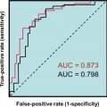
The 2017 ACC/AHA guidelines recommend measuring plasma renin activity (PRA) although newer direct renin measurements are available . Rossi et al. found that the direct renin measurement (e.g., renin mass) was diagnostically equivalent to the PRA measurement . It is important to recognize that many factors besides the analytes that are measured can affect the ratio . Aldosterone (in nanograms per deciliter)/renin (as an activity measurement, nanograms per milliliter per hour) ratios of >20–25 [ng/dL]/[ng/mL per hour] signify hyperaldosteronism with relative suppression of renin (e.g., primary hyperaldosteronism). Ratios of ≥50 [ng/dL]/[ng/mL per hour] are essentially diagnostic for renin-independent primary hyperaldosteronism.
Analytical problems with both the aldosterone and the renin assays have been emphasized that affect the sensitivity and specificity of the ratio for the diagnosis of primary hyperaldosteronism . One editorial cynically recommended that “the ratio should be repeated until it unmistakably is, or is not, raised…” .
Because the aldosterone to renin ratio is not diagnostically infallible, other tests differentiating primary hyperaldosteronism versus primary (essential) hypertension are available. These tests include (1) fludrocortisone suppression test, (2) oral salt loading or IV saline infusion, and (3) the captopril challenge test . The treating physician can also consider these tests as confirmatory measures of primary hyperaldosteronism. Indeed the Endocrine Society’s 2016 guidelines recommend that one confirmatory test be performed to diagnose primary hyperaldosteronism when the aldosterone to renin ratio is elevated. These confirmatory tests are discussed in the next paragraphs.
Fludrocortisone suppression test
The activity of fludrocortisone (9α-fluorohydrocortisone) as a mineralocorticoid should reduce aldosterone concentrations in normal individuals. Evidence of primary hyperaldosteronism would be that aldosterone is not suppressed after fludrocortisone is administered. In normal, nonhypertensive subjects who are not affected by hyperaldosteronism, urinary aldosterone excretion declines to <5 µg/24 h following salt loading or administration of fludrocortisone.
Oral salt loading or IV saline infusion
In normal individuals, salt administration (PO or IV) should lower aldosterone concentrations. However if aldosterone concentrations do not decline following salt administration, primary hyperaldosteronism is possible. If the patient’s BP is extremely elevated, salt loading could be dangerous by further raising BP .
If the patient is volume expanded (e.g., “salt loaded”) via the infusion of 0.9% NaCl (normal saline) at the rate of 500 mL/h for 4 h (i.e., the saline infusion test) or is given 10 g/day of extra NaCl for 3 days, and the aldosterone is not suppressed, the diagnosis of primary hyperaldosteronism is supported. If the aldosterone is suppressed, the diagnosis is essential hypertension. During the oral salt-loading test, aldosterone can be measured in a 24-h urine collected following the 3 days of salt administration. If aldosterone excretion exceeds 10–14 µg/day (28–39 nmol/day) when the urinary sodium excretion is >250 mmol/day, the diagnosis of primary hyperaldosteronism is supported. During the intravenous salt-loading test, plasma aldosterone levels >10 ng/dL (>280 pmol/L) also support the diagnosis of primary hyperaldosteronism.
Captopril challenge test
The normal action of the drug captopril as an ACE inhibitor is to decrease the conversion of ANG I to ANG II. Therefore aldosterone levels should fall. Failure of captopril to reduce aldosterone concentrations can be consistent with primary hyperaldosteronism with autonomous aldosterone secretion. Procedures for these tests are provided in the 2016 Endocrine Society guidelines .
Laboratory notes
A recent Endocrine Society guideline recommends pursuit of the diagnosis of primary hyperaldosteronism in persons with hypertension when: (1) BP exceeds 150/100 mmHg on 3 separate days, (2) the hypertension is resistant (e.g., BP is >140/90 when the patient is treated with three conventional antihypertensive medications (including a diuretic), (3) hypertension is controlled on four or more medications, (4) hypokalemia is detected, (5) an adrenal mass is incidentally detected from an abdominal imaging study, (6) sleep apnea is present, (7) the family history is positive for early-onset hypertension, or (8) a first-degree relative was diagnosed with primary hyperaldosteronism . The relationship between obstructive sleep apnea and hyperaldosteronism is curious: it may be that hyperaldosteronism causes fluid accumulation in the upper airway . Of importance, hypokalemia is not a requirement to consider primary hyperaldosteronism, as not all patients with hyperaldosteronism are hypokalemic. The Endocrine Society guideline reports that only 9%–37% of persons with primary hyperaldosteronism are hypokalemic. On the other hand, if hypokalemia is identified upon testing of a hypertensive individual, there is a high likelihood of hypermineralocorticoidism.
The 2017 ACC/AHA guideline recommends that the plasma aldosterone: renin activity ratio be determined when hypertensive adults (1) suffer from resistant hypertension, (2) are hypokalemic, (3) manifest an adrenal incidentaloma (e.g., an incidentally recognized adrenal mass), (4) have a positive family history of early-onset hypertension, or (5) suffered a stroke before age 40. Regarding incidentalomas, several pathologies should be considered including adrenal adenomas and pheochromocytomas .
Renin-dependent hyperaldosteronism (also known as hyperreninemic hyperaldosteronism) (mechanism 1a1)
In renin-dependent hyperaldosteronism (e.g., secondary hyperaldosteronism), hyperreninism drives hyperaldosteronism and aldosterone to renin ratio is 10 or less. Three disorders cause renin-dependent hyperaldosteronism: (1) primary hyperreninism (e.g., renin-secreting tumor, also known as reninoma), (2) renal artery stenosis, or (3) renal (parenchymal) disease. Renal artery stenosis causes renal hypoperfusion that elicits hyperreninism. Hyperreninism is a major cause of hypertension in renal (parenchymal) disease.
Renin-secreting tumors (mechanism 1a1)
Reninomas (juxtaglomerular cell tumors) are rare tumors that are most commonly diagnosed in adolescents or young adults . Usually there is a long history of headaches, and hypertension is recognized during the subsequent clinical evaluation. Hypokalemia and hypochloremic metabolic alkalosis then point the clinician to RAAS activation. Because aldosterone and renin are both elevated, the aldosterone to renin ratio will not be pathologically elevated.
Rarely in a paraneoplastic fashion, extrarenal tumors may secrete renin . Pathologic hyperreninism has been reported in rare cases of ovarian cancer, pancreatic adenocarcinoma, adenocarcinoma of the lung, small-cell tumor of the lung, adrenocortical carcinoma, and angiolymphoid hyperplasia with eosinophilia.
Renovascular hypertension (mechanism 1a1)
Renovascular hypertension (RVH) is hypertension that results from impaired macro or microvascular flow to the kidney . RVH has many causes as listed in Table 11.3 . Evidence of renovascular disease should be sought when there is resistant hypertension, hypertension of abrupt onset, or worsening or increasingly difficult to control hypertension, severe, acute-onset pulmonary edema, or early-onset hypertension. Renovascular disease should also be considered when the physical examination reveals abdominal bruits (a sound heard with a stethoscope produced by turbulent blood flow through an artery).
Renal ischemia then elicits elevated renin and subsequent elevations in ANG II and aldosterone. Secondary hyperaldosteronism can also be caused by nonvascular renal (parenchymal) disease.
Renal ultrasound is a screening test for renal (parenchymal) disease in pediatrics. For renovascular disease, renal duplex Doppler ultrasound, magnetic resonance angiography, and abdominal computed tomography (CT) scanning are screening tests. Renal angiography (bilateral selective renal intraarterial angiography) is a confirmatory test for renovascular disease. If renal artery stenosis is diagnosed, pharmacologic antihypertensive therapy should be initiated. If such medical therapy fails or the stenosis is not due to atherosclerosis (e.g., fibromuscular dysplasia), percutaneous renal artery angioplasty and/or stent placement can be considered . Surgical revascularization is not superior to medical therapy .
Obviously, RVH is not a primary endocrine disorder. However, because the mechanism of hypertension is renin-induced activation of the RAAS, the topic is included for completeness.
Renin-independent hyperaldosteronism (mechanism 1a1)
If the secretion of aldosterone is autonomous or is under the control of ACTH (e.g., glucocorticoid-remediable aldosteronism, GRA) and is excessive, hyperaldosteronism results. Hyperaldosteronism can cause vasoconstriction, and salt and water retention causing hypertension. In turn, elevated BP will suppress renin secretion. In the evaluation of hypertension, hypokalemia, alkalosis, an elevated aldosterone to renin ratio suggests primary hyperaldosteronism (e.g., renin-independent hyperaldosteronism, hyporeninemic hyperaldosteronism). As well, as noted earlier, hyperaldosteronism should be sought in cases of severe, resistant, or youth-onset hypertension not otherwise explained.
Of note, a depressed renin level is also observed with hyporeninemic hypoaldosteronism in association with diabetes mellitus, following bilateral nephrectomy, with glycyrrhizic acid ingestion (formerly found in black licorice) or mineralocorticoid ingestion, and with administration of certain drugs [e.g., β-adrenergic blockers, clonidine, and DOC].
Renin-independent hyperaldosteronism (also known as primary hyperaldosteronism) can be further divided into sporadic disorders and inherited disorders ( Table 11.7 ). The sporadic disorders are much more common than the genetic disorders. However, for those individuals and families with genetic disorders, proper diagnosis and treatment are extremely important.
Related posts:

Stay updated, free articles. Join our Telegram channel

Full access? Get Clinical Tree


