The epithelium of the breast ductal system is the site of origin for nearly all breast cancers.1,2 The intraductal environment and the significance of the secretions produced within the human mammary gland have been debated for centuries. The recognition of the role of carcinogens in the production of malignancies led to a renewed interest in the ductal anatomy and physiology in the 20th century. Today, the combination of technological enhancement providing direct and indirect access to the luminal milieu and advances in cellular methodology that hold the promise of reliable diagnostic and predictive markers of breast cancer are driving investigators to pursue a ductal approach to breast cancer diagnosis and therapy that will be preemptive and accurate with less toxicity and breast disfigurement.
The identification of high-risk individuals through statistical screening (ie, Gail and Claus models), histologic or cytologic identification of proliferative breast lesions (ie, atypical ductal or lobular hyperplasia and lobular carcinoma in situ), and recognition of genetic abnormalities (ie, BRCA1 and BRCA2) have resulted in an established paradigm for the management of high-risk individuals. Increased surveillance, chemoprevention, and breast extirpation are designed to either diagnose the disease at a treatable stage or prevent it entirely. If a malignancy is diagnosed, then a multidisciplinary approach, using some combination of local therapy with surgical intervention and radiation therapy and systemic therapy with cytotoxic chemotherapy, hormonal therapy, or targeted therapy is today’s standard. Despite dramatic advances, local therapy often results in alterations in the appearance of the breast, and the adverse events associated with systemic therapy remain discouraging.
In defining an ideal intraductal therapy, one seeks an approach that addresses all the tissue at risk, the mammary duct epithelium. Second, the technique for ductal access must be reliable and reproducible, for therapy as well as follow-up. Finally, the agents used intraluminally must exert their effect at a local level without systemic side effects or impact on the consistency or appearance of the breast. This is the challenge facing intraductal investigators. In this chapter, we review recent literature related to the breast ductal anatomy, the preclinical trials evaluating the efficacy of intraducal therapy, and the results of early clinical trials evaluating intraductal therapy.
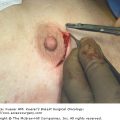
The anatomy of the breast ductal system has been the subject of much debate. At issue are questions regarding the number of ductal orifices on the surface of the nipple and the distribution of the arborizing ductal network. The literature divides into those that note 15 to 20 ducts and those that report 6 to 9. This discrepancy can be explained in part by the technique used for the study. Those reports that based their observation on cannulating intact nipples or imaging intact lactating or nonlactating breasts all report 6 to 9 functional ductal orifices.3-7 On the other hand, the studies that transected the nipple and described the ductal cross-sections concur on 15 to 20 ducts in the subareolar location.8-11 It is likely that some of the differences come from early bifurcations and trifurcations of the ductal network, as noted in ductoscopic examinations,12 whereas others may relate to additional subareolar structures that have yet to be characterized. These questions were addressed in an article evaluating the median number of ducts in the nipple, the volume of the complete duct system or lobes, and modeling of the collecting ducts in the nipple by using a digital 3-dimensional system.13,14 The authors described 3 distinct nipple duct populations that included ducts that maintained a wide communication with the surface of the nipple (type A), ducts that tapered to a minute lumen at their origin (type B), and a group of ducts that arose at the base of the nipple. Seven type A ducts were noted. These results and those of several other careful analyses suggest that further research is necessary to fully characterize this area of the breast.
Early work in prevention of mammary cancer in a chemically induced tumor model evaluated the impact of intraductal viral transduction of breast epithelial cells and subsequent intraductal therapy with gancyclovir.15 The authors postulated that the rapidly dividing epithelial cells in the terminal end buds of the glands were susceptible to chemical carcinogens, and therefore elimination of these cells should reduce malignant transformation. Although greater than 90% of the cells in question were eliminated, the remaining cells had a higher incidence of tumor formation.
The efficacy of triweekly intraductal administration of paclitaxel was contrasted with intraperitoneal treatment of rats with chemically induced mammary cancer.16 The animals were assessed for tumor burden, total number of mammary tumors, apoptosis, and microvessel density. None of the animals treated intraductally developed complications associated with paclitaxel. The intraductally treated animals had significantly reduced tumor burden with increased apoptosis and decreased microvessel density, suggesting an advantage to locally administered therapy with less toxicity.
A more recent study has taken a more comprehensive look at intraductal administration of hormonal and chemotherapeutic agents in the prevention and treatment of breast cancer. Sukumar et al17 used a chemically induced tumor model and an HER-2/neu transgenic spontaneous tumor model. They began by demonstrating they could access the glandular elements of the mouse and rat breasts. Unlike humans, mice and rats have a solitary duct that drains the breast tissue. Excellent distribution of the experimental agents was confirmed. They then evaluated the pharmacology of the intraductally versus intravenously administered pegylated liposomal doxorubicin (PLD) using high-powered liquid chromatography. The peak serum concentrations differed by a factor of 10 when the routes were compared. No myelosuppression was noted in the animals treated with intraductal PLD. They then showed that intraductally administered 4-hydroytamoxifen, the active metabolite of tamoxifen, was highly effective in preventing chemically induced breast tumors. This effect was comparable to subcutaneously administered tamoxifen. The intraductal administration of PLD reduced the size of established spontaneous tumors in transgenic mice. The therapeutic response had variable durability, but 4 of 10 tumors remained in regression throughout the study. With respect to prevention, intraductal PLD resulted in significant reduction of spontaneous development after intraductal therapy when compared with glands that went untreated or received placebo. This study provided comprehensive evidence that translational studies evaluating intraductally administered agents for the prevention and treatment of breast cancer were necessary and appropriate.
These studies raise a host of concerns that must be addressed in clinical trials. Is the accessibility of the human breast ductal system a reliable and reproducible route for the administration of intraductal agents? Is therapy through a single ductal orifice sufficient, or should therapy be administered through the entire ductal system? What are the pharmacokinetics and toxicity of chemotherapeutic agents administered through the human lactiferous duct? Will intraductal therapy result in unanticipated beneficial effects, such as tumor autoimmunity?
Stay updated, free articles. Join our Telegram channel

Full access? Get Clinical Tree


