Headache is the most common complaint of patients with increased ICP. In its classic form, the head pain is severe, resistant to common analgesics, and reaches maximum intensity on awakening in the morning.29 Decreased venous drainage in the supine position likely accounts for this observation. Patients frequently report immediate relief from their headache by vomiting. However, the majority have nonspecific tension-type or migraine-like headaches. The patient with increased ICP falls easily, particularly backward. With rising pressure, nausea and vomiting ensue. The patient becomes increasingly somnolent and ultimately lapses into a coma.
Funduscopic examination reveals papilledema in about half of patients with increased ICP. Absence of venous pulsations within the center of the optic disc is an early finding, whereas papilledema with blurring of the disc margins or small hemorrhages characterizes later stages. The Foster-Kennedy syndrome—optic nerve atrophy as a result of a sphenoid wing meningioma and contralateral papilledema from increased ICP—is rarely seen in the days of improved neuroimaging methods and earlier diagnosis.
Focal neurologic deficits can help localize the mass accounting for the pressure increase. Cognitive complaints such as slowness to respond and inattentiveness reflect frontal lobe dysfunction. Gaze paresis to the side opposite the lesion indicates involvement of the frontal eye field. Posterior frontal masses cause contralateral hemiparesis. Hemianesthesia or complex neglect syndromes reflect parietal lobe pathology. Temporal and occipital lobe disease causes visual field deficits. An upward gaze paresis occurs in patients with tumors of the tectal region such as pineal neoplasms or metastases. Paresis of extraocular muscles results from stretch injury of the fourth or sixth nerve or uncal herniation with compression of the third nerve. However, the clinician must be aware of “false” localizing signs. Temporal lobe tumors can cause compression of the cerebral peduncle at the tentorial notch on the opposite side, resulting in a hemiparesis on the same side as the mass lesion (Kernohan syndrome).30
Symptoms are aggravated by vasogenic edema surrounding intraparenchymal masses and partially or completely resolve with medical management. Hyponatremia as a result of inappropriate secretion of antidiuretic hormone is observed as a metabolic complication of increased ICP. Sphincter incontinence occurs in chronically elevated ICP. Patients with acute meningitis present with classic signs of meningeal irritation, including photophobia, phonophobia, and a Kernig or Brudzinski sign. In meningeal carcinomatosis, these signs are frequently absent.
Elevated ICP in infants results in increased head circumference. Chronic hydrocephalus can be recognized on plain radiographs of the skull as focal thinning of the tabula interna of the skull (Lückenschädel). This is accompanied by personality changes and loss of previously acquired motor skills. Herniation of one cerebellar tonsil causes a head tilt, neck stiffness, and unilateral forced eye closure.31 Tectal masses result in upgaze inhibition, light-near dissociation of pupillary response, and convergence-retraction nystagmus (Parinaud syndrome). Pressure on the mesencephalic tegmentum leads to pathologic lid retraction and an upward gaze palsy (setting sun sign).
Slowly progressive static ICP changes are accompanied by little or no symptoms. On the other hand, clinical deterioration is profound when dynamic pressure changes such as plateau waves occur or abnormal intracranial compartmentalization or herniation ensues.32 Signs and symptoms of increased ICP manifest earlier in patients with lesions of the posterior fossa because of the small size of this compartment. A brief bedside assessment including level of consciousness, pupillary size and reflexes, extraocular movements, blood pressure, heart rate, breathing pattern, and motor response to noxious stimuli enables the clinician to determine if herniation is present and which level of the central neuraxis is compromised. The triad of changes in breathing pattern, arterial hypertension, and bradycardia observed with rising ICP is known as the Kocher-Cushing reflex.33 In uncal herniation from temporal lobe masses or herpes encephalitis, ipsilateral compression of the third nerve and associated parasympathetic nerve fibers leads to pupillary dilatation before extraocular dysmotility. With progressive shift of brain substance, complete third nerve palsy ensues and signs of midbrain dysfunction appear. Patients develop contralateral hemiparesis from pressure on the cerebral peduncle and ultimately become stuporous. Increasing pressure from hemispheric or diencephalic mass lesions results in central (transtentorial) herniation. This leads to a progressive syndrome reflecting sequential damage to brainstem structures in a rostrocaudal fashion.32 At the early diencephalic stage, mild changes in the patient’s alertness are accompanied by periodic breathing, yawning, or hiccupping. Pupils are small but remain reactive to light. With further progression of central herniation, the patient becomes obtunded or stuporous. Roving eye movements reflect diffuse cortical dysfunction and preservation of lower brainstem gaze centers. Noxious stimuli elicit flexion of upper extremities and extension of lower extremities (decorticate posturing). Midsize pupils unresponsive to light indicate midbrain dysfunction. Damage to the mesencephalic reticular activating system produces coma. A fast and regular breathing pattern evolves (central neurogenic hyperventilation). Transition to the pontine stage of central herniation is accompanied by extensor posturing of all limbs to noxious stimulation (decerebrate posturing). Absence of the oculocephalic reflex (doll’s head maneuver) and horizontal eye movements to caloric stimulation of the vestibular system indicate damage to pontine structures. Breathing becomes apneustic with pontine compression. When the cerebellar tonsils herniate through the foramen magnum, ataxic breathing is observed and the blood pressure drops.32
The syndrome of raised ICP and cerebral herniation can evolve slowly over days to weeks or acutely over hours. Rapid progression usually indicates hemorrhage. Subdural hematomas in patients with coagulopathies can evolve so rapidly that signs of cerebral herniation are present before an imaging study can be obtained. Hemorrhage into a metastatic focus is typically characterized by the sudden onset of focal neurologic signs, including seizures. Intraparenchymal hemorrhage as a result of coagulopathy leads to slowly progressive neurologic deterioration.10
A peculiar syndrome is associated with tumors or developmental abnormalities causing a pressure valve effect, such as the colloid cyst of the foramen of Monro (Fig. 120.1B). Patients, typically in their late childhood or early adulthood, report sudden onset of severe imbalance, headache, and nausea that is brought on by positional changes (bending down) or Valsalva maneuvers. Sudden deaths have occurred, stressing the need for close observation of these patients until appropriate therapy can be provided.34,35
IIH (pseudotumor cerebri) is mostly characterized by nocturnal or hypnopompic headaches aggravated by Valsalva maneuver. Nonspecific visual changes, diplopia due to sixth nerve palsy, or transient visual obscuration are less frequent manifestations. Especially the latter is an ominous sign indicating the need for immediate intervention as the patient’s vision is at risk. On physical examination, papilledema is the most striking abnormality.36 The blind spot is enlarged.
Another characteristic clinical syndrome is recognized in patients with chronic disturbance of spinal fluid reabsorption. These patients, or more likely their family members on their behalf, report a combination of cognitive decline, precipitate micturition, and gait apraxia.37,38 Dementia is usually of the subcortical type. Precipitate micturition reflects dysfunction of the cortical center for bladder control (paracentral lobule). Minimal bladder filling results in the uncontrollable urge to urinate. The gait disturbance is characterized by difficulty initiating ambulation and postural instability with retropulsion. Strength is preserved.
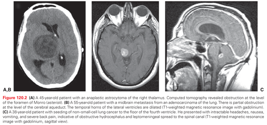
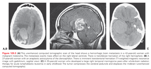
The history and clinical examination detect the presence of increased ICP. Imaging studies are helpful in determining its cause and confirming the clinical impression. The most readily available imaging study is unenhanced computed tomography. The study is adequate to determine the presence of intraventricular and subarachnoid CSF flow obstruction (Fig. 120.2), as well as uncal, transfalcian, and transtentorial herniation (Fig. 120.3). The presence of intracranial hemorrhage or a neoplastic or infectious mass lesion can be identified and emergency treatment initiated. Transependymal edema is seen as periventricular hypodensity and indicates CSF flow obstruction.


Stay updated, free articles. Join our Telegram channel

Full access? Get Clinical Tree


