(ICD‐O C01, C05.1, 2, C09, C10.0, 2, 3, C11–13)


PHARYNX (ICD‐O C01, C05.1, 2, C09, C10.0, 2, 3, C11–13)


Rules for Classification


The classification applies only to carcinomas. There should be histological confirmation of the disease.


Anatomical Sites and Subsites


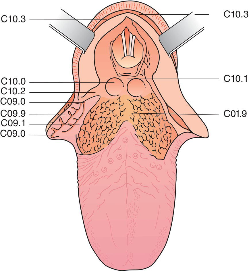
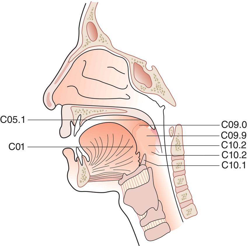
Oropharynx (C01, C05.1, 2, C09.0, 1, 9, C10.0, 2, 3) (Figs. 41, 42)


  1. Anterior wall (glosso‐epiglottic area)

    1. Base of tongue (posterior to the vallate papillae or posterior third) (C01)
    2. Vallecula (C10.0)

  2. Lateral wall (C10.2)

    1. Tonsil (C09.9)
    2. Tonsillar fossa (C09.0) and tonsillar (faucial) pillars (C09.1)
    3. Glossotonsillar sulci (tonsillar pillars) (C09.1)

  3. Posterior wall (C10.3)
  4. Superior wall

    1. Inferior surface of soft palate (C05.1)
    2. Uvula (C05.2)
Schematic illustration of Inferior surface of soft palate, Base of tongue (posterior to the vallate papillae or posterior third),Lateral wall, Tonsil, Tonsillar fossa (C09.0) and tonsillar (faucial) pillars, Glossotonsillar sulci (tonsillar pillars)

Fig. 41

Schematic illustration of Posterior wall, Vallecula, Posterior wall, Tonsil, Glossotonsillar sulci (tonsillar pillars), Tonsillar fossa and tonsillar (faucial) pillars

Fig. 42


Note


The lingual (anterior) surface of the epiglottis (C10.1) is included with the larynx, suprahyoid epiglottis (see pages 35–36).


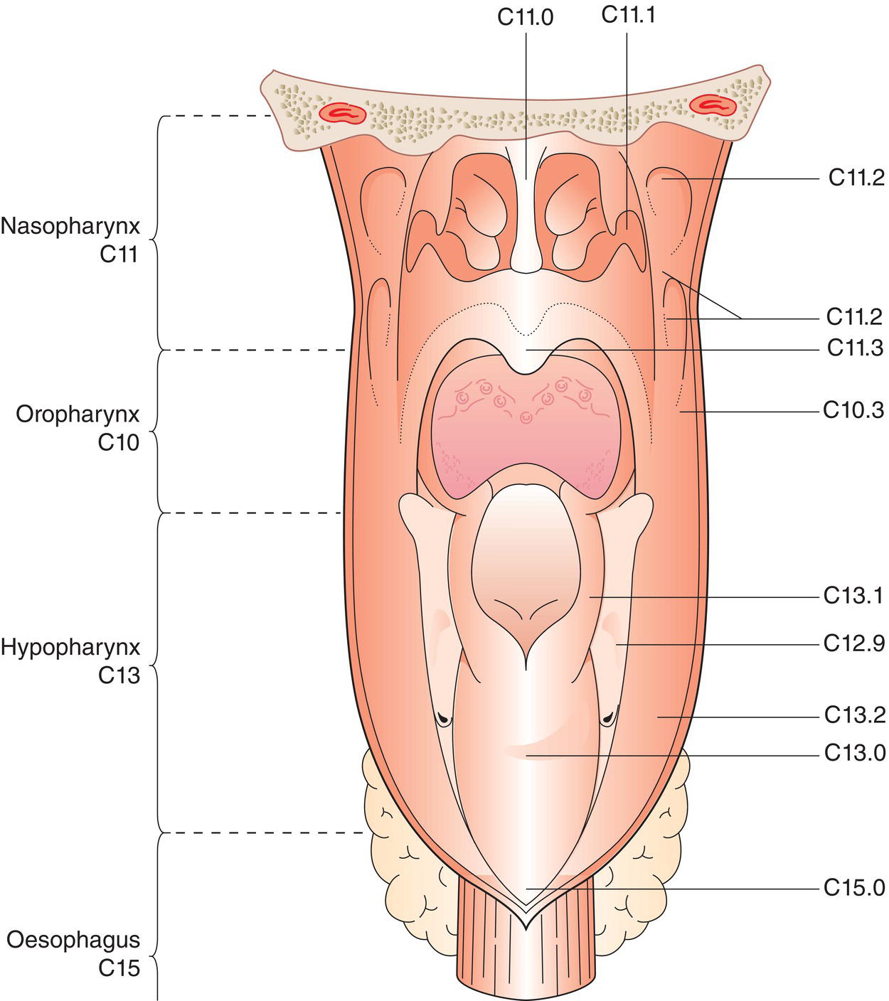
Nasopharynx (Fig. 43)


  1. Postero‐superior wall: extends from the level of the junction of the hard and soft palates to the base of the skull (C11.0, 1)
  2. Lateral wall: including the fossa of Rosenmüller (C11.2)
  3. Inferior wall: consists of the superior surface of the soft palate (C11.3)

Note


The margin of the choanal orifices, including the posterior margin of the nasal septum, is included with the nasal fossa.


Hypopharynx (C12, C13) (Fig. 43)


  1. Pharyngo‐oesophageal junction (postcricoid area) (C13.0): extends from the level of the arytenoid cartilages and connecting folds to the inferior border of the cricoid cartilage, thus forming the anterior wall of the hypopharynx
  2. Piriform sinus (C12.9): extends from the pharyngoepiglottic fold to the upper end of the oesophagus. It is bounded laterally by the thyroid cartilage and medially by the hypopharyngeal surface of the aryepiglottic fold (C13.1) and the arytenoid and cricoid cartilages
  3. Posterior pharyngeal wall (C13.2): extends from the superior level of the hyoid bone (or floor of the vallecula) to the level of the inferior border of the cricoid cartilage and from the apex of one piriform sinus to the other
Schematic illustration of Postero‐superior wall that extends from the level of the junction of the hard and soft palates to the base of the skull, Lateral wall inluding the fossa of Rosenmüller, Inferior wall consists of the superior surface of the soft palate.

Fig. 43


Regional Lymph Nodes


The regional lymph nodes are the cervical nodes.


The supraclavicular fossa (relevant to classifying nasopharyngeal carcinoma) is the triangular region defined by three points:


  1. the superior margin of the sternal end of the clavicle;
  2. the superior margin of the lateral end of the clavicle;
  3. the point where the neck meets the shoulder. This includes caudal portions of Levels IV and V (classification according to Robbins et al.2).

TN Clinical Classification


T – Primary Tumour


TX Primary tumour cannot be assessed
T0 No evidence of primary tumour
Tis Carcinoma in situ

Oropharynx


p16 negative cancers of the oropharynx, or oropharyngeal cancers without a p16 immunohistochemistry performed


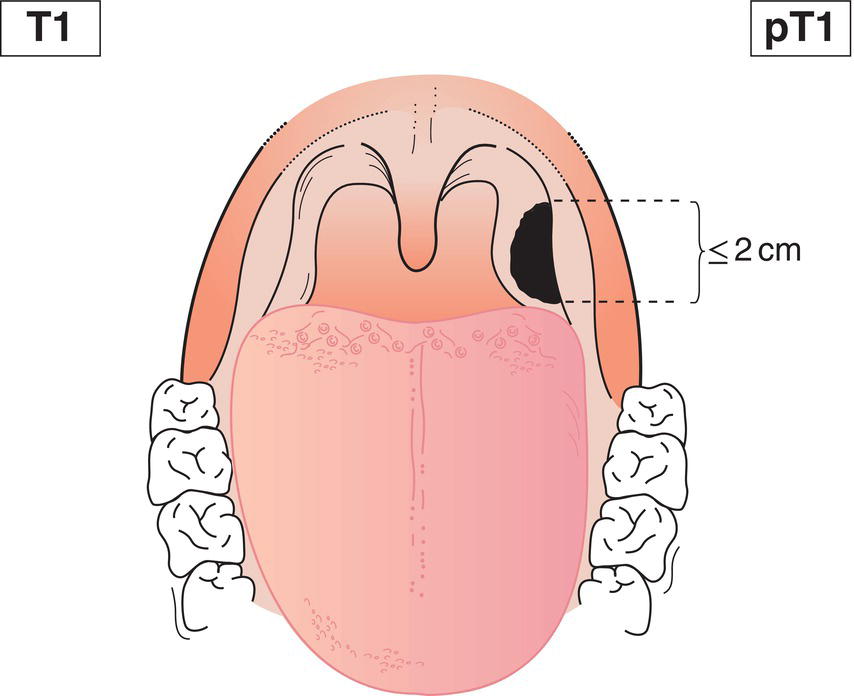
T1 Tumour 2 cm or less in greatest dimension (Fig. 44)
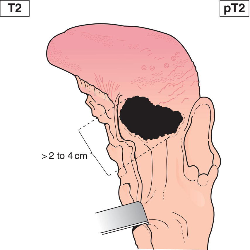
T2 Tumour more than 2 cm but not more than 4 cm in greatest dimension (Fig. 45)
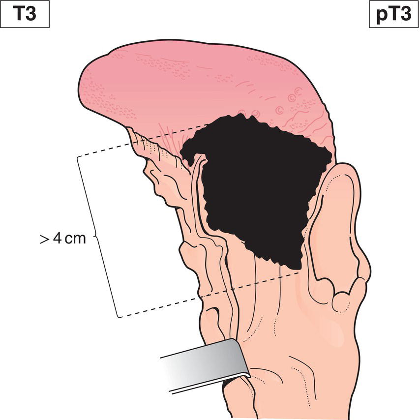
T3 Tumour more than 4 cm in greatest dimension or extension to lingual surface of epiglottis (Fig. 46)
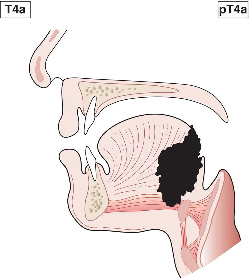
T4a Tumour invades any of the following: larynx, deep/extrinsic muscle of tongue (genioglossus, hyoglossus, palatoglossus and styloglossus), medial pterygoid, hard palate or mandible* (Fig. 47)
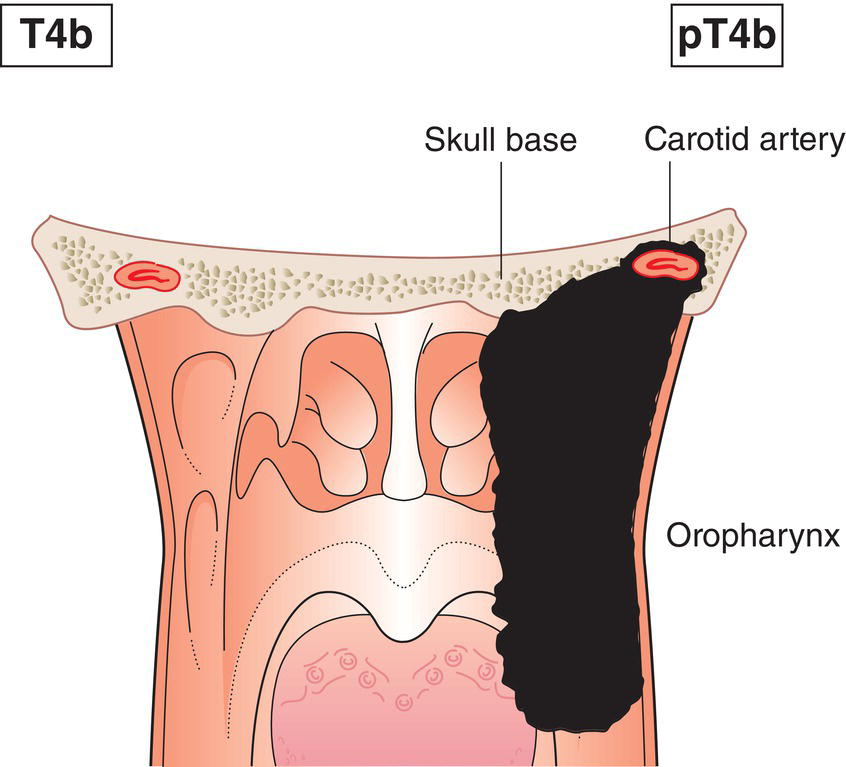
T4b Tumour invades any of the following: lateral pterygoid muscle, pterygoid plates, lateral nasopharynx, skull base; or encases carotid artery (Fig. 48)
Note


* Mucosal extension to lingual surface of epiglottis from primary tumours of the base of the tongue and vallecula does not constitute invasion of the larynx.

Schematic illustration of tumour 2 cm or less in greatest dimension.

Fig. 44

Schematic illustration of tumour more than 2 cm but not more than 4 cm in greatest dimension.

Fig. 45

Schematic illustration of tumour more than 4 cm in greatest dimension or extension to lingual surface of epiglottis.

Fig. 46

Schematic illustration of tumour invades any of the following: larynx, deep or extrinsic muscle of tongue, medial pterygoid,
hard palate or mandible.

Fig. 47

Schematic illustration of tumour invades lateral pterygoid muscle, pterygoid plates, lateral nasopharynx, skull base; or encases carotid artery.

Fig. 48


Oropharynx – p16‐Positive Tumours


Tumours that have positive p16 immunohistochemistry overexpression.


T1 Tumour 2 cm or less in greatest dimension (Fig. 44)
T2 Tumour more than 2 cm but not more than 4 cm in greatest dimension (Fig. 45)
T3 Tumour more than 4 cm in greatest dimension or extension to lingual surface of epiglottis (Fig. 46)
T4 Tumour invades any of the following: larynx, deep/extrinsic muscle of tongue (genioglossus, hyoglossus, palatoglossus and styloglossus), medial pterygoid, hard palate, mandible*, lateral pterygoid muscle, pterygoid plates, lateral nasopharynx, skull base; or encases carotid artery (Figs. 47, 48, 49)
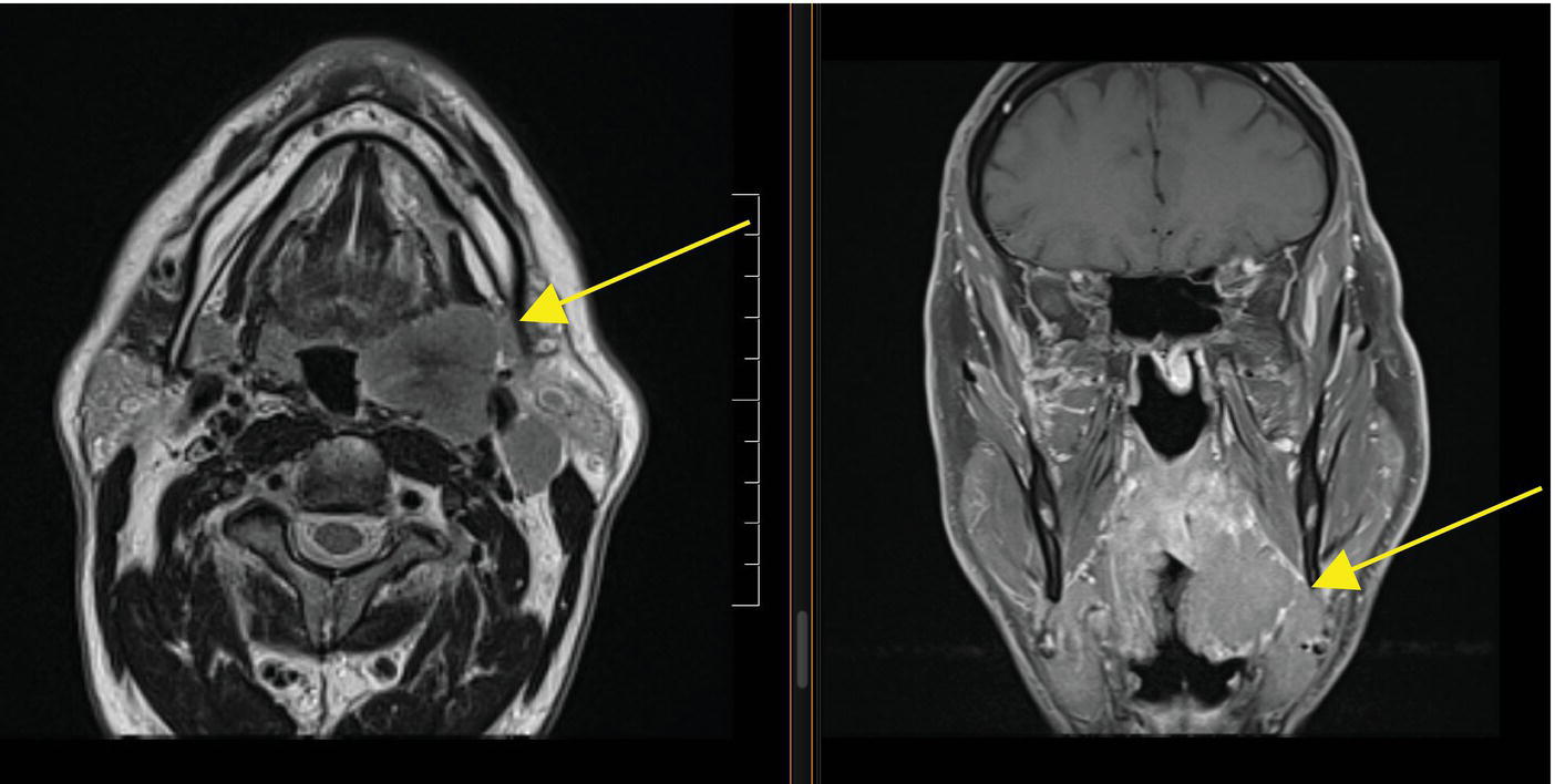
Photo depicts an MRI of an HPV-positive T4 tonsil lesion extending into the parapharyngeal space.

Fig. 49 MRI of an HPV‐positive T4 tonsil lesion extending into the parapharyngeal space (arrow).


Nasopharynx


T1 Tumour confined to nasopharynx, or extends to oropharynx and/or nasal cavity (Fig. 50)
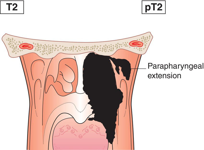
T2 Tumour with extension to parapharyngeal space and/or infiltration of the medial pterygoid, lateral pterygoid and/or prevertebral muscles (Figs. 51, 52)
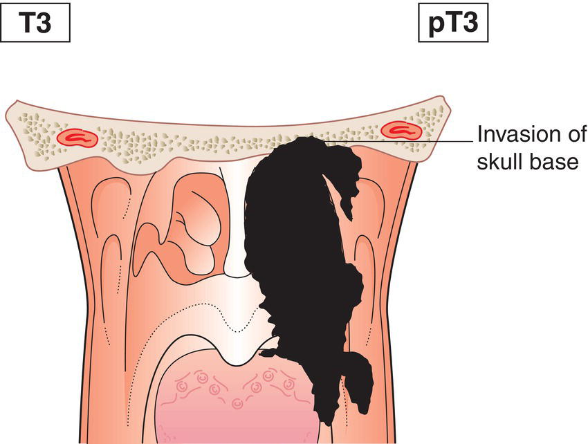
T3 Tumour invades bony structures of skull base cervical vertebra, pterygoid structures and/or paranasal sinuses (Fig. 53)
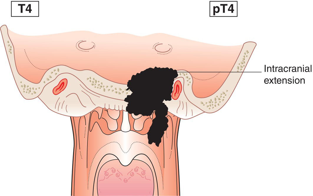
T4 Tumour with intracranial extension and/or involvement of cranial nerves, hypopharynx, orbit, parotid gland and/or infiltration beyond the lateral surface of the lateral pterygoid muscle (Fig. 54)
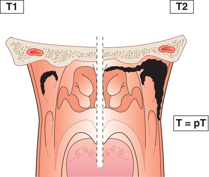
Schematic illustration of tumour confined to nasopharynx, or extends to oropharynx and nasal cavity.

Fig. 50

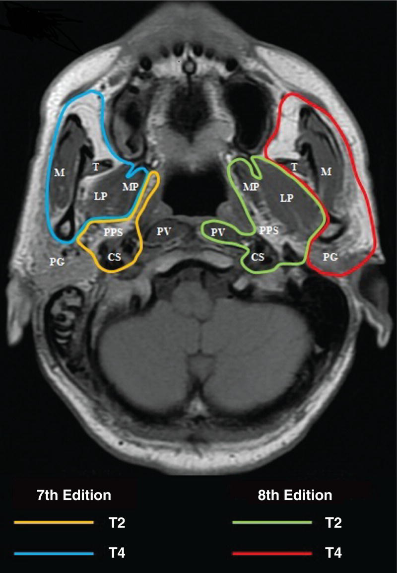
Photo depicts the Differences in defining criteria between the previous 7th edition and the current 8th edition: changing the extent of soft tissue involvement as T2 and T4 criteria.

Fig. 51 Differences in defining criteria between the previous 7th edition and the current 8th edition: changing the extent of soft tissue involvement as T2 and T4 criteria. CS indicates carotid space; LP, lateral pterygoid muscle; M, masseter muscle; MP, medial pterygoid muscle; PG, parotid gland; PPS, parapharyngeal space; PV, prevertebral muscle; T, temporalis muscle.


Source: Modified from Pan JJ, Ng WT, Zong JF, et al. (2016) Proposal for the 8th edition of the AJCC/UICC staging system for nasopharyngeal cancer in the era of intensity‐modulated radiotherapy. Cancer 122(4):546–558.

Schematic illustration of tumour with extension to parapharyngeal space and infiltration of the medial
pterygoid, lateral pterygoid and prevertebral muscles.

Fig. 52

Schematic illustration of tumour invades bony structures of skull base cervical vertebra, pterygoid structures
and paranasal sinuses.

Fig. 53

Schematic illustration of tumour with intracranial extension and involvement of cranial nerves,
hypopharynx, orbit, parotid gland and infiltration beyond the lateral surface of
the lateral pterygoid muscle.

Fig. 54


Hypopharynx


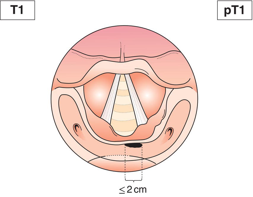
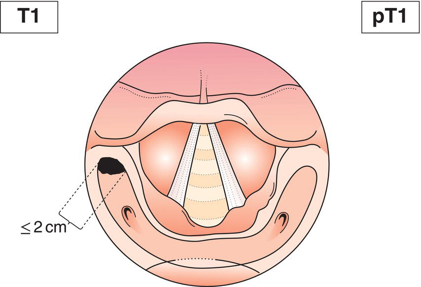
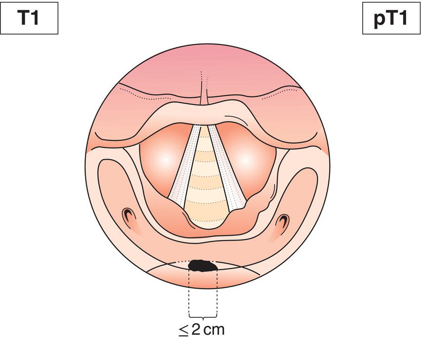
T1 Tumour limited to one subsite of hypopharynx (see Fig. 43) and/or 2 cm or less in greatest dimension (Figs. 55, 56, 57)
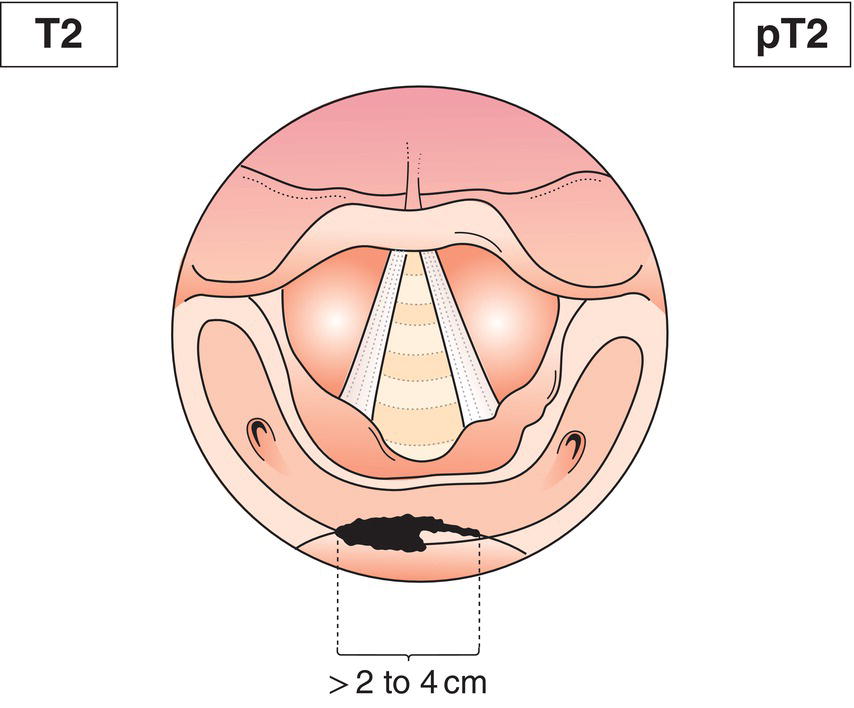
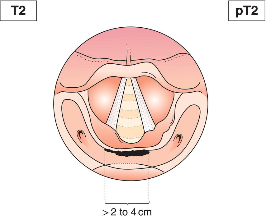
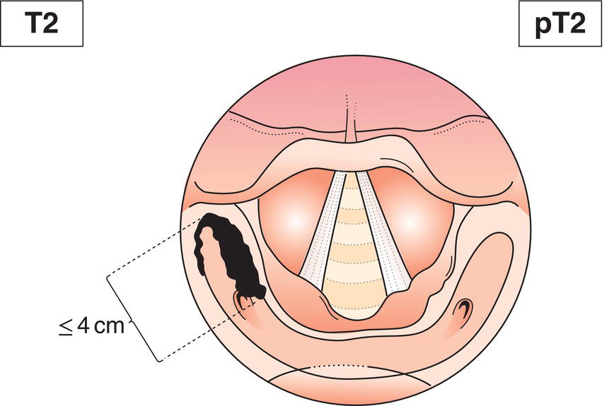
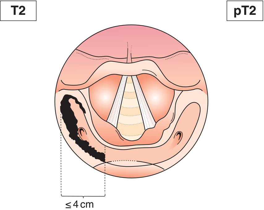
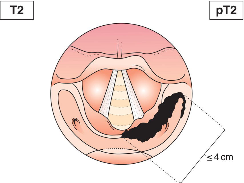
T2 Tumour invades more than one subsite of hypopharynx or an adjacent site, or measures more than 2 cm but not more than 4 cm in greatest dimension, without fixation of hemilarynx (Figs. 58, 59, 60, 61, 62)
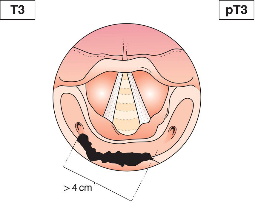
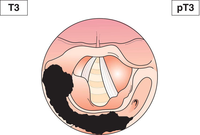
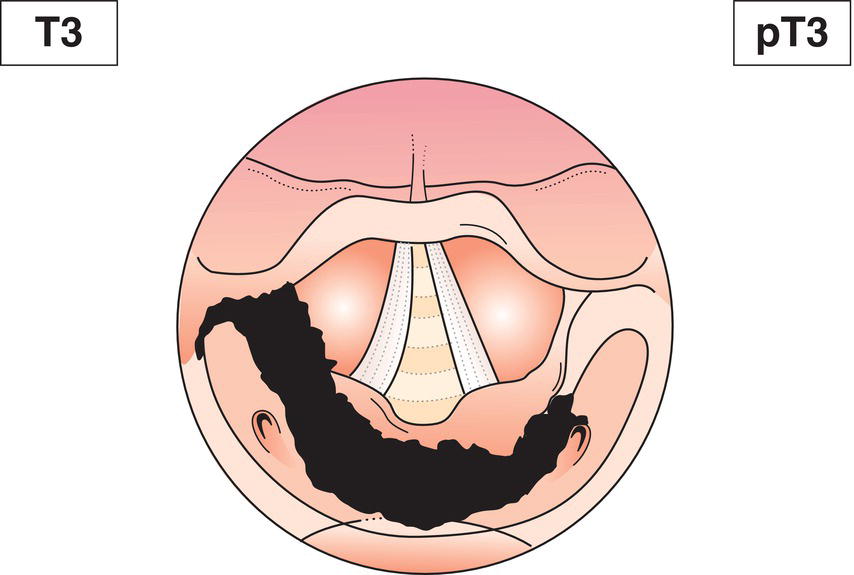
T3 Tumour more than 4 cm in greatest dimension, or with fixation of hemilarynx or extension to oesophagus (Figs. 63, 64, 65)
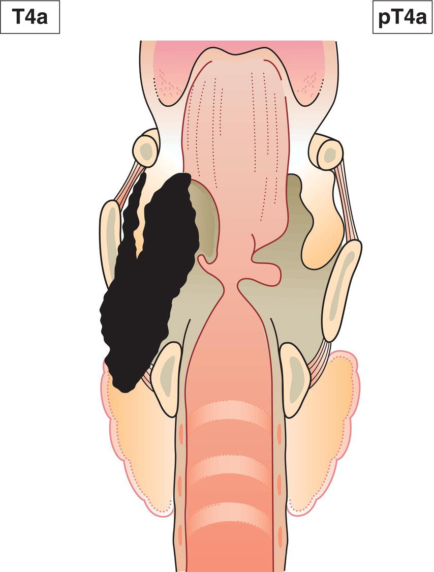
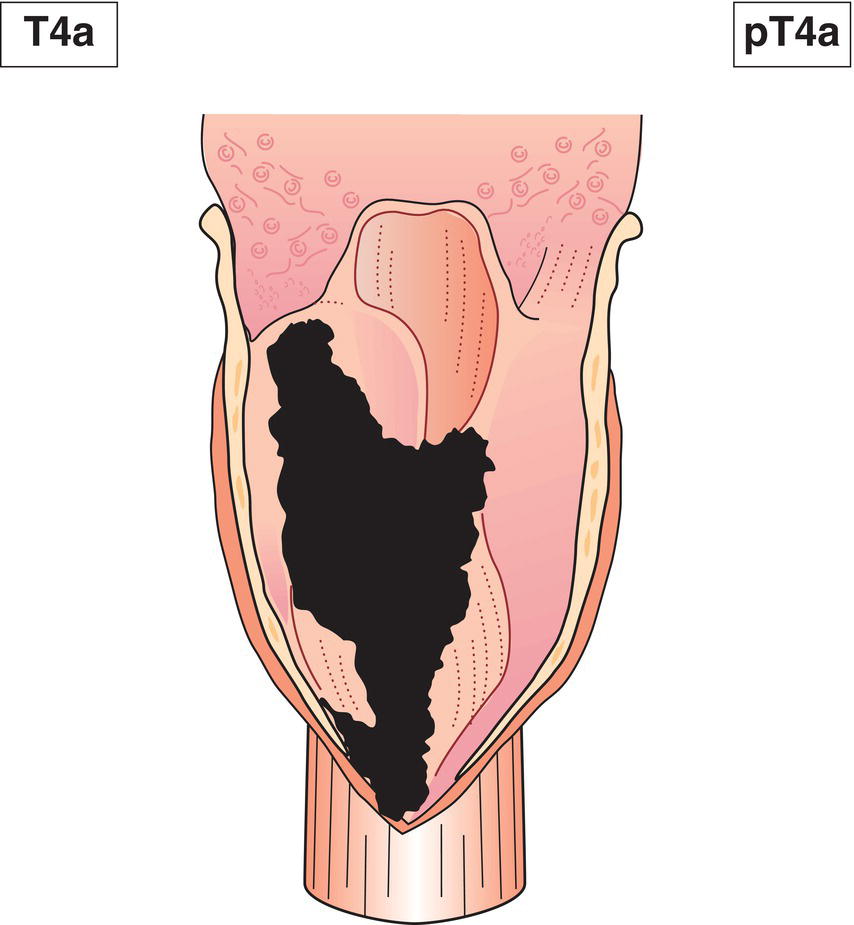
T4a Tumour invades any of the following: thyroid/cricoid cartilage, hyoid bone, thyroid gland, oesophagus, central compartment soft tissue* (Figs. 66, 67)
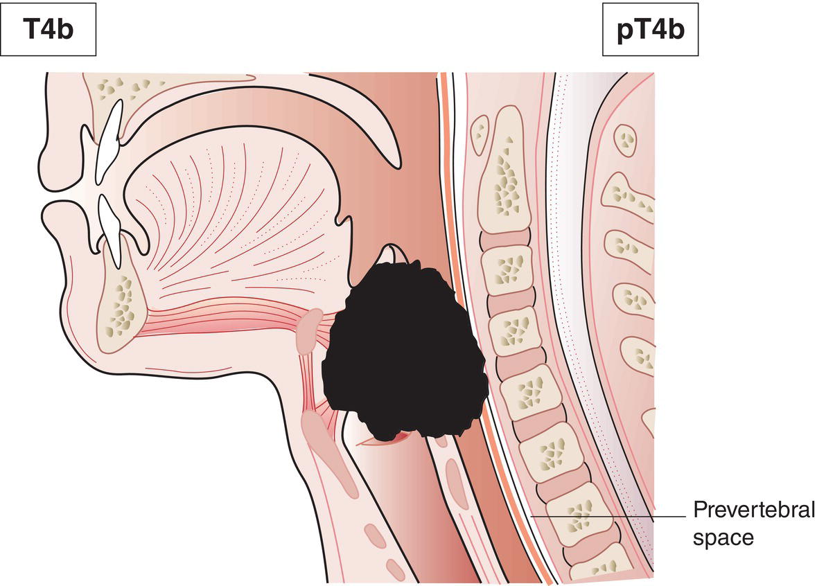
T4b Tumour invades prevertebral fascia (Fig. 68), encases carotid artery or invades mediastinal structures
Note


* Central compartment soft tissue includes prelaryngeal strap muscles and subcutaneous fat.

Schematic illustration of tumour limited to one subsite of hypopharynx and 2 cm or less
in greatest dimension.

Fig. 55

Schematic illustration of tumour limited to one subsite of hypopharynx and 2 cm or less
in greatest dimension.

Fig. 56

Schematic illustration of tumour limited to one subsite of hypopharynx and 2 cm or less
in greatest dimension.

Fig. 57

Schematic illustration of tumour invades more than one subsite of hypopharynx or an adjacent site, or measures more than 2 cm but not more than 4 cm in greatest dimension, without fixation of hemilarynx.

Fig. 58

Schematic illustration of tumour invades more than one subsite of hypopharynx or an adjacent site, or measures more than 2 cm but not more than 4 cm in greatest dimension, without fixation of hemilarynx.

Fig. 59

Schematic illustration of tumour invades more than one subsite of hypopharynx or an adjacent site, or measures more than 2 cm but not more than 4 cm in greatest dimension, without fixation of hemilarynx.

Fig. 60

Schematic illustration of tumour invades more than one subsite of hypopharynx or an adjacent site, or measures more than 2 cm but not more than 4 cm in greatest dimension, without fixation of hemilarynx.

Fig. 61

Schematic illustration of tumour invades more than one subsite of hypopharynx or an adjacent site, or measures more than 2 cm but not more than 4 cm in greatest dimension, without fixation of hemilarynx.

Fig. 62

Schematic illustration of tumour more than 4 cm in greatest dimension, or with fixation of hemilarynx or extension to oesophagus.

Fig. 63

Schematic illustration of tumour more than 4 cm in greatest dimension, or with fixation of hemilarynx or extension to oesophagus.

Fig. 64

Schematic illustration of tumour more than 4 cm in greatest dimension, or with fixation of hemilarynx or extension to oesophagus.

Fig. 65

Schematic illustration of tumour invades any of the following: thyroid or cricoid cartilage, hyoid bone, thyroid gland, oesophagus, central compartment soft tissue.

Fig. 66

Schematic illustration of tumour invades any of the following: thyroid/cricoid cartilage, hyoid bone, thyroid gland, oesophagus, central compartment soft tissue.

Fig. 67

Schematic illustration of tumour invades prevertebral fascia, encases carotid artery or invades mediastinal structures.

Fig. 68


p16‐Negative Oro‐ and Hypopharynx


N – Regional Lymph Nodes


See Head and Neck Tumours for p16‐negative oropharynx tumours and hypopharynx.


p16‐Positive Oropharynx


Clinical


NX Regional lymph nodes cannot be assessed
N0 No regional lymph node metastasis
N1 Unilateral metastasis, in lymph node(s), all 6 cm or less in greatest dimension (Fig. 69)
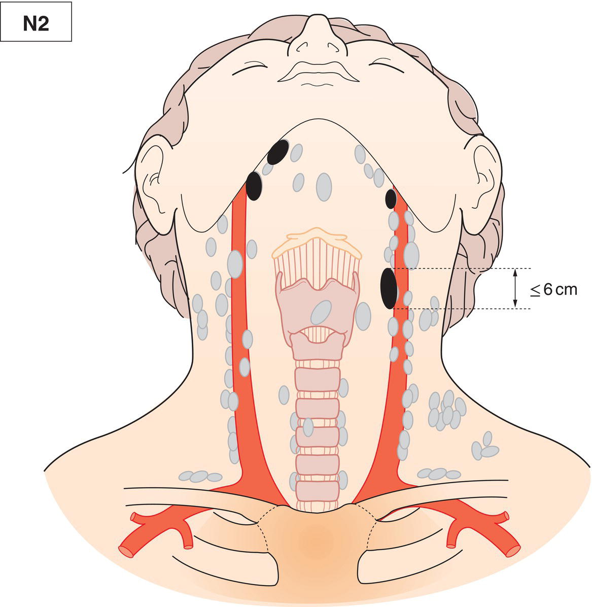
N2 Contralateral or bilateral metastasis in lymph node(s), all 6 cm or less in greatest dimension (Fig. 70)
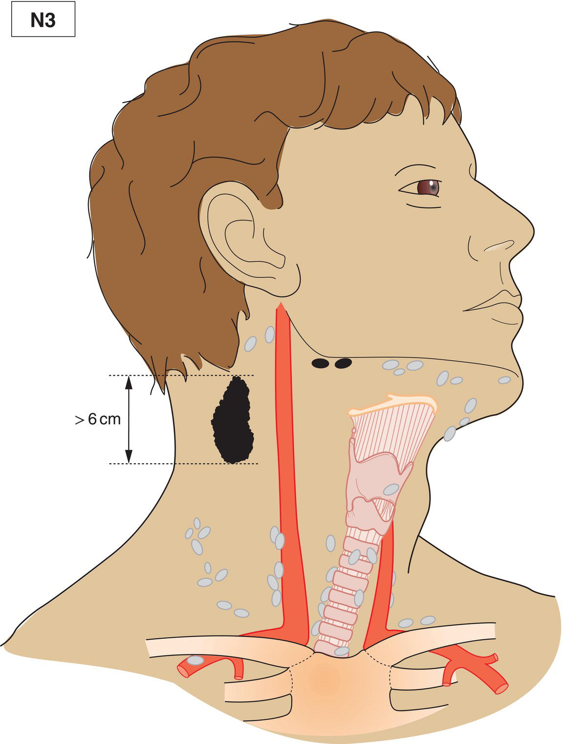
N3 Metastasis in lymph node(s) greater than 6 cm in dimension (Fig. 71)
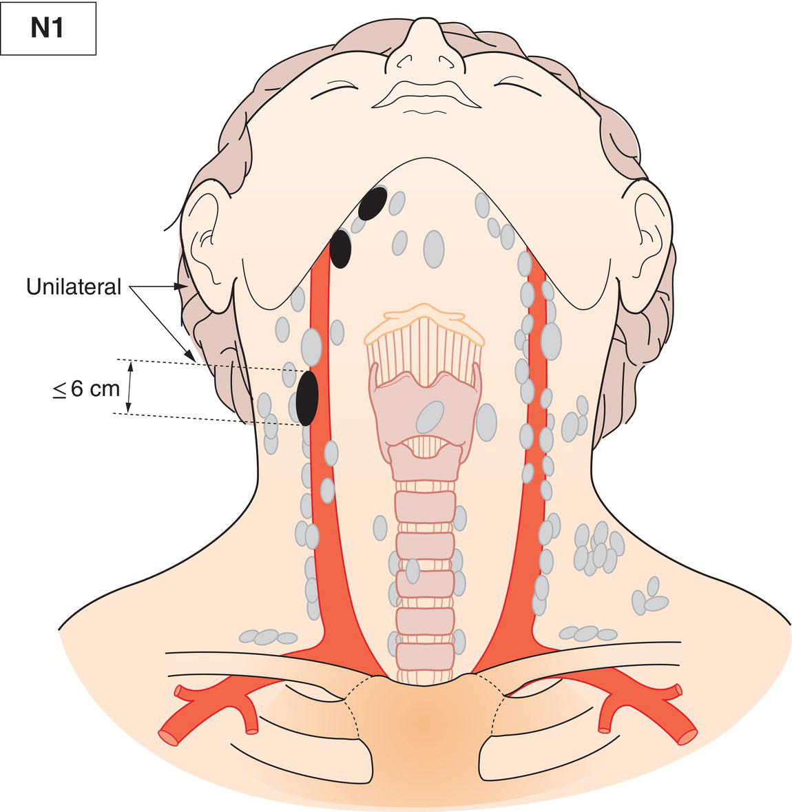
Schematic illustration of Unilateral metastasis, in lymph node(s), all 6 cm or less in greatest dimension.

Fig. 69

Schematic illustration of Contralateral or bilateral metastasis in lymph node(s), all 6 cm or less in greatest
dimension.

Fig. 70

Schematic illustration of Metastasis in lymph node(s) greater than 6 cm in dimension.

Fig. 71


Pathological


pNX Regional lymph nodes cannot be assessed
pN0 No regional lymph node metastasis
pN1 Metastasis in 1 to 4 lymph node(s) (Fig. 72)
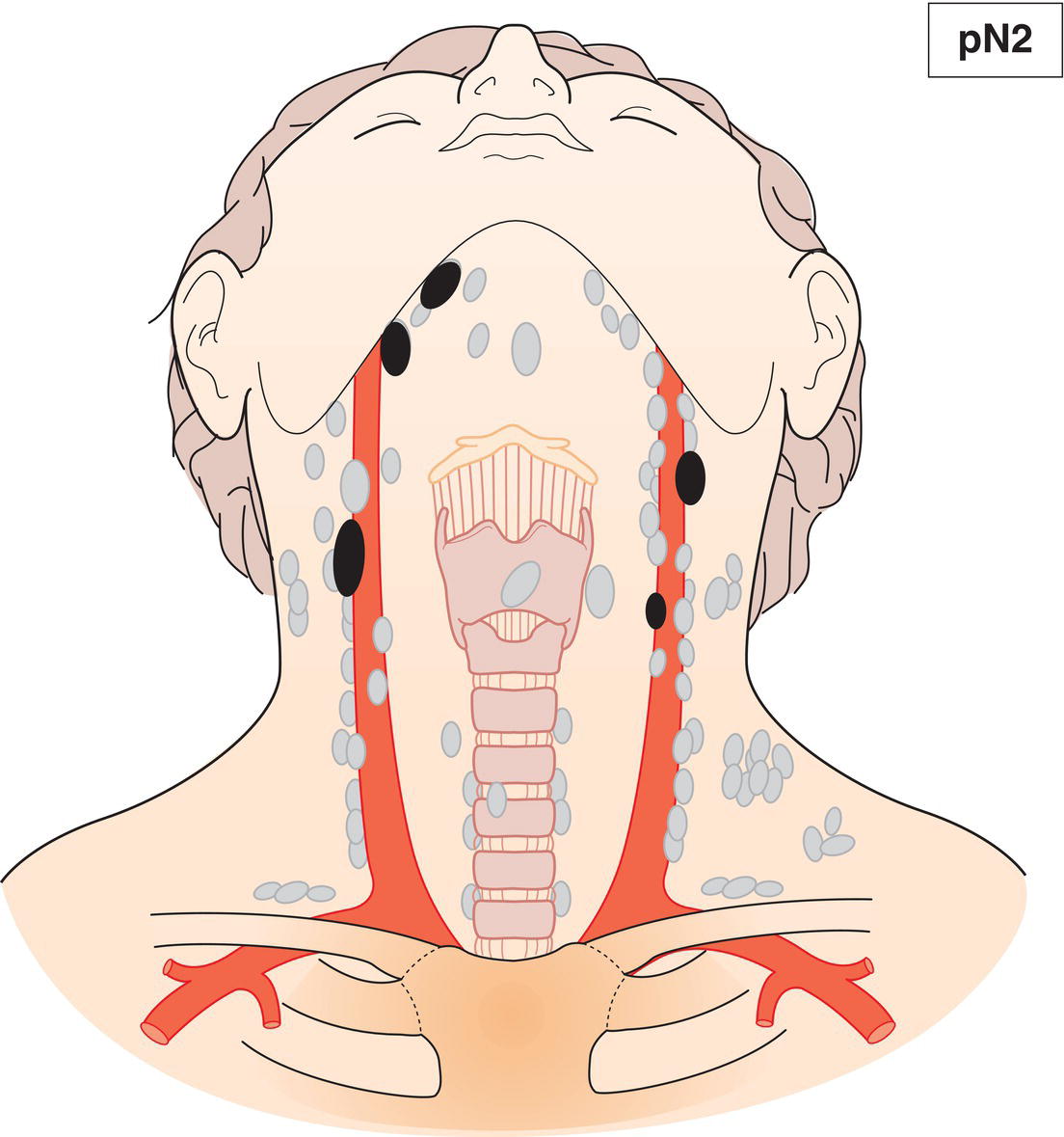
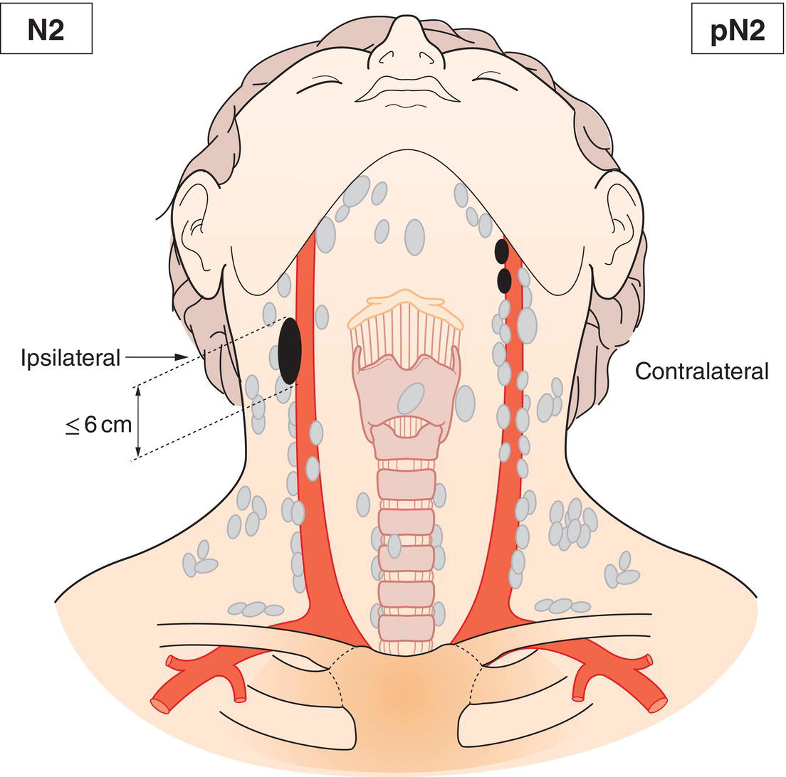
pN2 Metastasis in 5 or more lymph nodes (Fig. 73)
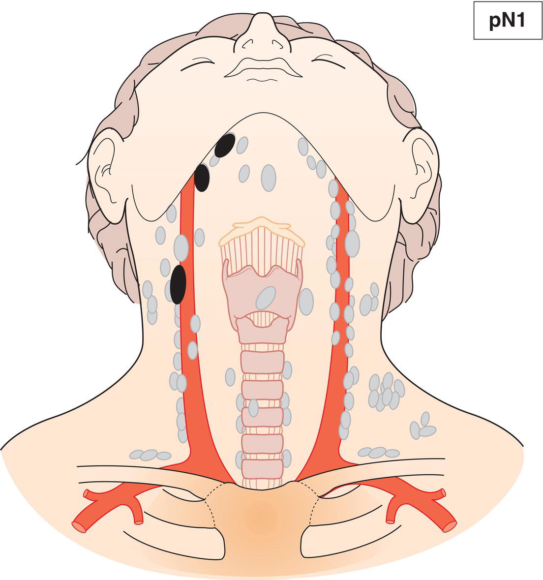
Schematic illustration of Metastasis in 1 to 4 lymph nodes.

Fig. 72

Schematic illustration of Metastasis in 5 or more lymph nodes.

Fig. 73


Nasopharynx


N – Regional Lymph Nodes


NX Regional lymph nodes cannot be assessed
N0 No regional lymph node metastasis
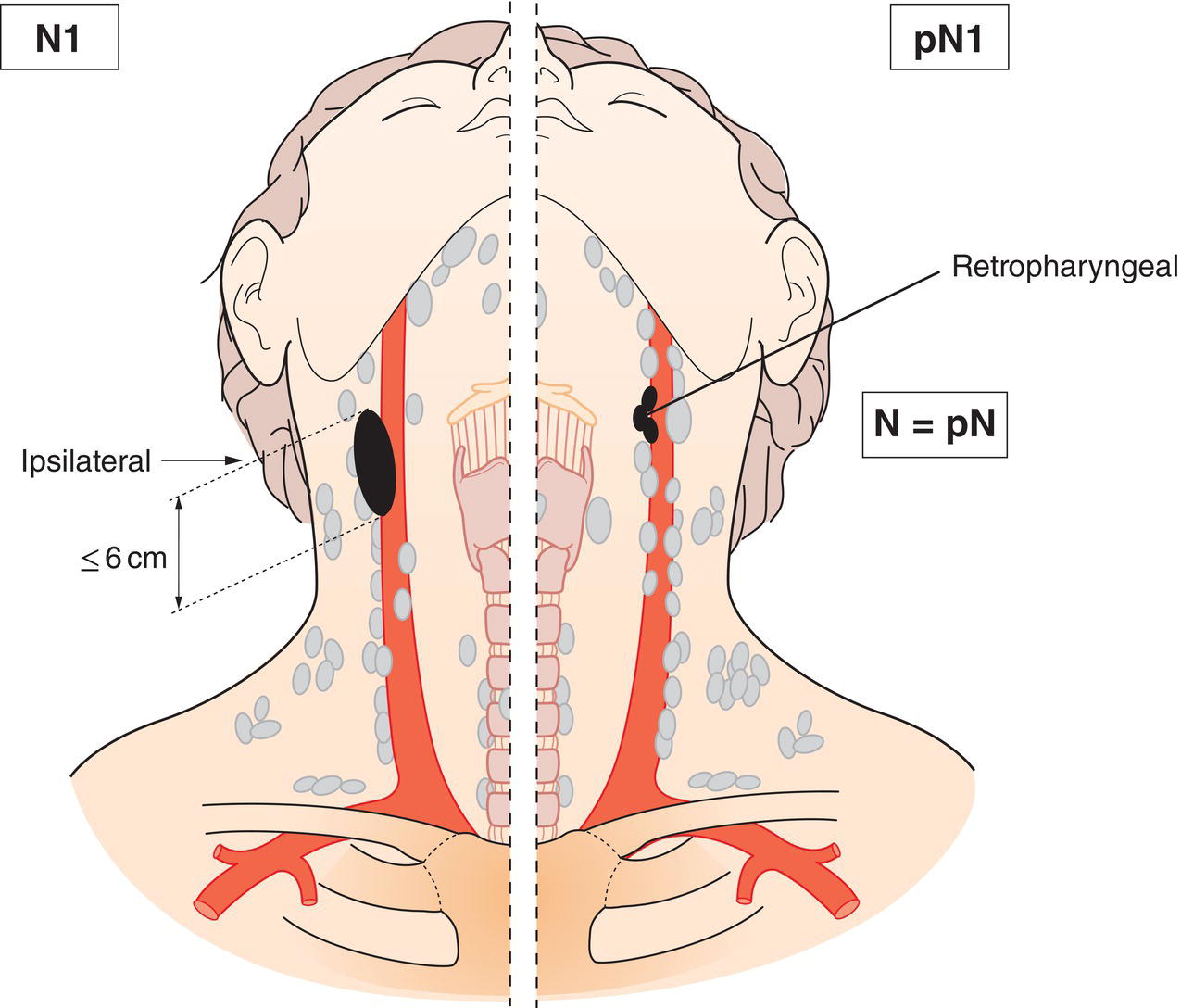
N1 Unilateral metastasis, in cervical lymph node(s), and/or unilateral or bilateral metastasis in retropharyngeal lymph nodes, 6 cm or less in greatest dimension, above the caudal border of cricoid cartilage (Fig. 74)
N2 Bilateral metastasis in cervical lymph node(s), 6 cm or less in greatest dimension, above the caudal border of cricoid cartilage (Fig. 75, 76)
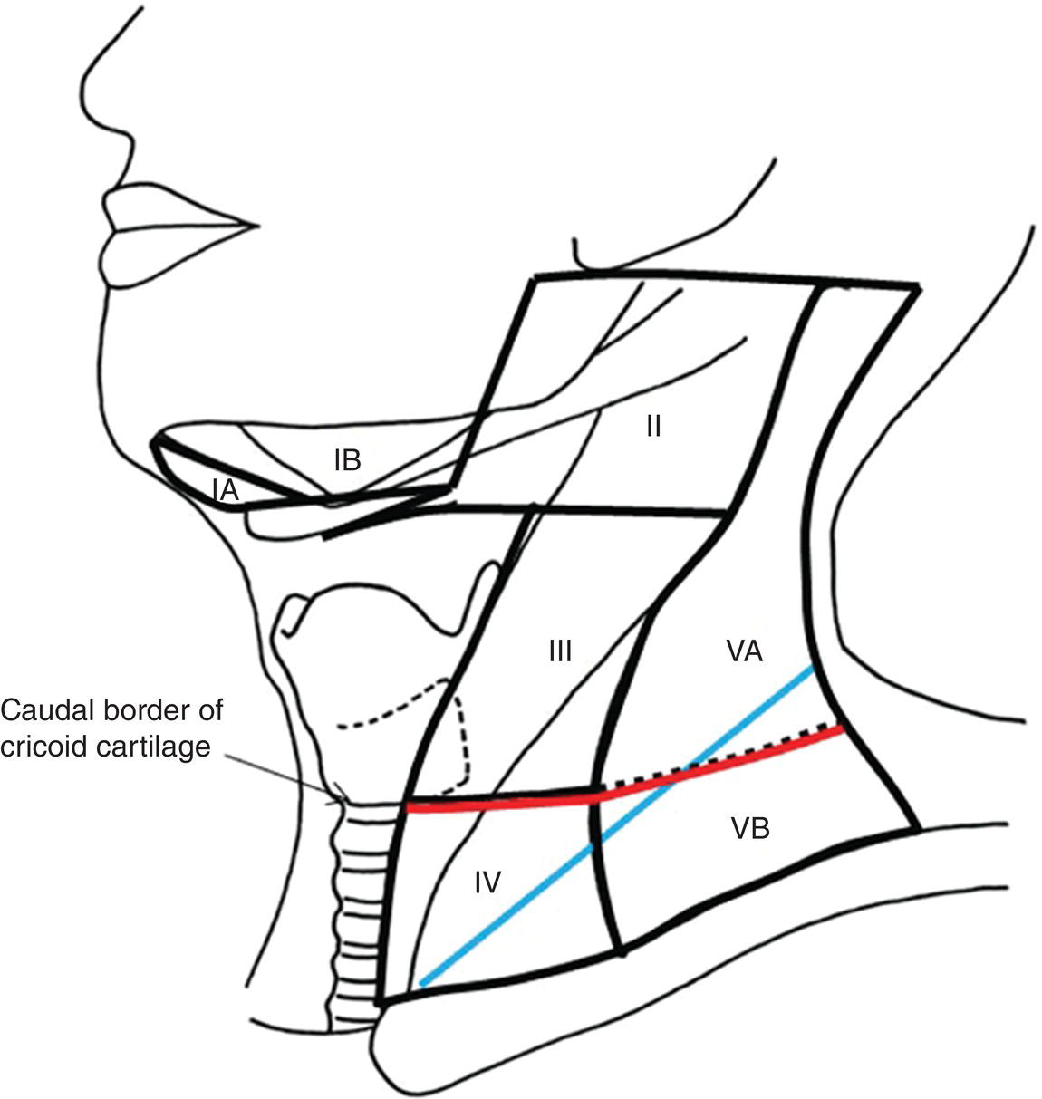
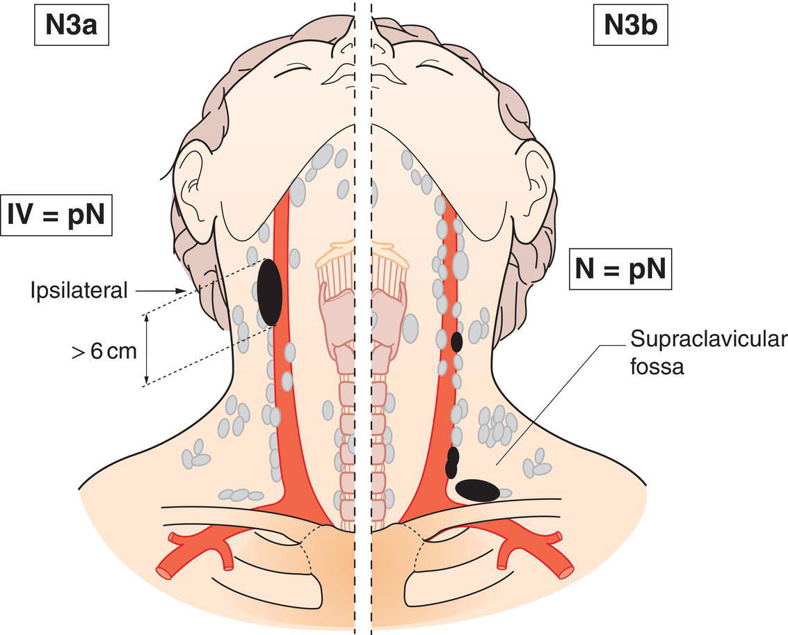
N3 Metastasis in cervical lymph node(s) greater than 6 cm in dimension and/or extension below the caudal border of cricoid cartilage (Fig. 76, 77)
Note


Midline nodes are considered ipsilateral nodes.

Schematic illustration of Unilateral metastasis, in cervical lymph nodes, and unilateral or bilateral
metastasis in retropharyngeal lymph nodes, 6 cm or less in greatest dimension,
above the caudal border of cricoid cartilage.

Fig. 74

Schematic illustration of Bilateral metastasis in cervical lymph nodes, 6 cm or less in greatest dimension,
above the caudal border of cricoid cartilage.

Fig. 75

Schematic illustration of Differences in defining criteria between the previous 7th edition and the current 8th edition: replacing the supraclavicular fossa with the lower neck as N3 criteria.

Fig. 76 Differences in defining criteria between the previous 7th edition and the current 8th edition: replacing the supraclavicular fossa (blue) with the lower neck (i.e., below the caudal border of cricoid cartilage; red) as N3 criteria.


Source: Modified from Pan JJ, Ng WT, Zong JF, et al. (2016) Proposal for the 8th edition of the AJCC/UICC staging system for nasopharyngeal cancer in the era of intensity‐modulated radiotherapy. Cancer 122(4):546–558.

Schematic illustration of Metastasis in cervical lymph nodes greater than 6 cm in dimension and extension below the caudal border of cricoid cartilage.

Fig. 77


pTN Pathological Classification


The pT and pN categories correspond to the T and N categories.


Summary

Tags:
Dec 15, 2022 | Posted by in ONCOLOGY | Comments Off on (ICD‐O C01, C05.1, 2, C09, C10.0, 2, 3, C11–13)

Full access? Get Clinical Tree
Get Clinical Tree app for offline access