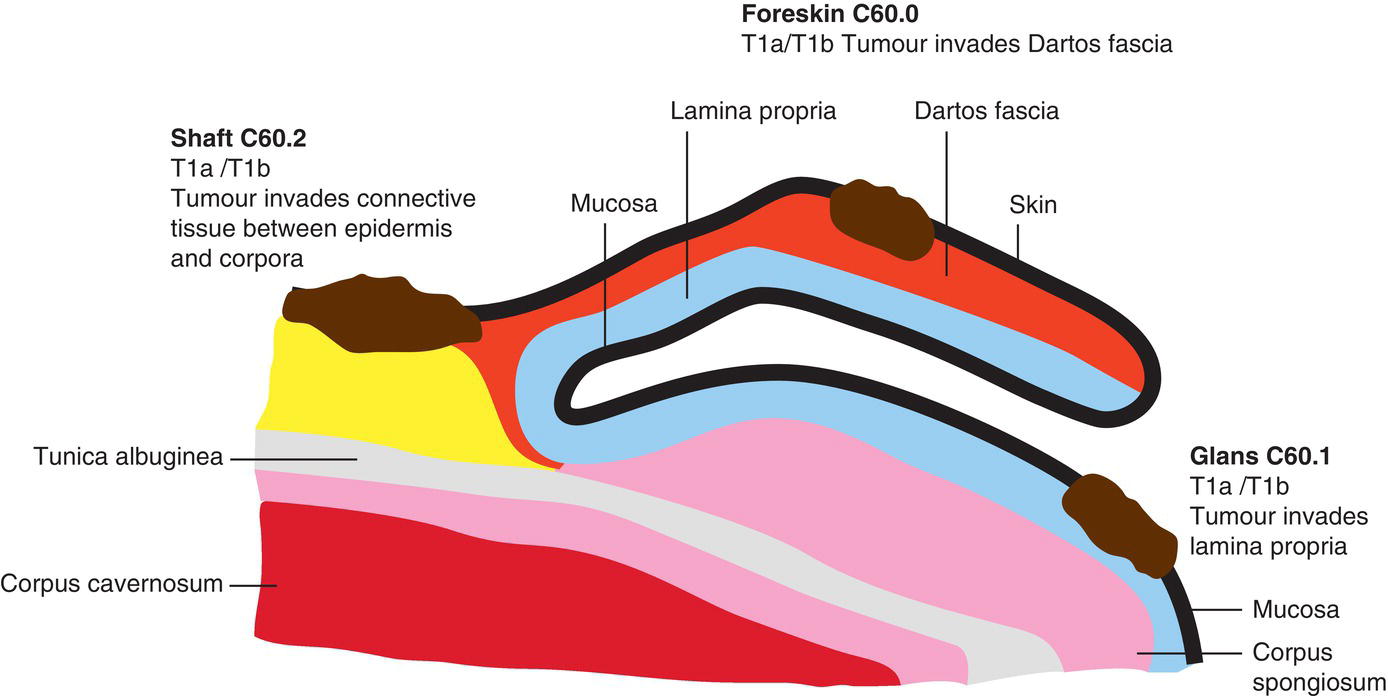
The classification applies only to carcinomas. There should be histological confirmation of the disease. The regional lymph nodes are the superficial and deep inguinal and the pelvic nodes. Notes 1 Including verrucous carcinoma. 2Glans: Tumour invades lamina propria.Foreskin: Tumour invades dermis, lamina propria or dartos fascia.Shaft: Tumour invades connective tissue between epidermis and corpora and regardless of location. The pT categories correspond to the T categories. The pN categories are based upon biopsy, or surgical excision. Note
PENIS (ICD‐O‐3 C60)
Rules for Classification
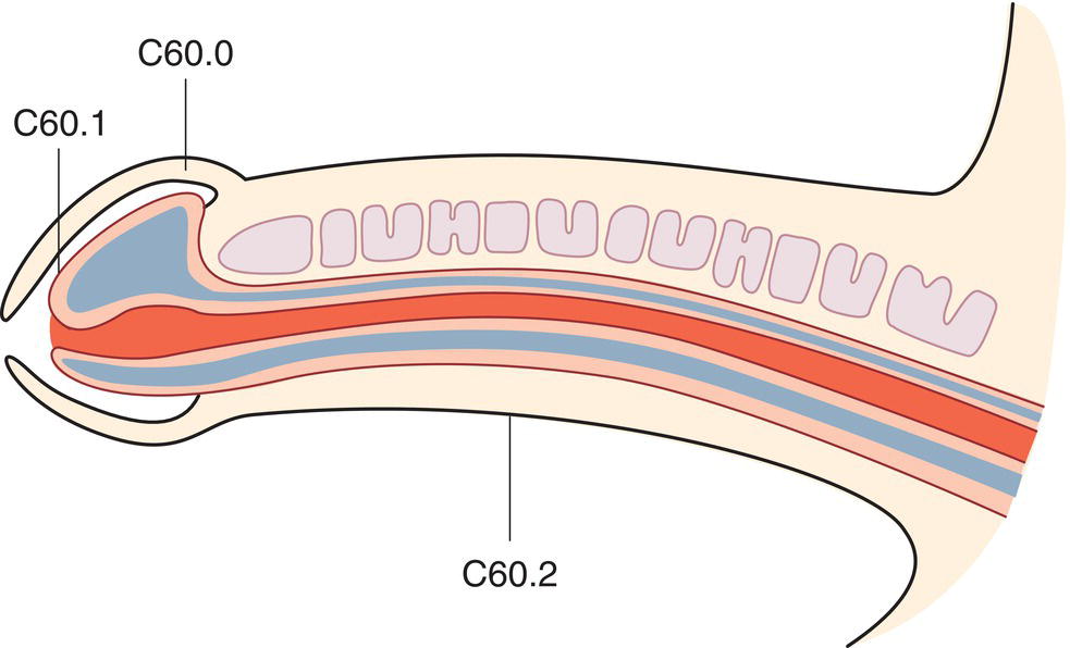
Anatomical Subsites (Fig. 463)
Regional Lymph Nodes
TNM Clinical Classification
T – Primary Tumour
TX
Primary tumour cannot be assessed
T0
No evidence of primary tumour
Tis
Carcinoma in situ (penile intraepithelial neoplasia – PeIN)
Ta
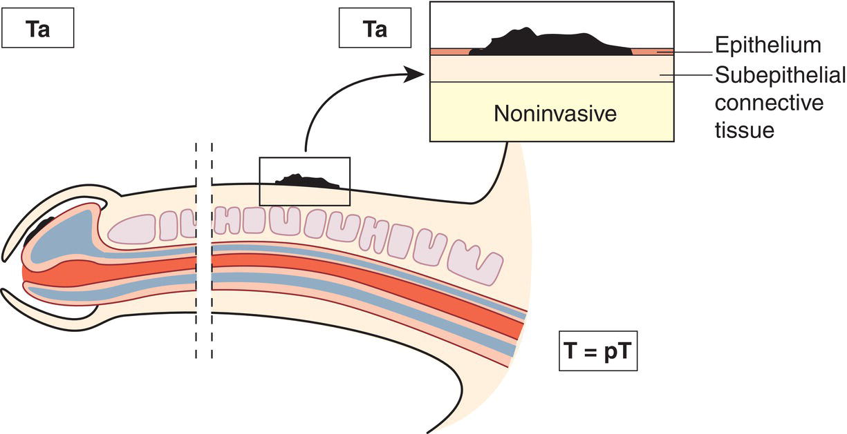
Noninvasive localized squamous cell carcinoma1 (Fig. 464)
T1
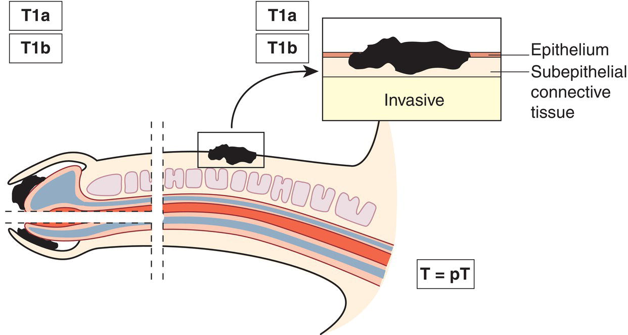
Tumour invades subepithelial connective tissue2 (Figs. 465, 466)
T1a
Tumour invades subepithelial connective tissue without lymphovascular invasion or perineural invasion and is not poorly differentiated
T1b
Tumour invades subepithelial connective tissue with lymphovascular invasion or perineural invasion or is poorly differentiated
T2
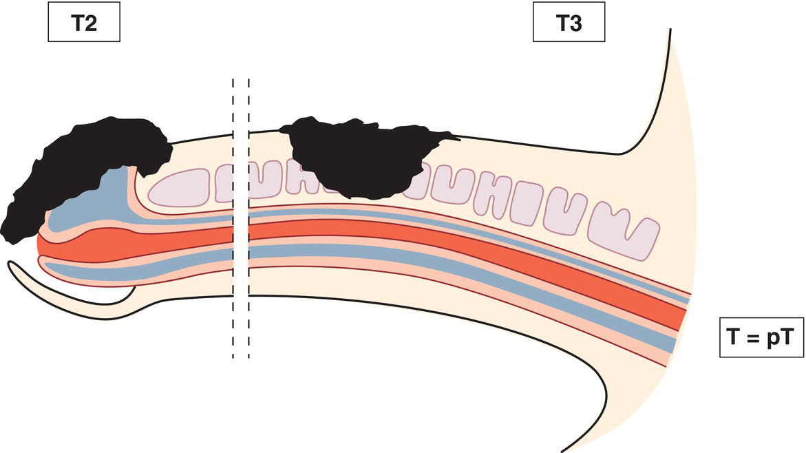
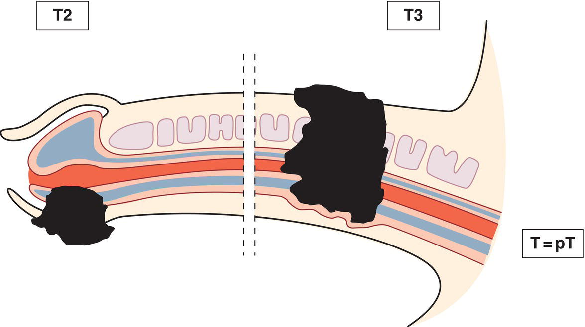
Tumour invades corpus spongiosum with or without invasion of the urethra (Figs. 467, 468)
T3
Tumour invades corpus cavernosum with or without invasion of the urethra (Figs. 467, 468)
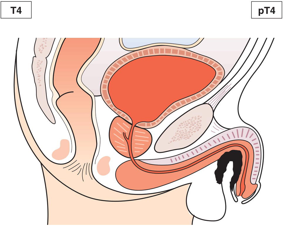
T4
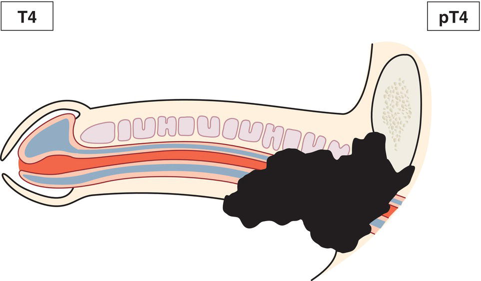
Tumour invades other adjacent structures (Figs. 469, 470)
N – Regional Lymph Nodes
NX
Regional lymph nodes cannot be assessed
N0
No palpable or visibly enlarged inguinal lymph nodes
N1
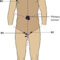
Palpable mobile unilateral inguinal lymph node (Fig. 471)
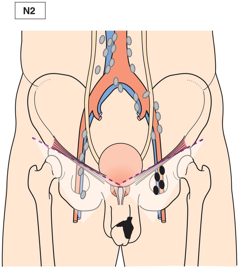
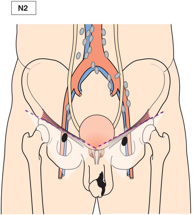
N2
Palpable mobile multiple (Fig. 472) or bilateral inguinal lymph nodes (Fig. 473)
N3
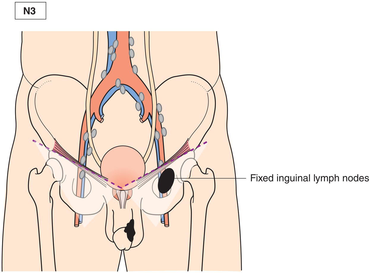
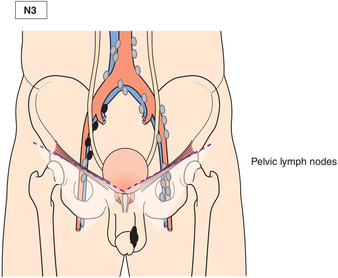
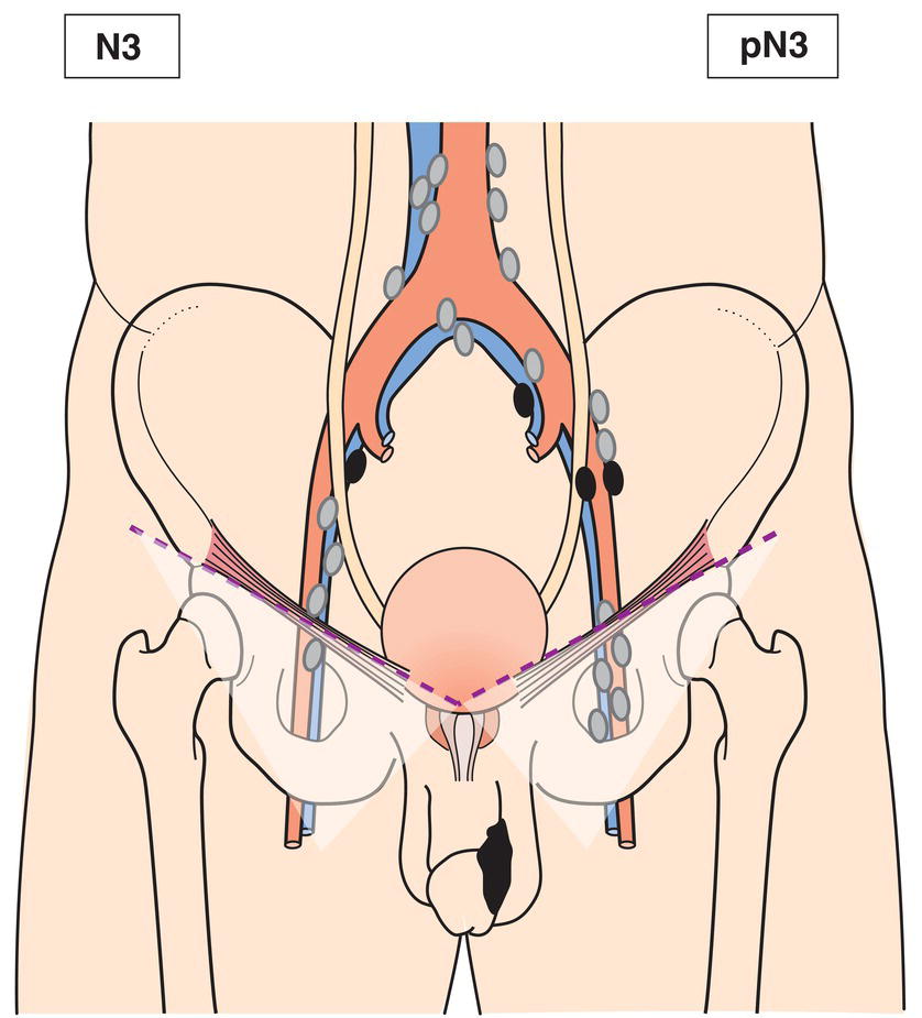
Fixed inguinal nodal mass (Fig. 474) or pelvic lymphadenopathy unilateral (Fig. 475) or bilateral (Fig. 476) 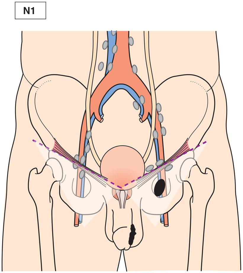
M – Distant Metastasis
M0
No distant metastasis
M1
Distant metastasis
pTNM Pathological Classification
pNX
Regional lymph nodes cannot be assessed
pN0
No regional lymph node metastasis
pN1
Metastasis in one or two inguinal lymph nodes
pN2
Metastasis in more than two unilateral or bilateral inguinal lymph nodes
pN3
Metastasis in pelvic lymph node(s), unilateral or bilateral or extranodal extension of regional lymph node metastasis
pM1
Distant metastasis microscopically confirmed
pM0 and pMX are not valid categories.
Summary
Stay updated, free articles. Join our Telegram channel

Full access? Get Clinical Tree


